Mădălina-Giorgiana Murariu*![]() | Florica-Ramona Dorobanțu
| Florica-Ramona Dorobanțu![]() | Daniela Tărniceriu
| Daniela Tărniceriu![]()
© 2024 The authors. This article is published by IIETA and is licensed under the CC BY 4.0 license (http://creativecommons.org/licenses/by/4.0/).
OPEN ACCESS
The rising occurrence of epilepsy along with the intricate nature of focal epilepsy and its potential to progress to generalized epilepsy requires the advancement of intelligent systems capable of delivering precise diagnoses. This paper developed a novel approach for the classification of patients with focal and generalized epilepsy based on the analysis of EEG signals using the Ensemble Empirical Mode Decomposition (EEMD) and Complete Ensemble Empirical Mode Decomposition with Adaptive Noise (CEEMDAN) techniques. These adaptive methods are applied to a comprehensive database consisting of EEG signals captured during sleep from patients with focal and generalized epilepsy. Feature extraction is performed on the resulting Intrinsic Mode Functions (IMFs) obtained from EEMD and CEEMDAN methods, four statistical features being computed for each extracted IMF. Finally, the K-nearest neighbors (KNN) and Naïve Bayes (NB) classifiers are employed to accurately classify the EEG signals into focal and generalized categories. The combination of CEEMDAN-based approach and KNN classifier achieved the highest classification rate of 94.54%, exceeding the EEMD-based approach and KNN classifier, which attained a maximum classification rate of 93.32%. By means of the proposed methods, we aim to contribute to the development of effective and efficient diagnostic systems for epilepsy.
adaptive methods, classifiers, diagnostic systems, electroencephalography signals, epilepsy, focal, generalized
Epilepsy is a predominant chronic neurological disease represented by seizures affecting approximately 1.5% of the global population. Seizures are episodic and unpredictable events resulting from abnormal electrical activity of the brain. They can be present in various forms, such as convulsions, loss of consciousness, or abnormal movements [1].
Seizures are represented through distinctive waveforms, known as epileptiform discharges, exhibiting pronounced spikes and sharp wave complexes [2]. A spike refers to a brief and transient signal observed in the EEG, lasting from 20-70 ms, while sharp-waves have durations of 70-200 ms [3].
The electroencephalogram (EEG) is a technique employed to capture the electrical activity of the brain [4]. By monitoring and analyzing complex and non-stationary EEG signals, epilepsy seizures can be detected and identified, contributing to a better understanding of the brain condition and its impact on the quality of life of the patients [5, 6].
The detection of epileptic seizure types consists of the analysis of the collected EEG signals. Epilepsy can take two forms: focal and generalized.
In focal epilepsy, the seizures are manifested and can only be observed in a specific area of the brain. The EEG signals are recorded from the brain zone where the epileptic activity originates, allowing us to locate the specific area of seizure onset. In generalized epilepsy, the seizures involve the entire brain and the generalized EEG signals are detectable across all EEG channels [7].
These signals often exhibit distinct patterns and frequency characteristics in various brain regions, providing valuable information for the accurate diagnosis and classification of epilepsy cases.
Experienced neurologists usually analyze these EEG signals through visual inspection, considering various waveform characteristics such as amplitude, frequency, reactivity to stimuli, and spatial-temporal abnormalities [8]. Visual observation for seizure detection has certain limitations: analysis is laborious, time-consuming, expensive, and can be prone to fatigue-induced errors. Therefore, it is a need for a reliable computerized algorithm to automatically detect the type of epilepsy [5].
Over the years, different techniques based on Fourier transform and parametric methods have been used for the automatic analysis of epileptic EEG signals [9, 10]. These approaches are built on previous findings indicating that epileptic seizures lead to alterations in specific frequency bands, including the $\delta(0.5-4 \mathrm{~Hz}),\ \theta(4-8 \mathrm{~Hz}),\ \alpha(8-12 \mathrm{~Hz}),\ \beta(12-30 \mathrm{~Hz})$ and $\gamma(30-60 \mathrm{~Hz})$ bands [11, 12].
In the field of EEG epilepsy recognition, researchers have developed numerous algorithms. Previous studies in the literature have used several methods such as Empirical Mode Decomposition (EMD), proposed by Huang et al. [13], which is an adaptive signal processing technique that decomposes a signal into a finite number of intrinsic mode functions (IMFs). Despite its data adaptivity, the EMD method encounters the challenge of mode mixing [8]. Further, extensions of EMD method have been proposed to address multivariate data [5, 14], and mitigate the issue of mode mixing. Mode mixing occurs when signals with significantly different amplitudes coexist within the same intrinsic mode function, or when a signal of similar scale is divided into different IMF components [14]. Recent advancements in signal decomposition have significantly contributed to the use of Empirical Wavelet Transform (EWT) [15], Variational Mode Decomposition (VMD), EEMD [14], Complete Ensemble Empirical Mode Decomposition (CEEMD) [16], and Complete Ensemble EMD with Adaptive Noise (CEEMDAN) techniques [8].
In study [14], the EEMD method is developed in order to avoid the EMD mode mixing. The proposed approach transforms the EMD method into a really dyadic filter bank applicable to various types of data.
In study [17], both EEMD and CEEMDAN methods are employed. The reconstructed signal is used to approximate the skewness, crest factor, and root mean square value parameters to analyze their characteristics and behavior. Results show that CEEMDAN technique produces higher statistical values for the reconstructed signal compared to EMD and EEMD ones.
In study [5], CEEMDAN is applied to a database with epileptic EEG recordings to detect the type of seizure. The mode functions are extracted from the analysis and are subsequently modeled using parameters from the normal inverse Gaussian (NIG) probability density function. The experimental findings demonstrate that the proposed approach shows promising performances for all clinically significant cases.
In this study, two novel and effective approaches are proposed for the classification of focal and generalized epilepsy. These methods involve two adaptive decomposition techniques based on EEMD and CEEMDAN, along with a combination of four features: median, kurtosis, skewness, and fluctuation index. By extracting relevant features from the decomposed signals, a robust and accurate classification algorithm is developed. These approaches have the potential to overcome the limitations of existing methods and to improve the efficiency and reliability of epilepsy diagnosis [18]. The current research uses a property database consisting of interictal focal and generalized EEG epileptic signals recorded during the sleep state. To classify EEG signals, the K- nearest neighbors (KNN) and Naive Bayes (NB) classifiers are employed.
The paper is structured as follows: Section II provides an overview of the database employed in the study. Section III outlines the methodology used, including the feature extraction process and the classification stage. Section IV describes the experimental results. Section V provides the conclusion of the paper.
The current study is conducted using our own database obtained in the EEG Epilepsy and Monitoring Center in Cluj-Napoca, Romania [18]. The database comprises interictal epileptiform EEG recordings obtained from 16 patients ranging in age from 7 to 66 years, diagnosed with focal and generalized epilepsy. The non-invasive acquisition of EEG signals is accomplished by employing the Nicolet Clinical EEG Natus System, featuring 21 channels. The positions of the electrodes adhered to the guidelines of the international 10-20 system.
The study received approval of the Ethics Committee, and all participants provided informed consent, allowing access to anonymized raw data for analysis purposes. Specifically, this research used a subset of 16 EEG recordings achieved in sleep state. The diagnosis of focal and generalized epilepsy was established by a qualified neurologist for the patients involved in the study. To simplify the analysis process, the data was segmented into 40 segments, each of them consisting of 6 seconds of recorded data. The sampling frequency was 1000 Hz.
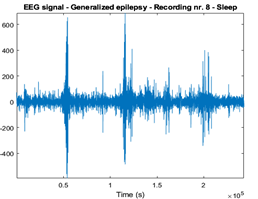
Figure 1 illustrates an EEG recording from a person with focal epilepsy, while Figure 2 shows an EEG recording from a patient diagnosed with generalized epilepsy.
Figure 1. Focal EEG signals
Figure 2. Generalized EEG signals
3.1 Adaptive decomposition approaches
Ensemble empirical mode decomposition is an extension of the empirical mode decomposition method, designed to overcome the issue of mode mixing [13].
The principle of EEMD method can be briefly defined as follows:
In the process of EEMD, during each trial, white noise is introduced into the original signal. The act of employing EMD on the input signal affected by noise is known as a trial. The trials are executed multiple times to obtain the mode functions of EEMD [5].
The relation between the EEG signal $X\left( t \right)$ and its corrupted versions is given below:
$X^i(t)=X(t)+\varepsilon_0 \omega^i(t)$, for $i=1,2, \ldots, K$ (1)
where, ${{X}^{i}}\left( t \right)$ represent the signal corrupted by white noise, the white noise is denoted by ${{\omega }^{i}}\left( t \right)$, ${{\varepsilon }_{0}}$ is the standard deviation of the white noise, and K represents the trials [16].
In this method, the white noise has zero mean and unit variance, consequently, ${{\varepsilon }_{0}}=1$.
EMD is applied to the noisy signal ${{X}^{i}}\left( t \right)$ and decomposes it into N IMF components noted $IMF_{j}^{i}\left( t \right)$ and $r\left( t \right)$ residuals, ($j=$1, 2, …, $N$).
$X^i(t)=\sum_{j=1}^N I M F_j^i(t)+r_i(t)$ (2)
where, $IMF_{j}^{i}\left( t \right)$ represents the j-th IMF obtained through the decomposition process after addition of the Gaussian white noise for the i-th trial.
The addition of noise in the EEMD method helps to evenly distribute the signal’s components across different scales or frequencies [5].
The EEMD modes noted $\overline{IM{{F}_{j}}\left( t \right)}$ are obtained by averaging the IMFs obtained throughout the ensemble number of trials - $IMF_{j}^{i}\left( t \right)$ [16].
$\overline{I M F_j(t)}=\frac{1}{K} \sum_{i=1}^K I M F_j^i(t),\ r(t)=\frac{1}{K} \sum_{i=1}^K r_i(t)$ (3)
The quantity of intrinsic mode function generated through the EEMD method depends on the number of trials conducted during the procedure. By employing the ensemble number of trials and averaging the IMFs, EEMD enhances the separation of different oscillations and improves the accuracy of mode decomposition. EEMD is characterized by a high computational complexity [17].
While the EEMD method effectively addresses the issue of mode mixing, it still has certain limitations. These include the presence of residual noise in the reconstructed signal and varying numbers of modes in different trials [18].
The enhanced complete ensemble empirical mode decomposition with adaptive noise method represents a significant advancement over EMD and EEMD techniques by addressing the above-mentioned issues [19].
The CEEMDAN method involves several key steps, as follows:
$\widetilde{I M F_1(t)}=\frac{1}{K} \sum_{i=1}^K I M F_1^i(t)=\overline{I M F_1(t)}$ (4)
$r_1(t)=X(t)-\widetilde{I M F}_1(t)$ (5)
The operator $EM{{D}_{j}}\left( \cdot \right)$produces the j-th IMF attained by EMD method.
The signal ${{r}_{1}}\left( t \right)+{{\varepsilon }_{1}}EM{{D}_{1}}\left( {{\omega }^{i}}\left( t \right) \right)$, i=1, 2, …, K, undergoes additional decomposition using EMD to calculate the second IMF mode noted $\widetilde{IM{{F}_{2}}\left( t \right)}$:
$\widetilde{I M F_2(t)}=\frac{1}{K} \sum_{i=1}^K E M D_1\left(r_1(t)+\varepsilon_1 E M D_1\left(\omega^i(t)\right)\right)$ (6)
and the related residual ${{r}_{2}}\left( t \right)$:
$r_2(t)=r_1(t)-\widetilde{I M F_2(t)}$ (7)
$r_j(t)=r_{(j-1)}(t)-\widetilde{I M F_j(t)}$ (8)
$\widetilde{I M F_{(j+1)}(t)}=\frac{1}{K} \sum_{i=1}^K E M D_1\left(r_j(t)+\varepsilon_j E M D_j\left(\omega^i(t)\right)\right)$ (9)
where, ${{\varepsilon }_{j}}$ is the standard deviation of white noise in the j-th iteration, $\widetilde{IM{{F}_{\left( j+1 \right)}}\left( t \right)}$ represents the $\left( j+1 \right)$-th IMF obtained by CEEMDAN method.
When steps 4 and 5 are iterated, at a certain stage, the residues form a strictly monotonic function. Consequently, it becomes unfeasible to further extract IMFs. The algorithm stops when the following stop condition is met:
$\sum_{t=0}^T \frac{\left|r_{j-1}(t)-r_j(t)\right|^2}{r_{j-1}^2(t)} \leq S D_K$ (10)
where, T represents the length of the signal X(t). After performing the j-th decomposition, the resulting signal is referred to as ${{r}_{j}}\left( t \right)$, and the standard deviation -$S{{D}_{K}}$ is set to 0.2 [20].
If ${{r}_{j}}\left( t \right)$ represents the ultimate residue, and N is the total number of modes beyond which no additional mode extraction is achievable, the original input signal $X\left( t \right)$ can be reconstructed using the following relationship from all the CEEMDAN mode functions.
$X(t)=\sum_{j=1}^N\left(\widetilde{I M F_j(t)}+r_j(t)\right)$ (11)
In our study, we have used the first five EEMD and CEEMDAN IMFs from each EEG signal for feature extraction. Then, four features were computed from each extracted IMFs.
By incorporating the adaptive noise term, CEEMDAN improves the removal of noise contamination during the mode decomposition process, resulting in cleaner and more accurate IMFs [8].
Compared to EMD and EEMD, CEEMDAN has notable advantages. It ensures complete reconstruction without residual noise interference and requires a reduced number of trials, leading to enhanced computational efficiency [20].
3.2 Features extraction
For each IMF obtained through EEMD and CEEMDAN methods, the parameters: median, skewness, kurtosis, and fluctuation index were calculated.
The median value of the extracted intrinsic mode functions represents a significant feature that can be used to identify the central point in the distribution of signal amplitudes. This measure can be a valuable tool in detecting and characterizing the abnormal patterns in EEG epilepsy [21].
The kurtosis is a reliable indicator in terms of tail extremity in a distribution.
The kurtosis is given as follows:
$Kurtosis=\frac{E\{(X-\mu)\}^4}{\sigma^4}$ (12)
In this context, $\mu $ represents the mean of the variable, $\sigma $ is the standard deviation, and $E\left[ \cdot \right]$ represents the expected value [22].
The skewness evaluates the symmetry of the data distribution relative to the sample mean. It helps in establishing whether the distribution is symmetrical or skewed towards one side [23]. The skewness is computed as:
$Skewness=\frac{E\{(X-\mu)\}^3}{\sigma^3}$ (13)
The fluctuation index provides a numerical value that reflects the degree of variation in a signal over a given time period, allowing the quantification of signal intensity changes [24]. The fluctuation index is defined as follows:
$Fluctuation\text{_}index=\frac{1}{l-1} \sum_{j=1}^{l-1}|I M F(j+1)-I M F(j)|$ (14)
where, IMF denotes the extracted IMFs with a length of $l$.
3.3 Classifiers
This paper uses the K-nearest neighbors and Naïve Bayes classifiers to classify the data. To evaluate the performance of these classifiers, a ten-fold cross-validation procedure is employed.
Previous studies [18, 25] have conducted comprehensive evaluations of various classifiers and concluded that the KNN and NB classifiers are particularly suitable for distinguishing between the two different classes of EEG epileptic data.
The KNN classifier identifies the majority class among its closest training examples in the feature space. On the other hand, the NB classifier applies the theorem of Bayes to calculate the probability of each class given the input features, making assumptions of feature independence.
The classifiers have been employed to classify various types of EEG epileptic signals, including normal and abnormal [26], focal and non-focal, focal and generalized [18], as well as different stages of seizures: ictal, interictal, or postictal [27].
The performances of the classifiers used in this paper were estimated employing the following metrics: sensitivity, specificity, classification rate, and F1-Score, with detailed computations provided below.
The sensitivity (recall) of the proposed methods indicates their ability to accurately classify focal EEG signals, while the specificity demonstrates their capability to correctly classify generalized EEG signals.
$Sensitivity=\frac{T P}{TP+F N} \times 100$ (15)
$Specificity=\frac{T N}{T N+F P} \times 100$ (16)
Classification rate (Class_rate) represents the percentage of accurately classified EEG signals.
$Class\text{_}rate=\frac{T P+T N}{T N+F P+F N+F P} \times 100$ (17)
The F1-Score [28] assesses the accuracy of a model by considering both precision and recall. The harmonic mean gives equal weight to precision and recall, making the F1-Score a balanced measure that considers both true positives and true negative instances in classification tasks.
$F 1-$ Score $=2 \times \frac{Precision\times Recall}{Precision+Recall}$ (18)
$Precision=\frac{TP}{TP+FP}\times 100$ (19)
True Positives (TP) represent the number of instances (in this study, focal EEG signals) that are correctly categorized as positive (focal). False Positives (FP) are the number of instances (focal EEG signals) that are incorrectly categorized as positive (generalized EEG signals). True Negatives (TN) comprise the number of instances (in this study, generalized EEG signals) that are correctly categorized as negative (generalized). False Negatives (FN) are the number of instances (generalized EEG signals) that are incorrectly categorized as negative (focal EEG signals).
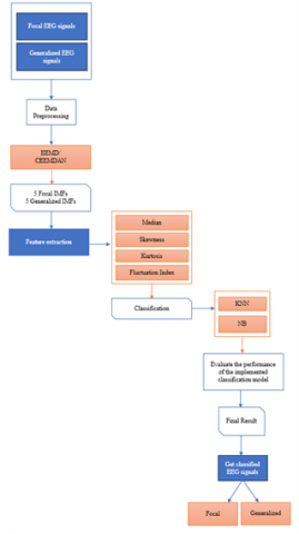
A flowchart with the proposed methods is presented in Figure 3.
Figure 3. A schematic outline of the proposed methods
Two different offline approaches based on adaptive mode decomposition techniques for classifying epileptic EEG signals were proposed. To assess their individual performance, the EEG signals were decomposed into intrinsic mode functions using two different approaches: EEMD and CEEMDAN.
After several attempts to set the ensemble size, testing values both higher and lower than 500, the best results for data classification were obtained with an ensemble of 500, leading to a good balance between accuracy and computational efficiency. In both proposed methods, the ensemble size was set to 500 trials and the standard deviation was set to 0.2.
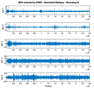
A number of five IMFs were extracted. Each IMF includes 40 segments and 6000 samples. The order of IMFs is from highest to lowest frequency.
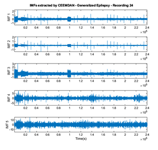
Figure 4 and Figure 5 represent the extracted IMFs with both methods: EEMD and CEEMDAN, and they are given to illustrate the differences between their representation. The variations observed in the IMFs obtained from EEMD and CEEMDAN method illustrate the distinct decomposition characteristics and signal features captured by each method.
Further, the above-mentioned feature extractions were computed for each of the five extracted IMF components, separately for each proposed method. Based on the data collected from these features, the EEG signals were classified.
The results get with the proposed approaches for categorizing focal and generalized EEG epileptic signals are presented in the following tables.
Standard formulas were used to calculate the minimum, maximum, mean, and median values. These metrics provide a descriptive overview of the performance of the proposed methods.
Figure 4. IMFs extracted through the EEMD method from a generalized EEG recording
Figure 5. IMFs extracted through the CEEMDAN method from a generalized EEG recording
Table 1. The results with EEMD method using KNN classifier
|
|
EEMD |
||
|
|
Classification Rate (%) |
Sensitivity (%) |
Specificity (%) |
|
Min |
63.78 |
65.10 |
57.87 |
|
Max |
93.32 |
92.89 |
96.31 |
|
Mean |
79.57 |
79.27 |
79.88 |
|
Median |
80.01 |
79.06 |
80.16 |
Table 2. The results with EEMD method using NB classifier
|
|
EEMD |
||
|
|
Classification Rate (%) |
Sensitivity (%) |
Specificity (%) |
|
Min |
57.80 |
60 |
55.63 |
|
Max |
90.60 |
90 |
94.38 |
|
Mean |
75.99 |
75.73 |
76.24 |
|
Median |
75.90 |
75.63 |
74.38 |
Table 3. The results with CEEMDAN method using KNN classifier
|
|
CEEMDAN |
||
|
|
Classification Rate (%) |
Sensitivity (%) |
Specificity (%) |
|
Min |
66.83 |
64.28 |
59.74 |
|
Max |
94.54 |
92.73 |
96.94 |
|
Mean |
82.14 |
80.78 |
83.75 |
|
Median |
85.15 |
81.96 |
87.19 |
Table 4. The results with CEEMDAN method using NB classifier
|
|
CEEMDAN |
||
|
|
Classification Rate (%) |
Sensitivity (%) |
Specificity (%) |
|
Min |
61.30 |
59.38 |
60 |
|
Max |
91.40 |
92 |
93.75 |
|
Mean |
79.48 |
77.56 |
81.46 |
|
Median |
80.60 |
77.5 |
84.38 |
Using the KNN classifier, the EEMD approach achieved a classification rate ranging from 63.78% to 93.32%, as shown in Table 1. Similarly, when using the NB classifier, the classification rate varied between 57.80% to 90.60%, as presented in Table 2.
The KNN classifier obtained a maximum sensitivity of 92.89% in recognizing focal EEG data, while the NB classifier demonstrated a maximum sensitivity of 90% in recognizing focal EEG data.
For the identification of generalized EEG data, the KNN classifier demonstrated a maximum specificity of 96.31%, while the NB classifier obtained a maximum specificity of 94.38%.
The results achieved with the CEEMDAN approach are presented in Table 3 and Table 4. The KNN classifier achieved a classification rate between 66.83% and 94.54%, while the NB classifier achieved a classification rate between 61.30% and 91.40%.
The analysis of sensitivity in the classification results showed that the signals were correctly identified as focal, with a maximum sensitivity of 92.73% using the KNN classifier, and 92% using the NB classifier.
The attained maximum specificity is 96.94% using the KNN classifier and 93.75% using the NB classifier. This indicates that the classification algorithm is capable of accurately distinguishing generalized EEG signals.
The performances of the classifiers are also assessed using F1-Score and are presented in Table 5.
Table 5. Performances of the classifiers based on F1-Score
|
F1-Score |
||||
|
|
EEMD |
CEEMDAN |
||
|
|
KNN |
NB |
KNN |
NB |
|
Min |
0.40 |
0.40 |
0.30 |
0.30 |
|
Max |
0.90 |
0.90 |
0.90 |
0.90 |
|
Mean |
0.60 |
0.59 |
0.63 |
0.60 |
|
Median |
0.70 |
0.60 |
0.70 |
0.60 |
CEEMDAN combined with the used four features, KNN and NB classifiers, works as a powerful tool for achieving high accuracy and robustness in the classification of EEG epileptic signals.
Table 6 presents a comparison between these approaches and other methods. Only methods that are evaluated using the Cluj-Napoca Database are involved. The results are reported according to the median values obtained for the data collected during sleep.
Table 6. Performances obtained with the proposed methods and other works using the same database
|
Work |
Method |
Classifier |
Results |
|
[18] |
EMD with power spectral density feature extraction |
KNN NB |
Classification Rate: 95.79% KNN/ 95.83%NB Sensitivity: 95.29% KNN/ 95.14% NB Specificity: 96.29% KNN/ 99.51% NB F1-Score: 0.70 KNN / 0.70 NB |
|
[29] |
EMD with median, skewness, kurtosis, and fluctuation index feature extraction |
KNN |
Classification Rate: 74.21% Sensitivity: 73.77% Specificity: 77% F1-Score: 0.70 Classification Rate: 80.01% KNN/ 75.90% NB |
|
This paper |
EEMD with median, skewness, kurtosis, fluctuation, and index feature extraction |
KNN NB |
Sensitivity: 79.06% KNN/ 75.60% NB Specificity: 80.16% KNN/ 74.38% NB F1-Score: 0.70 KNN/ 0.60 NB |
|
CEEMDAN with median, skewness, kurtosis, and fluctuation index feature extraction |
KNN NB |
Classification Rate:85.15 % KNN/ 80.60% NB Sensitivity: 81.96% KNN/ 77.50% NB Specificity: 87.19% KNN/ 84.38% NB F1-Score: 0.70 KNN/ 0.60 NB |
The proposed algorithms are developed in an original way, being applied on our own database and the results confirm their applicability in epilepsy diagnosis domain.
The comparison between the attained results from the application of the proposed methods and the established diagnosis by the neurologist revealed accurate and consistent identification of focal and generalized epilepsy.
In all classification scenarios, the proposed methods have correctly estimated the diagnosis, aligning closely with the expert assessments provided by the neurologist. This agreement further validates the accuracy and effectiveness of the proposed methods in the classification of epilepsy cases.
This study successfully achieved its aim by implementing the proposed EEMD and CEEMDAN methods, which led to higher accuracy in classifying the data compared to EMD approach developed in study [19].
The application of the adaptive decomposition methods in the classification of focal and generalized EEG signals produced superior performances than other methods reported in the literature.
The implementation of the EEMD method combined with the four-feature extraction mentioned parameters has shown higher results in the identification of focal and generalized EEG signals compared to the algorithm based on EMD method.
The results obtained highlight the superiority of CEEMDAN approach over EMD and EEMD methods, in capturing the intricate characteristics of epileptic EEG signals. The application of the chosen four features provides valuable insights into the underlying patterns and abnormalities associated with focal and generalized epilepsy leading to more accurate and robust classification of EEG signals.
In the case of CEEMDAN method, the adaptive noise reduction mechanism helps mitigate the impact of noise on the decomposition process. By adaptively estimating and removing the noise components from the signal during decomposition, CEEMDAN approach demonstrated that it is able to extract more accurate and reliable intrinsic mode functions to be used in further analysis.
The classification outcomes for differentiating between focal and generalized EEG epileptic sleep signals using KNN and NB classifiers showcased diverse classification rates, sensitivities, and specificities across both EEMD and CEEMDAN methodologies. Notably, the CEEMDAN combined with the KNN classifier demonstrated superior classification rates in comparison to the CEEMDAN with NB approach, and the EEMD with KNN, and NB approaches.
The F1-Score analysis revealed a remarkable level of performance for both methods, indicating their effectiveness in classifying EEG epileptic signals.
The achieved results demonstrate the capacity of CEEMDAN approach in precisely distinguishing between different types of EEG epileptic signals. This mark an important starting point in the development of automated systems capable not only of identifying the type of epilepsy, but also of being utilized in designing an epilepsy prediction system.
The agreement between the results obtained from the proposed methods and the established diagnosis by neurologists further validates their accuracy in classifying epilepsy cases.
The future research directions aim to expand the number of classifiers, features, and the database involved in the study.
The authors would like to acknowledge the contributions of Prof. Dr. Eng. Anca-Mihaela Lazăr for the valuable support and guidance.
[1] Fiest, K.M., Sauro, K.M., Wiebe, S., Patten, S.B., Kwon, C.S., Dykeman, J., Pringsheim, T., Lorenzetti, D.L., Jetté, N. (2017). Prevalence and incidence of epilepsy: A systematic review and meta-analysis of international studies. Neurology, 88(3): 296-303. https://doi.org/10.1212/WNL.0000000000003509
[2] Slimen, I.B., Boubchir, L., Seddik, H. (2020). Epileptic seizure prediction based on EEG spikes detection of ictal-preictal states. Journal of Biomedical Research, 34(3): 162-169. https://doi.org/10.7555/JBR.34.20190097
[3] Dingle, A.A., Jones, R.D., Carroll, G.J., Fright, W.R. (1993). A multistage system to detect epileptiform activity in the EEG. IEEE Transactions on Biomedical Engineering, 40(12): 1260-1268. https://doi.org/10.1109/10.250582
[4] Chaddad, A., Wu, Y., Kateb, R., Bouridane, A. (2023). Electroencephalography signal processing: A comprehensive review and analysis of methods and techniques. Sensors, 23(14): 6434. https://doi.org/10.3390/s23146434
[5] Hassan, A.R., Subasi, A., Zhang, Y.C. (2020). Epilepsy seizure detection using complete ensemble empirical mode decomposition with adaptive noise. Knowledge-Based Systems, 191: 105333. https://doi.org/10.1016/j.knosys.2019.105333
[6] Mohan, R., Perumal, S. (2023). Classification and detection of cognitive disorders like depression and anxiety utilizing deep convolutional neural network (CNN) centered on EEG signal. Traitement Du Signal, 40(3): 971-979. https://doi.org/10.18280/ts.400313
[7] Tzallas, A.T., Tsipouras, M.G., Fotiadis, D.I. (2009). Epileptic seizure detection in EEGs using time–frequency analysis. IEEE Transactions on Information Technology in Biomedicine, 13(5): 703-710. https://doi.org/10.1109/TITB.2009.2017939
[8] Carvalho, V.R., Moraes, M.F., Braga, A.P., Mendes, E.M.A.M. (2020). Evaluating five different adaptive decomposition methods for EEG signal seizure detection and classification. Biomedical Signal Processing and Control, 62: 102073. https://doi.org/10.1016/j.bspc.2020.102073
[9] Baykara, M., Abdulrahman, A. (2021). Seizure detection based on adaptive feature extraction by applying extreme learning machines. Traitement du Signal, 38(2): 331-340. https://doi.org/10.18280/ts.380210
[10] Subasi, A., Ercelebi, E. (2005). Classification of EEG signals using neural network and logistic regression. Computer Methods and Programs in Biomedicine, 78(2): 87-99. https://doi.org/10.1016/j.cmpb.2004.10.009
[11] Bhattacharyya, A., Sharma, M., Pachori, R.B., Sircar, P., Acharya, U.R. (2018). A novel approach for automated detection of focal EEG signals using empirical wavelet transform. Neural Computing and Applications, 29: 47-57. https://doi.org/10.1007/s00521-016-2646-4
[12] Akbari, H., Sadiq, M.T. (2021). Detection of focal and non-focal EEG signals using non-linear features derived from empirical wavelet transform rhythms. Physical and Engineering Sciences in Medicine, 44: 157-171. https://doi.org/10.1007/s13246-020-00963-3
[13] Huang, N.E., Shen, Z., Long, S.R., Wu, M.C., Shih, H.H., Zheng, Q., Liu, H.H. (1998). The empirical mode decomposition and the Hilbert spectrum for nonlinear and non-stationary time series analysis. Proceedings of the Royal Society of London. Series A: Mathematical, Physical and Engineering Sciences, 454(1971): 903-998. https://doi.org/10.1098/rspa.1998.0193
[14] Wu, Z., Huang, N.E. (2009). Ensemble empirical mode decomposition: A noise-assisted data analysis method. Advances in Adaptive Data Analysis, 1(1): 1-41. https://doi.org/10.1142/S1793536909000047
[15] Liu, T., Luo, Z., Huang, J., Yan, S. (2018). A comparative study of four kinds of adaptive decomposition algorithms and their applications. Sensors, 18(7): 2120. https://doi.org/10.3390/s18072120
[16] Purba, H., Musu, J.T., Diria, S.A., Permono, W., Sadjati, O., Sopandi, I., Ruzi, F. (2018). Completed ensemble empirical mode decomposition: A robust signal processing tool to identify sequence strata. In IOP Conference Series: Earth and Environmental Science, Lampung, Coimbatore, Indonesia, p. 012033. https://doi.org/10.1088/1755-1315/132/1/012033
[17] Teja, K., Tiwari, R., Mohanty, S. (2020). Adaptive denoising of ECG using EMD, EEMD and CEEMDAN signal processing techniques. In Journal of Physics: Conference Series, India, p. 012077. https://doi.org/10.1088/1742-6596/1706/1/012077
[18] Murariu, M.G., Dorobanțu, F.R., Tărniceriu, D. (2023). A novel automated empirical mode decomposition (EMD) based method and spectral feature extraction for epilepsy EEG signals classification. Electronics, 12(9): 1958. https://doi.org/10.3390/electronics12091958
[19] Torres, M.E., Colominas, M.A., Schlotthauer, G., Flandrin, P. (2011). A complete ensemble empirical mode decomposition with adaptive noise. In 2011 IEEE international conference on acoustics, speech and signal processing (ICASSP), Prague, Czech Republic, pp. 4144-4147. https://doi.org/10.1109/ICASSP.2011.5947265
[20] Gu, D., Lin, A., Lin, G. (2023). Detection of attention deficit hyperactivity disorder in children using CEEMDAN-based cross frequency symbolic convergent cross mapping. Expert Systems with Applications, 226: 120105. https://doi.org/10.1016/j.eswa.2023.120105
[21] Schwilden, H. (1989). Use of the median EEG frequency and pharmacokinetics in determining depth of anaesthesia. Baillière's Clinical Anaesthesiology, 3(3): 603-621. https://doi.org/10.1016/S0950-3501(89)80021-2
[22] Alam, S.S., Bhuiyan, M.I.H. (2013). Detection of seizure and epilepsy using higher order statistics in the EMD domain. IEEE Journal of Biomedical and Health Informatics, 17(2): 312-318. https://doi.org/10.1109/JBHI.2012.2237409
[23] Sriraam, N., Raghu, S. (2017). Classification of focal and non focal epileptic seizures using multi-features and SVM classifier. Journal of Medical Systems, 41: 160. https://doi.org/10.1007/s10916-017-0800-x
[24] Li, S.F., Zhou, W.D., Yuan, Q., Geng, S.J., Cai, D.M. (2013). Feature extraction and recognition of ictal EEG using EMD and SVM. Computers in Biology and Medicine, 43(7): 807-816. https://doi.org/10.1016/j.compbiomed.2013.04.002
[25] Murariu, M.G., Tărniceriu, D. (2022). Discrimination of focal and non-focal epileptic EEG signals using different types of classifiers. In International Conference of the Doctoral School, Romania, 68: 61-79, https://doi.org/10.2478/bipie-2022-0011
[26] Escobar-Ipuz, F.A., Torres, A.M., García-Jiménez, M.A., Basar, C., Cascón, J., Mateo, J. (2023). Prediction of patients with idiopathic generalized epilepsy from healthy controls using machine learning from scalp EEG recordings. Brain Research, 1798: 148131. https://doi.org/10.1016/j.brainres.2022.148131
[27] Wang, Z.P., Na, J.Y., Zheng, B.Y. (2020). An improved kNN classifier for epilepsy diagnosis. IEEE Access, 8: 100022-100030. https://doi.org/10.1109/ACCESS.2020.2996946
[28] Chicco, D., Jurman, G. (2020). The advantages of the Matthews correlation coefficient (MCC) over F1 score and accuracy in binary classification evaluation. BMC Genomics, 21: 6. https://doi.org/10.1186/s12864-019-6413-7
[29] Murariu, M.G., Hrişcă-Eva, O.D., Tărniceriu, D. (2023). Classification of focal and generalized epilepsy using multi-features and empirical mode decomposition method. In 2023 International Symposium on Signals, Circuits and Systems (ISSCS), Iasi, Romania, pp. 1-4. https://doi.org/10.1109/ISSCS58449.2023.10190929