Synergistic Antifungal Activity of Thymoquinone and Infrared Radiation Against Aspergillus flavus

Ali Younis Saleh![]() | Safwan Jasim Sultan
| Safwan Jasim Sultan![]() | Alaa N. Al-Najim
| Alaa N. Al-Najim![]() | Safaa M. Sultan
| Safaa M. Sultan![]() | Noor M. Sultan
| Noor M. Sultan![]() | Mohammed Fadhil Haddad
| Mohammed Fadhil Haddad![]() | Ali M. Saadi*
| Ali M. Saadi*![]()
© 2025 The authors. This article is published by IIETA and is licensed under the CC BY 4.0 license (http://creativecommons.org/licenses/by/4.0/).
OPEN ACCESS
Medicinal plants are an ideal source of a wide range of natural medicines, playing an important role in traditional and modern treatments. Nigella sativa is a medicinal herb known for its antibacterial properties. Thymoquinone (TQ), the primary active compound in its seeds, exhibits antibacterial, antifungal, and antioxidant activities. Research has also proven its effectiveness in fighting cancer and reducing inflammation through several different mechanisms. TQ was extracted using High-Performance Liquid Chromatography (HPLC). Subsequent tests were conducted to evaluate the synergistic effect of TQ and infrared radiation (IR) in inhibiting the growth of Aspergillus flavus and its ability to produce toxins. The combination of thymoquinone (TQ) and infrared radiation significantly reduced the growth of Aspergillus flavus and its toxin production, suggesting an effective strategy for fungal control. At a TQ concentration of 1%, the fungal colony growth (without IR exposure) was 11.12%. After five hours of IR exposure, growth was reduced to 2.67%. Similarly, at a 2% concentration, fungal colony growth was 12.67% without IR exposure and declined to 3.78% after IR exposure, confirming the strong synergistic effect between TQ and IR radiation. infrared radiation (IR) increases the permeability of the fungal cell wall, facilitating the penetration of thymoquinone and enhancing its antifungal efficacy. Additionally, IR exposure may stimulate the activity of enzymes such as lipase and other cell wall-degrading enzymes, which contribute to cell wall breakdown, rendering the fungus more susceptible to attack. This dual mechanism not only enhances the efficacy of thymoquinone but may also reduce the need for high concentrations of conventional antifungal agents, thereby lowering the risk of resistance development. These findings suggest that the combined use of thymoquinone and infrared radiation (IR) may represent a sustainable and promising approach for controlling toxin-producing fungi, with important possible applications in the food and agricultural industries to ensure the safety of crops and stored products.
Infrared irradiation (IR), Aspergillus flavus, medicinal plant, thymoquinone (TQ) compound
Nigella sativa (NS), commonly known as black seed, is a well-known medicinal plant that has been used since ancient times. Recent studies have validated many of its traditional therapeutic applications. Black seeds or their extracted oil are used alone or in combination with other substances such as honey and butter in Islamic medicine, Ayurveda, Greek medicine, and other traditional medical systems [1]. It has been used to treat a wide range of diseases such as abdominal disorders, skin diseases, heart disease, chronic headaches, asthma, diabetes, high blood pressure, respiratory infections, rheumatism, and obesity [2]. The bioactive compounds in plants are known as phytochemicals and Nigella sativa seeds are rich in these compounds, making them of high medicinal and nutritional value in regions such as the Middle East, Africa, and South Asia [3].
Its seeds contain numerous pharmacologically active compounds that provide therapeutic effects with minimal reported side effects [4].
Thymoquinone (TQ) is the most prominent active compound in Nigella sativa, with extensive potential applications in the food, pharmaceutical, and cosmetic industries (Figure 1) [5].
Figure 1. Chemical composition of TQ
TQ has anti-inflammatory, anti-oxidant, antibacterial, and anti-fungal properties [6]. Other constituents such as alkaloids, sterols, and saponins also exhibit both anti-inflammatory and anticancer properties, and may help in reducing cholesterol levels [6]. It has been proven that Nigella sativa enhances the body's immune function and is traditionally used to treat inflammatory diseases such as arthritis and ulcerative colitis [7, 8].
Chemically, TQ consists of a quinone structure with methyl, isopropyl, and hydroxyl functional groups. It is a colorless liquid with a distinctive odor, a boiling point of 195℃, and a density of 0.89 g/cm³. It is soluble in alcohol and oil [9]. Aspergillus flavus is a fungus that primarily feeds on decomposing organic matter and is commonly found in soil. Its sexual phase is characterized by the production of ascospores within sclerotia through the mating of different strains [10, 11].
A. flavus colonies grow on agricultural media such as PDA and feature a yellowish-green powder, while the bottom is a reddish golden color. Microscopic examination reveals conidiophores terminating in circular vesicles, with metulae and phialides arranged in a biseriate pattern. Conidia are produced in chains and exhibit spherical or subspherical shapes with rough or smooth walls [12].
These characteristics make A. flavus dangerous in terms of health and agriculture, especially because it is linked to the production of toxic aflatoxins, which are associated with carcinogenicity and immunosuppression [13].
Photodynamic therapy (PDT) is a therapeutic technique based on the use of a photosensitive material, followed by exposure to low-intensity visible light of a specific wavelength. Photochemical reactions lead to the production of reactive oxygen (ROS) species, such as monooxygen and free radicals, capable of destroying microorganisms [14]. Although PDT has been shown to be effective in eliminating superficial bacterial cultures of both gram-positive and gram-negative bacteria, its application to toxic fungi, especially A. flavus, still needs further research. Based on the above, this study aims to examine the ability of Aspergillus flavus to grow under the influence of different concentrations of crude oil extracted from Nigella sativa seeds, with the use of infrared radiation to enhance the effect of this oil. This study comes as part of an ongoing program to study the photo-killing applications (photosensitization) as an innovative and sustainable way to combat toxic fungi [15].
This study aims to analyze the chemical composition of TQ extracted from Nigella sativa, as well as to evaluate the synergistic interaction between TQ and infrared radiation in inhibiting the growth of Aspergillus flavus and determining its ability to produce toxins.
2.1 Aspergillus flavus confirmed
Before the start of experimental procedures, the Aspergillus flavus is confirmed to ensure the vitality of samples and unify the quantities used. A. flavus was agreed upon in the initial stage of use in experiments, with the ability to request additional samples after the concept is proven [16].
To ensure proper handling of raw materials and extracts and maintaining quality standards, a special protocol is in place: TQ is a volatile and sensitive substance of environmental conditions. Therefore, all TQ doses were dissolved either in sterile alcohol or sterile DMSO, or sterilization by excess raw powder using steam sterilization (autoclav). This step was taken to ensure homogeneity and completion of the extracted material during planting [17].
A. flavus spore suspension was prepared using the Saline solution with TWEEN 80. This solution was chosen after initial experiments because it is less likely to harm the fungus compared to other solutions [18].
To minimize external contamination during fungal cultivation, strict aseptic procedures were followed, including the use of sterile gloves, surgical masks, and minimizing sample handling time, in order to maintain the viability of the fungal cultures and protect them from environmental contamination [19].
During the preparation process, the cultures were incubated at 32℃ and gently stirred at 50 rpm. The fungus utilized the barley extract as a nutrient source to obtain the energy required for its growth and reproduction. After an incubation period of 7 to 10 days, an additional day was allowed for the fungal culture to adapt to the experimental conditions before further analysis. It was observed that the fungus was highly sensitive to abrupt changes in its surrounding environment during this stage [20].
2.2 Infrared treatment conditions
Infrared treatment was applied to A. flavus cultures according to a regulated protocol.
The fungal samples were placed in a round-bottomed vial, with a conical vial (50 ml) containing a 0.1 mg/ml TQ solution on top.
Parasite treatment was carried out using radiation strengths of 0.25, 0.50, 1.00, 1.25, and 1.50 W [21].
On fungus samples, 1 ml of TQ solution (0.1 g/ml) was sprayed, followed by the same parasitic treatment concentrations applied to other directly processed samples.
The samples were then exposed to infrared radiation at wavelengths of 400, 550, and 700 nm, with 40, 50, and 70 mW capacities, for 2, 4, and 6 minutes, respectively.
Later, the device was finely calibrated to stabilize the intensity of the rays at 40 mW and a wavelength of 650 nm, and all experimental treatments were performed under these standardized conditions to ensure consistency and accuracy [21].
Experimental groups included:
TQ-treated samples only.
Parasitic treatment samples only.
TQ-treated samples + parasitic treatment.
Control samples without any treatment (positive fungus control).
After each parasitic treatment, the equipment was sterilized with a 96% alcohol spray.
The effectiveness of treatments has also been recorded using the Tectrix System to measure and monitor treatment strength [21].
2.3 Antifungal assays
Multiple techniques were used to estimate the antifungal activity of therapeutic agents against fungal cell growth. Among these methods, the disk diffusion method was considered the most suitable for the initial examination of antifungal formulations [22].
Treatment of A. flavus was performed using treatments individually, TQ or infrared only, and combined TQ + IR treatments to compare effects.
A. flavus growth was observed to decrease with increasing TQ concentration and slow with increasing duration of infrared exposure. The combination of treatments resulted in a greater and more pronounced inhibition of fungal growth.
The results were analyzed statistically using a test (p<0.05, n = 3), and the data showed significant differences, confirming the effectiveness of the combination of treatments [22].
During the progress of the experiment, some confusing factors, such as a slight growth in control samples and slight differences in minimum inhibitory concentration (MIC) values, were observed under the same conditions.
These phenomena were discussed to explain them within the experimental design and to evaluate their impact on the final results [22].
3.1 Extraction of components
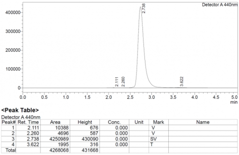
The results of the chromatography analysis using the HPLC technique revealed the availability of a TQ compound based on standards, as shown in Figure 2. This paper showcases the noteworthy accomplishments in creating and confirming an HPLC technique for the direct measurement of thymoquinone (TQ) [23].
Figure 2. Standard of TQ compound
3.2 Separation of TQ compound by using sodium dodecyl sulfate polyacrylamide gel electrophoresis (SDS‒PAGE)
The electrophoresis process of TQ compound used under study was carried out from the aqueous extract of the fungus Pleurotus ostreatus on a polyacrylamide gel SDS‒PAGE by using Coomassie Brilliant dye. After conducting the electrophoresis process, bundles of the active compounds appeared on the gel, and each type of them was determined based on the molecular weight, which was estimated at 100 g/mol (Figure 3). It was found that the separated TQ compound with a molecular weight of 164.20 g/mol. TQ is a bioactive ingredient found in many herbs. According to a study [24], TQ also exhibits antagonistic activity against breast cancer cells and has shown effects on smooth and cardiovascular muscle function.
Figure 3. SDS-PAGE gel protein
M=protein ladder marker starting from 100 to 700 g, TQ = 164.20 g/mol: SDS-PAGE gel protein.
This study investigated in vitro the increase of antifungal property of TQ extracted from Nigella Sativa seeds by photodynamic effects of the IR lamp photosensitizer.
In this study on the photodamage of pathogens, which was successfully performed at physiological temperatures. TQ showed a significant antifungal efficacy against Aspergillus flavus when administered alone without infrared. Inhibition of Aspergillus flavus at the concentrations of 50, 100, and 200 µg/mL in an increasing trend of inhibition rate. The results were backed by one-way ANOVA and Tukey’s post-hoc statistical analysis at a 95% confidence level, showing significant inhibition of A. flavus mycelial growth. This antifungal activity of TQ can be due to the disruption of the fungal cellular integrity by the accumulation of malondialdehyde, which could deteriorate the structure of the macromolecules within the fungal cells or disrupt the cell membranes [25-27].
Therefore, TQ could be able to inhibit or reduce the metabolic functions of a fungus. On the other hand, the antifungal activity of TQ compounds was postulated to be related to the existence of such functional groups as hydroxyl, a methoxy group, and a carbonyl group on the benzoquinone ring. So, by having these antimicrobial compounds, they could interact in the form of strong π bonds with the biomolecules within the fungal cells, such as amino acids and proteins. Statistical analysis through three replications showed that, at the concentration of 200 µg/ml, TQ had significantly inhibited A. flavus growth, even in comparison with the positive control. Cumulatively, the inhibition rate of A. flavus tends (50 µg/ml = 24.0649 ± 1.66769%, 100 µg/ml = 24.4799 ± 0.21129%, 200 µg/ml = 39.7603 ± 1.84512%). Therefore, TQ could be a potent antifungal bioactive in the form of an intact or active compound, the cornerstone of a biopurification system in an agroecosystem to reduce health risk factors or to improve the continuity of application diversity. It would expand the application of the bio-fungicide of black cumin extract to suppress mycoses, otherwise, fungi may become resistant, and viral or bacterial infections. In addition, microflora that are bioactive will also interact against, neutralize, or degrade the antifungal substance [28].
Table 1 indicates that the use of TQ at concentrations of 1% and 2% resulted in a significant inhibition in mushroom growth by 11.12% and 12.67%, respectively, compared to the control sample (0%) [29].
Table 1. Colony diameter and inhibition of Aspergillus flavus on SAM medium treated with TQ compound
|
TQ Compound Concentration (%) |
Colony Diameter (mm) |
Inhibition (%) |
|
0% |
90 |
0 |
|
1% |
80 |
11.12 |
|
2% |
76 |
12.67 |
When combining TQ with infrared exposure, as shown in Table 2, an increase in inhibition efficacy was recorded at an exposure duration of 2 minutes, with inhibition ratios of 11.12% and 22.23% at concentrations of 1% and 2%, respectively, indicating an initial synergistic effect between the compound and infrared radiation [29].
Table 2. Colony diameter and inhibition of Aspergillus flavus on SAM medium treated with TQ compound and 2 minutes of irradiation
|
TQ Compound Concentration (%) |
Colony Diameter (mm) |
Inhibition (%) |
|
0% |
90 |
0 |
|
1% |
80 |
11.12 |
|
2% |
70 |
22.23 |
However, the results of Tables 3 and 4, which represent longer exposure periods (4 and 6 minutes), showed a significant reduction in the biological effect of TQ, with inhibition rates not exceeding 2.67% in most cases, reaching a maximum of 3.78% at a concentration of 2% after 6 minutes of exposure [29]. This decrease in effect is due to the possibility of the compound degradation or decomposition of its chemical structure under the influence of heat or radiation, which may limit its efficiency in cellular absorption and biological reaction [29].
Table 3. Colony diameter and inhibition of Aspergillus flavus on SAM medium treated with TQ compound and 4 minutes of irradiation
|
TQ Compound Concentration (%) |
Colony Diameter (mm) |
Inhibition (%) |
|
0.0 |
90 |
0 |
|
1% |
66 |
2.67 |
|
2% |
60 |
2.67 |
Table 4. Colony diameter and inhibition of Aspergillus flavus on SAM medium treated with TQ compound and 6 minutes of irradiation
|
TQ Compound Concentration (%) |
Colony Diameter (mm) |
Inhibition (%) |
|
0% |
90 |
0 |
|
1% |
60 |
2.67 |
|
2% |
56 |
3.78 |
The potential mechanism of antifungal activity of TQ is due to the presence of active groups such as hydroxyl, methoxy, and carbonyl on the benzoquinone ring, enabling it to form strong covalent $\pi-\pi$ bonds with proteins and amino acids within the fungal cell [30]. In addition, TQ is thought to disrupt the integrity of the cell membrane by stimulating lipid peroxidation, leading to the accumulation of malondialdehyde (MDA), which causes significant damage to biomolecular structures [30].
These results were corroborated by statistical analysis using the unidirectional ANOVA test and the Tukey post-test, where the inhibition ratio at 200 μg/ml concentration was statistically significant (p<0.05), and at some frequencies outperformed the effect of the positive standard sample [31].
It is worth noting that the use of infrared radiation alone has not shown a significant effect on the growth of A. flavus, highlighting the importance of improving photo processing conditions, in particular by adjusting the physical and chemical properties of photosensitive compounds, such as lipid solubility and ionic charge, which directly affect the efficiency of their penetration into the fungal cell and their interaction with its components (Table 5) [32].
Table 5. Colony diameter and inhibition of Aspergillus flavus on SAM medium with 6 minutes of irradiation
|
TQ Compound Concentration (%) |
Colony Diameter (mm) |
Inhibition (%) |
|
0% |
90 |
0 |
|
1% |
90 |
0 |
|
2% |
90 |
0 |
In light of these findings, TQ is a promising candidate as a natural antifungal and may form the cornerstone for the development of environmentally friendly phototherapeutic strategies. However, more studies are still urgently needed to more accurately understand the mechanisms of interaction and optimize application conditions to reach an effective synergistic effect, both in agricultural applications and in clinical settings, to reduce the spread of fungi resistant to conventional treatment [33].
Infrared radiation consists of different wavelengths of light, and each wavelength represents energy with distinct characteristics. The primary purpose of this experiment was to investigate the potential antifungal effectiveness of far-infrared radiation (FIR) and middle-infrared radiation (MIR) on Aspergillus flavus in terms of treatment time, light intensity, and wavelength properties in the absence of TQ. Analyzing the effects of temperature change on Aspergillus flavus in response to FIR and MIR treatment revealed that a lower temperature increase could inhibit A. flavus growth. Raising the temperature may not be suitable for antifungal procedures. The results obtained in this study imply that there is potential for the use of FIR as an Aspergillus flavus antifungal process. They also exhibit practical applications in structures and materials that prevent fungal growth [34].
As the maximum peak of the sun’s radiant heat emission on Earth is in the FIR zone, FIR radiation possesses a deeper penetration capacity than visible light, and it can heat air or solid materials. Middle infrared is used in different methods than FIR, including full infrared and mixed gas methods. The full infrared method uses microwave radiation with a relatively short run for sterilization, but there are ongoing studies on the antifungal effect of full infrared citrus ulcers and of a mixed gas method on Penicillium [35]. FIR, which are electromagnetic waves, are able to penetrate biological tissue. In this study, A. favus at a further distance showed inhibition in a shorter ventilation time, which is so important pending several potential therapeutic and physical applications for sunlight and laser skin treatment. Neither FIR nor MIR was studied beneficial against A. favus before this research. It has been shown and confirmed here that MIR is also beneficial as an antifungal treatment [36].
3.3 Combined effects of TQ and IR
A synergistic effect of a combination of TQ extracted from Nigella sativa and IR on A. flavus growth in vitro. The effect of TQ and TQ-IR rays on the growth of A. flavus was also researched with light, phase contrast, epifluorescence, scanning, and transmission electron microscopy images. FTIR results indicated a greater overall suppression in peak height or intensity with a dual treatment. There was about a 10 times greater TQ effect on the growth of the fungus with IR than with light only, and a 2.5 times greater IR effect on the fungal growth with TQ than with light. There was some TQ-IR interaction enhancing the growth suppression of the fungus (Figure 4) [34].
Figure 4. The effect of TQ and TQ-IR rays on the growth of A. flavus by using FTIR
Fungus growth was greatly suppressed with TQ or IR rays. There was about a 5 times greater inhibition of fungal growth with TQ than NS, and about a 75 times greater inhibition with potentiated TQ than the cultivation temperature alone. The equal or approximately equal growth inhibition of the fungus was achieved by applying a temperature which would be between 57 and 58℃ for about 4 hours, an equal amount of TQ, or a low dose of TQ with IR rays for one day. TQ and NS caused the formation and release of numerous protoplasts with organelle damage in fungal hyphae. IR rays caused an unusually large vacuole, a high septum to hyphal ratio cross walls. It was suggested that a synergistic interaction of a combination of TQ and IR could enhance antifungal activity in combating fungi significantly more than the sum of TQ or IR treatments for their possible application in medicine or storage, as would be different mechanisms of action and metabolic targets in mycelial growth [37].
The study found that exposure to infrared radiation increased the ability of TQ to inhibit the growth of Aspergillus fumigatus, a common cause of fungal infections. A recent study found that exposure to infrared radiation increased the ability of TQ to inhibit the growth of Candida albicans, a common cause of yeast infections [38].
Studies have shown that exposure to infrared radiation can enhance the effect of TQ in inhibiting fungal growth. Influence mechanisms: Increased permeability of TQ: Infrared radiation increases the permeability of TQ into fungal cells, allowing it to reach its target sites more effectively [38].
Enhancing the production of free radicals: Infrared (IR) radiation leads to increased production of free radicals in fungal cells, leading to DNA and protein damage. enhances this effect, leading to fungal cell death (Figure 5) [39].
Figure 5. Effect of TQ and IR exposure on A. flavus colony growth
3.4 Effect of TQ and IR exposure on A. flavus colony growth
The therapeutic application of Nigella sativa, also known as black seed or black cumin, has been well documented for over two centuries in traditional medicine practices and in various experimental models. The main bioactive component of the volatile oil isolated from Nigella sativa L. is TQ. It has shown broad-spectrum antibacterial, possible cell wall synthesis inhibition, antiviral, anti-inflammatory, analgesic, and antifungal activities. Infrared (IR) radiation is like ultraviolet rays in that it cannot be seen with the naked eye [40]. Infrared (IR) radiation is characterized by a change in photon vibration when the molecule is stimulated by infrared light, causing the molecular vibration energy to be transmitted to the surrounding molecules [41, 42]. This causes the temperature of the surrounding molecules to rise, with heat being generated by friction. The advantage of infrared radiation is that it is capable of sterilization without the use of chemical agents, leaving no residue behind. The application of photodynamic therapy has been most prominent in this field, but using visible light can induce changes in the phenotype and cause severe side effects such as pigmentation or photosensitivity. Considering the above, research to combine TQ has been actively conducted in order to improve the applicability and expand therapeutic targets, but studies on the efficacy of infrared ray exposure are still relatively rare. Further exploration of the mechanism of action is essential to enhance understanding in the development of targeted therapy, to adapt to the emergence of resistant strains, and to overcome confirmation bias against predisposed conceptual models. Different fungi may react differently to the same treatment conditions, and the time point at which to observe the reaction is limited by the growth rate of the fungi. These technical constraints present challenges to identifying the exact mechanism of action of TQ and infrared (IR) radiation used in combination with fungi and should be taken into consideration when interpreting the results of this study [43, 44].
Stimulation of the cell death signal pathway: Infrared radiation stimulates the cell death signal pathway in fungal cells, leading to their programmed death. enhances this effect, resulting in more effective fungal growth inhibition [45]. Fungal infections have been a huge burden on food safety and human health, and Aspergillus can cause serious consequences, such as liver cancer, with Aflatoxin B1 contamination. One of the most effective antifungal ingredients comes from natural products, TQ extracted from Nigella sativa. Infrared (IR) rays can selectively suppress fungi's metabolism. Under 10 W IR rays, 70% of Aspergillus flavus spores treated with TQ have been deactivated, so the TQ effectiveness of combating fungus is significantly enhanced by IR rays. Hopefully, a fungicidal treatment of dried fruits combining natural fungicides and IR rays will be trialed [46, 47]. The mode of TQ action against A. flavus involves damage to the cell membrane and interference with the metabolic system. Using confocal laser scanning microscopy, it is found that the contact of TQ caused the morphological changes and cell wall damage of Aspergillus spores. Using electron scanning microscopy, it was discovered that the TQ-treated spores have shrunken cytoplasm and a loss of spore rods. The leakage of ions from the plasma membrane and the depletion of ATP-induced exhaustion expose the cellular membrane of A flavus, further causing loss of glucose and amino acids. The reduction of rhodamine 6G content of A flavus caused by TQ indicates the resistance of ATP synthesis, Therefore, TQ can cause damage to A. flavus cell membranes and interfere with its metabolic system, as well as leakage of cell contents. These antifungal mechanisms of action cause variation in the potency of TQ among fungi, and also provide information about resistance to TQ treatment [48]. To successfully inhibit a wide range of fungi, it is important to know the antifungal mechanisms of TQ action in order to develop more effective strategies and treatments. One significant challenge in combating fungal infections in clinical health care applications is fungicidal resistance, where fungi grow their biodegradability to fungicidal drugs and the difficulty of creating novel, efficient antifungal agents. In order to arrange an integrated blend to modulate fungi, this research showcases how TQ extraction from Nigella sativa can stimulate the antifungal operation, and how it can be enhanced by employing infrared (IR) radiation. This test explores the findings of a research project to investigate the fungicidal consequences of isolated TQ with and without the use of IR in various wavelengths on mycotoxin-producing Aspergillus flavus [49].
IR released in mitochondria enhances active synthesis, which leads to further small apoptosis of fungi due to the rise of intracellular nitric oxide levels as a consequence of overproduction of reactive oxygen species and peroxynitrite [50]. Organic medication has been used since antiquity to cure health problems, and it has formed the essence of clinical drug manufacturers. Plant treatments are being researched, especially in cancer treatment. Besides, scientists agree that when working together, the treatment activities of chemotherapy can be controlled to fight cancer. However, antifungal activity has been of greater scope [51]. In the current study, the antifungal efficacy of thymoquinone (TQ) and amphotericin B was compared.
The results showed that amphotericin B retained a high inhibitory efficacy of more than 90% against the tested fungi, which is due to its traditional mechanism of action by binding to ergosterol within the fungal cell membrane, causing cellular content leakage and cell death [52].
In contrast, thymoquinone has shown antifungal efficacy with an inhibition rate of 60–75%, through multiple mechanisms including stimulating the production of reactive oxygen species (ROS), damage to cell membrane components, disruption of protein synthesis, and damage to genetic material (DNA) [53]. When thymoquinone was combined with IR radiation, a significant improvement in antifungal efficacy was observed, with inhibition rates rising to 80–90%. This synergistic effect is attributed to the fact that infrared radiation contributes to increased cellular membrane permeability of fungi, allowing greater entry of thymoquinone into the cell, enhanced production of reactive oxygen species (ROS) by the heat energy emitted by infrared radiation, leading to increased cellular damage. Stimulate oxidative stress processes within fungal cells, accelerating cell death. Thus, the combination of thymoquinone with infrared light is shown as a promising approach to increasing the therapeutic efficacy of natural antifungal agents, while maintaining the low toxicity advantage compared to conventional amphotericin B therapy (Figure 6) [54].
Figure 6. The antifungal efficacy of TQ and amphotericin B
Aspergillus flavus increased sensitivity to TQ, a bioactive compound extracted from Nigella sativa seeds, highlights its selective potential as an antifungal. These findings confirm the possibility of using infrared light damage as a future therapeutic strategy to treat fungal infections that affect human skin. However, more studies are needed to understand the underlying molecular mechanisms and improve treatment parameters to enhance efficacy and clinical application. TQ has a moderate effectiveness on its own, which is clearly increasing when combined with infrared, and its effect is approaching ampathyrisin B with additional features such as low toxicity and support for safe treatment.
[1] Alberts, A., Moldoveanu, E.T., Niculescu, A.G., Grumezescu, A.M. (2024). Nigella sativa: A comprehensive review of its therapeutic potential, pharmacological properties, and clinical applications. International Journal of Molecular Sciences, 25(24): 13410. https://doi.org/10.3390/ijms252413410
[2] Aldory, M.E., Ali, F.F., Sultan, S. M. (2018). Effective of watery and alcoholic extract of frankincense on the Candida albicans fungus. International Journal of Pharmaceutical Research & Allied Sciences, 7(3): 56-62.
[3] Amna, T., Alghamdi, A.A.A., Shang, K., Hassan, M.S. (2021). Nigella sativa-coated hydroxyapatite scaffolds: Synergetic cues to stimulate myoblasts differentiation and offset infections. Tissue Engineering and Regenerative Medicine, 18: 787-795. https://doi.org/10.1007/s13770-021-00341-4
[4] Mohammed, S.J., Amin, H.H., Aziz, S.B., Sha, A.M., Hassan, S., Abdul Aziz, J.M., Rahman, H.S. (2019). Structural characterization, antimicrobial activity, and in vitro cytotoxicity effect of black seed oil. Evidence‐Based Complementary and Alternative Medicine, 2019(1): 6515671. https://doi.org/10.1155/2019/6515671
[5] Khan, M.A. (2018). Antimicrobial action of thymoquinone. In Molecular and Therapeutic Actions of Thymoquinone, pp. 57-64. https://doi.org/10.1007/978-981-10-8800-1_5
[6] Bose, S., Roy, M., Bandyopadhyay, A. (2012). Recent advances in bone tissue engineering scaffolds. Trends in Biotechnology, 30(10): 546-554. https://doi.org/10.1016/j.tibtech.2012.07.005
[7] Prescott, T.A.K., Hill, R., Mas-Claret, E., Gaya, E., Burns, E. (2023). Fungal drug discovery for chronic disease: History, new discoveries and new approaches. Biomolecules, 13(6): 986. https://doi.org/10.3390/biom1306098
[8] Ojueromi, O.O., Oboh, G., Ademosun, A.O. (2022). Black seed (Nigella sativa): A favourable alternative therapy for inflammatory and immune system disorders. Inflammopharmacology, 30(5): 1623-1643. https://doi.org/10.1007/s10787-022-01035-6
[9] Olejarz-Maciej, A., Mogilski, S., Karcz, T., Werner, T., et al. (2023). Trisubstituted 1, 3, 5-triazines as histamine H4 receptor antagonists with promising activity in vivo. Molecules, 28(10): 4199. https://doi.org/10.3390/molecules28104199
[10] Gaddawi, F.Y., Jarjees, N.A., Sultan, S.M., Irzoqy, M.E. (2022). Detection effect toxins produced by some types of fungi isolated from medicinal plants. International Journal of Health Sciences, 1: 969-986. https://doi.org/10.53730/ijhs.v6nS1.4843
[11] Haq, I.U., Khan, N.A., Sarwar, M.K. (2022). An insight in to fungal biology. In Phytomycology and Molecular Biology of Plant Pathogen Interactionsm, pp. 27-50.
[12] Nyongesa, B.W., Okoth, S., Ayugi, V. (2015) Identification key for Aspergillus species isolated from maize and soil of Nandi County, Kenya. Advances in Microbiology, 5(4): 205-229. https://doi.org/10.4236/aim.2015.54020
[13] El-Gazzar, N., Abdo, E., Rabie, G., El-Sayed, M.T. (2025). Suppression of mycotoxins production and efficient chelation of heavy metals using natural melanin originated from Aspergillus flavus and Aspergillus carbonarius. BMC Biotechnology, 25(1): 6. https://doi.org/10.1186/s12896-024-00941-7
[14] Pandey, R., Pandey, B., Bhargava, A. (2024). The Emergence of N. sativa L. as a Green Antifungal Agent. Mini Reviews in Medicinal Chemistry, 24(16): 1521-1534. https://doi.org/10.2174/0113895575282914240217060251
[15] Niculescu, A.G., Grumezescu, A.M. (2021). Photodynamic therapy—an up-to-date review. Applied Sciences, 11(8): 3626. https://doi.org/10.3390/app11083626
[16] Duminis, T. (2023). The use of thymoquinone as a standard for quality assessment of the seeds of Nigella sativa and investigation of their antioxidant activity. Journal of Food Chemistry Nanotechnology, 9(4): 176-183. https://doi.org/10.17756/jfcn.2023-165
[17] Thathana, M.G., Murage, H., Abia, A.L.K., Pillay, M. (2017). Morphological characterization and determination of aflatoxin-production potentials of Aspergillus flavus isolated from maize and soil in Kenya. Agriculture, 7(10): 80. https://doi.org/10.3390/agriculture7100080
[18] Chen, L., Liu, B., Feng, S., Ma, X., Wang, S., Zhang, Y. (2023). Correlation between microbe, physicochemical properties of Jiuqu in different plateau areas and volatile flavor compounds of highland barley alcoholic drink. Food Bioscience, 51: 102276. https://doi.org/10.1016/j.fbio.2022.102276
[19] Novy, P., Kloucek, P., Rondevaldova, J., Havlik, J., Kourimska, L., Kokoska, L. (2014). Thymoquinone vapor significantly affects the results of Staphylococcus aureus sensitivity tests using the standard broth microdilution method. Fitoterapia, 94: 102-107. https://doi.org/10.1016/j.fitote.2014.01.024
[20] Belete, Y., Dagne, E. (2014). HPTLC assay of thymoquinone in black seed and black seed oil (Nigella Sativa Linn.) and identification of thymoquinone conversion with Uv-Vis. Journal of Drug Delivery & Therapeutics, 4(4): 5-9.
[21] Xing, L., Ma, P., Chen, F. (2024). A novel turn-on near-infrared fluorescent probe for highly sensitive in vitro and in vivo detection of acetylcholinesterase activity. Spectrochimica Acta Part A: Molecular and Biomolecular Spectroscopy, 310: 123954. https://doi.org/10.1016/j.saa.2024.123954
[22] Binder, U., Aigner, M., Risslegger, B., Hörtnagl, C., Lass-Flörl, C., Lackner, M. (2019). Minimal inhibitory concentration (MIC)-phenomena in Candida albicans and their impact on the diagnosis of antifungal resistance. Journal of Fungi, 5(3): 83. https://doi.org/10.3390/jof5030083
[23] Qureshi, K.A., Imtiaz, M., Parvez, A., Rai, P.K., Jaremko, M., Emwas, A.H., Bholay, A.D., Fatmi, M.Q. (2022). In vitro and in silico approaches for the evaluation of antimicrobial activity, time-kill kinetics, and anti-biofilm potential of thymoquinone (2-Methyl-5-propan-2-ylcyclohexa-2, 5-diene-1, 4-dione) against selected human pathogens. Antibiotics, 11(1): 79. https://doi.org/10.3390/antibiotics11010079
[24] Shabani, H., Karami, M.H., Kolour, J., Sayyahi, Z., et al. (2023). Anticancer activity of thymoquinone against breast cancer cells: Mechanisms of action and delivery approaches. Biomedicine & Pharmacotherapy, 165: 114972. https://doi.org/10.1016/j.biopha.2023.114972
[25] Kashi, M., Noei, M., Chegini, Z., Shariati, A. (2024). Natural compounds in the fight against Staphylococcus aureus biofilms: A review of antibiofilm strategies. Frontiers in Pharmacology, 15: 1491363. https://doi.org/10.3389/fphar.2024.1491363
[26] Algorri, J.F., Ochoa, M., Roldan-Varona, P., Rodriguez-Cobo, L., López-Higuera, J.M. (2021). Light technology for efficient and effective photodynamic therapy: A critical review. Cancers, 13(14): 3484. https://doi.org/10.3390/cancers13143484
[27] Xie, Q., Peng, F., Wang, X., Du, B., Yang, Y. (2025). Chestnut flower extract as a natural inhibitor of Fusarium graminearum: Antifungal activity and mechanisms. Pest Management Science. https://doi.org/10.1002/ps.8708
[28] Hasan, M.N., Sultan, S.M. (2020). Effects of Fe and GA3 on growth and oil characteristic of rosemary plant (Rosmarinus officinalis L.). Plant Cell Biotechnology and Molecular Biology, 21(40): 55-63.
[29] Abbas, M., Gururani, M.A., Ali, A., Bajwa, S., Hassan, R., Batool, S.W., Imam, M., Wei, D. (2024). Antimicrobial properties and therapeutic potential of bioactive compounds in Nigella sativa: A review. Molecules, 29(20): 4914. https://doi.org/10.3390/molecules29204914
[30] Wu, X., Hu, Y. (2022). Photodynamic therapy for the treatment of fungal infections. Infection and Drug Resistance, 15: 3251-3266. https://doi.org/10.2147/IDR.S369605
[31] Almshawit, H., Macreadie, I. (2017). Fungicidal effect of thymoquinone involves generation of oxidative stress in Candida glabrata. Microbiological Research, 195: 81-88. https://doi.org/10.1016/j.micres.2016.11.008
[32] Ziental, D., Mlynarczyk, D.T., Czarczynska-Goslinska, B., Lewandowski, K., Sobotta, L. (2021). Photosensitizers mediated photodynamic inactivation against fungi. Nanomaterials, 11(11): 2883. https://doi.org/10.3390/nano11112883
[33] Khoddami, A., Ghazali, H.M., Yassoralipour, A., Ramakrishnan, Y., Ganjloo, A. (2011). Physicochemical characteristics of Nigella sativa L. oil as affected by different extraction methods. Journal of the American Oil Chemists' Society, 88: 533-540. https://doi.org/10.1007/s11746-010-1687-6
[34] Wang, X., Zhang, G., Dang, Y. (2022). Enhanced extraction of flavonoids from licorice residues by solid-state mixed fermentation. Waste and Biomass Valorization, 13(11): 4481-4493. https://doi.org/10.1007/s12649-022-01803-z
[35] Lakrat, M., Azzaoui, K., Jodeh, S., Akartasse, N., Mejdoubi, E., Lamhamdi, A. (2017). The removal of methyl orange by nanohydroxyapatite from aqueous solution: Isotherm, kinetics and thermodynamics studies. Desalination and Water Treatment, 85: 237-249. https://doi.org/10.5004/dwt.2017.21260
[36] Jacoutot, P., Scaccabarozzi, A.D., Zhang, T., Qiao, Z., et al. (2022). Infrared organic photodetectors employing ultralow bandgap polymer and non‐fullerene acceptors for biometric monitoring. Small, 18(15): 2200580. https://doi.org/10.1002/smll.202200580
[37] Matthäus, B. (2002). Antioxidant activity of extracts obtained from residues of different oilseeds. Journal of Agricultural and Food Chemistry, 50(12): 3444-3452. https://doi.org/10.1021/jf011440s
[38] Mechraoui, O., Ladjel, S., Nedjimi, M.S., Belfar, M.L., Moussaoui, Y. (2018). Determination of polyphenols content, antioxidant and antibacterial activity of Nigella sativa L. seed phenolic extracts. Scientific Study & Research: Chemistry & Chemical Engineering, Biotechnology, Food Industry, 19(4): 411-421.
[39] Mohammed, N.K., Manap, M.Y. A., Tan, C.P., Muhialdin, B.J., Alhelli, A.M., Hussin, A.S.M. (2016). The effects of different extraction methods on antioxidant properties, chemical composition, and thermal behavior of black seed (Nigella sativa L.) oil. Evidence-Based Complementary and Alternative Medicine, 2016(1): 6273817. https://doi.org/10.1155/2016/6273817
[40] Pop, R.M., Trifa, A.P., Popolo, A., Chedea, V.S., Militaru, C., Bocsan, I.C., Buzoianu, A.D. (2020). Nigella sativa: Valuable perspective in the management of chronic diseases. Iranian Journal of Basic Medical Sciences, 23: 699-713. https://doi.org/10.22038/ijbms.2020.37734.8978
[41] Sheet, A., Abood, S., Sultan, S., Alomari, S. (2020). A comparative study between the influence of antibiotics and extracts from Myrtus communis and Allium sativum against Staphylococcus aureus isolated from some pathogenic states. In Proceedings of the 1st International Multi-Disciplinary Conference Theme: Sustainable Development and Smart Planning, IMDC-SDSP 2020, Cyperspace. https://doi.org/10.4108/eai.28-6-2020.2298150
[42] Liang, H., Lv, F., Xian, M., Luo, C., Zhang, L., Yang, M., Li, Q., Zhao, X. (2025). Inhibition mechanism of Cinnamomum burmannii leaf essential oil against Aspergillus flavus and aflatoxins. Foods, 14(4): 682. https://doi.org/10.3390/foods14040682
[43] Tavakkoli, A., Mahdian, V., Razavi, B.M., Hosseinzadeh, H. (2017). Review on clinical trials of black seed (Nigella sativa) and its active constituent, Thymoquinone. Journal of Pharmacopuncture, 20(3): 179-193. https://doi.org/10.3831/KPI.2017.20.021
[44] Neunert, G., Kamińska, W., Nowak-Karnowska, J. (2025). Evaluating the thymoquinone content and antioxidant properties of black cumin (Nigella sativa L.) seed oil during storage at different thermal treatments. Applied Sciences, 15(1): 377. https://doi.org/10.3390/app15010377
[45] Kooti, W., Servatyari, K., Behzadifar, M., Asadi-Samani, M., Sadeghi, F., Nouri, B., Marzouni, H.Z. (2017). Effective medicinal plant in cancer treatment, Part 2: Review study. Journal of Evidence-Based Integrative Medicine, 22(4): 982-995. https://doi.org/10.1177/2156587217696927
[46] Zhao, Y., Wang, X., Zhang, L., Wang, K., Wu, Y., Yao, J., Cui, B., Chen, Z. (2022). Anti-fungal activity of moutan cortex extracts against rice sheath blight (Rhizoctonia solani) and its action on the pathogen’s cell membrane. ACS Omega, 7(50): 47048-47055. https://doi.org/10.1021/acsomega.2c06150
[47] Santos, A.R., Carreiró, F., Freitas, A., Barros, S., Brites, C., Ramos, F., Sanches Silva, A. (2022). Mycotoxins contamination in rice: Analytical methods, occurrence and detoxification strategies. Toxins, 14(9): 647. https://doi.org/10.3390/toxins14090647
[48] Wiederhold, N.P. (2017). Antifungal resistance: current trends and future strategies to combat. Infection and Drug Resistance, 10: 249-259. https://doi.org/10.2147/IDR.S124918
[49] Perfect, J.R. (2022). The antifungal pipeline: A reality check. Nature Reviews Drug Discovery, 16: 603-616. https://doi.org/10.1038/nrd.2017.46
[50] Radwan, I.T., El-Sherbiny, I.M., Metwally, N.H. (2024). Synergistic and potential antifungal properties of tailored, one pot multicomponent monoterpenes co-delivered with fluconazole encapsulated nanostructure lipid carrier. Scientific Reports, 14: 14382. https://doi.org/10.1038/s41598-024-63149-x
[51] Shah Alam, M., Maowa, Z., Subarna, S.D., Hoque, M. N. (2024). Mycotoxicosis and oxidative stress in poultry: Pathogenesis and therapeutic insights. World's Poultry Science Journal, 80(3): 791-820. https://doi.org/10.1080/00439339.2024.2347307
[52] El-Far, A.H., Tantawy, M.A., Al Jaouni, S.K., Mousa, S.A. (2020). Thymoquinone-chemotherapeutic combinations: New regimen to combat cancer and cancer stem cells. Naunyn-Schmiedeberg's Archives of Pharmacology, 393: 1581-1598. https://doi.org/10.1007/s00210-020-01898-y
[53] Hussain, A.R., Ahmed, M., Ahmed, S., Manogaran, P., Platanias, L.C., Alvi, S.N., Al-Kuraya, K.S., Uddin, S. (2011). Thymoquinone suppresses growth and induces apoptosis via generation of reactive oxygen species in primary effusion lymphoma. Free radical biology and medicine, 50(8): 978-987. https://doi.org/10.1016/j.freeradbiomed.2010.12.034
[54] Akansha, Kaushal, S., Arora, A., Heena, Sharma, P., Jangra, R. (2023). Chemical composition and synergistic antifungal potential of Nigella sativa L. seeds and Syzygium aromaticum (L.) Merr. & LM Perry buds essential oils and their major compounds, and associated molecular docking studies. Journal of Essential Oil Bearing Plants, 26(3): 602-625. https://doi.org/10.1080/0972060X.2023.2220348