Mohammed Sabah Ali*![]() | Auday Abid Mehatlaf
| Auday Abid Mehatlaf![]()
© 2025 The authors. This article is published by IIETA and is licensed under the CC BY 4.0 license (http://creativecommons.org/licenses/by/4.0/).
OPEN ACCESS
This study examines the impact of adding different ratios of alumina (Al2O3) and nano-copper (Cu) materials to dental amalgam alloy (Ag-Sn-Cu) on their physical, microstructural, and mechanical properties. The study utilized powder metallurgy and compaction techniques to prepare dental amalgam samples. The scanning electron microscope (SEM), and mechanical tests (compressive strength) were used to determine the microstructure, density, and mechanical properties. Various ratios (2.5, 5, and 10 wt.%) of nano-copper and alumina have been added to the amalgam capsules. The findings indicated that when the proportion of nano copper particles rose to 10 wt.%. The compressive strength of amalgam declined. This drop in characteristics could be linked to the malleability of copper. Conversely, dental amalgam specimens containing alumina (Al2O3) demonstrated strength, especially with 5wt.% alumina, in comparison to the amalgam sample. The enhancement in strength in these samples can be attributed to the high hardness and rigidity of alumina whereas the decline in strength in samples with 10 wt.% copper can be attributed to the flexibility of copper, versus alumina.
dental amalgam, reinforced, alumina, nanoparticles, copper, mechanical properties
Biomaterials play a role in creating tools that can replace or support body functions when needed, providing cost-effective and biologically suitable solutions. Various materials and tools are utilized in the management of injuries and illnesses [1]. For over 150 years, dental amalgam has been widely used to address cavities because of its robust mechanical characteristics and long-lasting nature [2]. Nevertheless, there is an increasing preference for materials such as resins, glass ionomer cement, ceramics and gold alloys. This shift may be influenced by the appealing characteristics of these materials or concerns about the potential health risks associated with dental amalgam use. The silver alloy used in dental amalgam, which is in the form of a fine powder, primarily consists of silver (Ag), tin (Sn), and copper (Cu), along with small amounts of zinc (Zn), palladium (Pd), or indium (In). Dental amalgam is commonly used in dental restorations, particularly on the surfaces of posterior teeth. It is known for its affordability, durability, and ease of use. However, it is worth noting that dental amalgam is typically not used on the lingual pits of front teeth due to its silvery-gray appearance. As a result, a significant portion of dental restoration is made using dental amalgam. Amalgam fillings in particular have been shown to be superior to other restorative materials in quality. But, the failures, the failures mainly come because of improper care of the material and inadequate cavity preparation. Therefore, all stages from the choice of alloy to the final polishing of repair may affect the appearance and mechanical properties of the restoration. The use of amalgam is not associated with any risks by other researchers. The selection of dental restorative materials based on numerous factors, including strength of restoration, improved performance of hardness, toughness, and wear resistance for a long period. These materials need to be able to withstand the forces exerted during chewing and have a high operational lifespan. On the other hand, they need to be highly compressive and tensile resistant to be ideal to fill the space inside the tooth structure. The performance of dental filling materials can be influenced by the forces generated during mastication, which can lead to impairment [3]. Compressive and tensile strength are essential attributes of dental restorative materials for withstanding chewing forces. Dental amalgam is exceptionally strong, with one of its advantages being its specific composition. Controlling it, affording it, and its compatibility with living organisms are all simple. Dental amalgam restorations are durable and somewhat resistant, when compared to the other restorative materials such as composite resins and have a longer lifespan. Mechanical properties assess how dental materials respond to applied forces, taking into account factors such as masticatory forces, deformation, and stress experienced by the body [4]. Several attempts have been made to improve the properties of amalgam alloy. According to a study conducted by Hasheminezhad et al. [5], the researchers examined the impact of copper on the compressive strength of dental amalgam. They used amalgam capsules containing different amounts of copper, along with mercury and alloy powders. the researchers mixed and amalgamated the materials and then created standard samples using a molding method. The study found that the hardness and compressive strength of the dental amalgam alloy increased with the addition of copper. Additionally, as the strain rate increased fracture stress for the amalgam with a specific weight percentage of copper also increased [5]. Cochran et al. [6] conducted a study where they replaced mercury in a copula with Ga-In liquid and prepared two samples, one with pure amalgam and another with a combination of steel fiber and amalgam. The weight of the powder was adjusted when fibers were included. The two samples were then compressed into cylindrical molds with dimensions of 7 mm in diameter and 10mm in length. The researchers determined the mechanical properties of the prepared cylindrical samples by exposing them to compressive loads until failure. They observed that the samples containing a 6% volume of fibers exhibited a significant increase in compression load to diametral tensile strength and failure. In the study, conducted by Al Sarraj and Atiyah [7], different weight percentages of copper (10-30 % wt.) and silver (40-60%) content were added to standard amalgam alloy powders. The composite powder was then triturated with a specific percentage of mercury. The findings of the study have shown that increasing the copper ratio affects the mechanical properties and microstructure of dental amalgam. In a study by Khodaei et al. [8], an amalgam/nano-hydroxyapatite composite was prepared with different weight percentages of hydroxyapatite. The results indicated that as the weight percentage of hydroxyapatite increased, the compressive strength of the composite increased by 18% with the addition of up to 1% hydroxyapatite but then decreased due to the agglomeration of the hydroxyapatite nanoparticles. Overall, the developed amalgam/nanohydroxyapatite composite appears to be a suitable option for filling anterior teeth with reduced harmful effects [6]. Ortega-Arroyo et al. [9] studied the effect of adding nanosilver particles to dental amalgam alloy. The results showed an improvement in the mechanical performance of the dental amalgam alloy. Moxon et al. [10] investigated the effect of adding titanium (Ti) in nanoscale particles on the properties of dental amalgam alloy. The results present an enhancement in the mechanical properties of around 70%. A Few researchers have used nano-copper (Cu) and alumina (Al2O3) to improve the mechanical properties of dental amalgam. Therefore, in this research, the nano-copper (Cu) and alumina (Al2O3) have been used to improve the properties of dental amalgam.
2.1 Materials
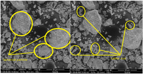
This study used dental amalgam capsules (specifically, NAIS Dental Amalgam Capsules manufactured by NAIS, Bulgaria) that were obtained from a commercial supplier to prepare the research samples. The compositions of these dental amalgam capsules, which are known for their high copper content and non-gamma properties, are detailed in Table 1. The capsules consist of a combination of lathe-cut and spherical particles.
Table 1. Composition of the amalgam capsule
|
Amalgam Powder Bag of Mercury Mixing Time |
Ratio of Alloy/Mercury |
Silver Ratio wt.% (Ag) |
Tin Ratio wt.% (Sn) |
Copper Ratio wt.% (Cu) |
|
26s |
1:1 |
43% |
32% |
25% |
To analyze the characterization of the amalgam powders, the researchers employed scanning electron microscopy (SEM) and energy dispersive X-ray spectroscopy (EDX) techniques. These techniques have been used to investigate the structure of the particles and the chemical composition of amalgam powder. The amalgam involves a spherical shape and lathe-cut with particles. The results of these analyses were presented in Figure 1 and Table 2, respectively.
Figure 1. SEM micrograph for amalgam powder (A) spherical shape, (B) lathe-cut
Table 2. EDX result of used alloy powders
|
Element |
Unn. C (wt.%) |
Norm. C (wt.%) |
Atom. C (wt.%) |
3 Sigma (wt.%) |
|
Silver |
57.39 |
36.73 |
40.72 |
13.26 |
|
Tantalum |
51.12 |
32.72 |
21.62 |
30.91 |
|
Tin |
35.40 |
22.66 |
22.83 |
10.66 |
|
Copper |
12.32 |
7.88 |
14.83 |
11.06 |
|
Hafnium |
0.00 |
0.00 |
0.00 |
0.00 |
|
|
156.23 |
100.00 |
100.00 |
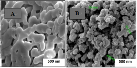
A commercial supplier was used to obtain alumina (Al2O3) with a particle size of 0.5 µm, a purity of 99.9%, and a density of 3.94 g/cm3. This alumina was utilized as a filler material in the study. Additionally, copper (Cu) with an average particle size of 50 nm, a purity of 99.9%, and a density of 7.59 g/cm3 was obtained from another commercial source. The morphology structure and size of the powders have been examined using the SEM examination, and the results can be seen in Figure 2.
Figure 2. (A) Alumina (Al2O3) and (B) Nano-copper (Cu) powders
2.2 Sample preparation
A total of nine series ratios of alloy powders and mercury were prepared in this study. These ratios involved varying proportions of nano-copper (Cu) and alumina (Al2O3) added to dental amalgam according to the weight of amalgam powder in the capsule (1.1540 g) and the weight of mercury (0.5588 g). The mixing time was 26 s. The amounts of (2.5,5, and 10 wt.%) nano-copper (Cu) and alumina (Al2O3) were added to the powder of amalgam alloy. The specific combinations and proportions of these alloy powders are detailed in Table 3.
Table 3. Weight percentages of samples with different ratios of Cu and Al2O3 per capsule
|
Sample Name |
Sample Code |
Cu wt.% (g) |
Al2O3 wt.% (g) |
|
(Standard) amalgam without additives (Al2O3 and Cu) |
S1 (0 wt.%) |
0 |
0 |
|
Amalgam with nano-copper (Cu) additive |
S2 (2.5 wt.% Cu) |
2.5 wt.% |
0 |
|
S3 (5 wt.% Cu) |
5 wt.% |
0 |
|
|
S4 (10 wt.% Cu) |
10 wt.% |
0 |
|
|
Amalgam with alumina (Al2O3) additive |
S5 (2.5 wt.% Al2O3) |
0 |
2.5 wt.% |
|
S6 (5 wt.% Al2O3) |
0 |
5 wt.% |
|
|
S7(10 wt.% Al2O3) |
0 |
10 wt.% |
|
|
Amalgam with Composite (Al2O3+Cu) additive |
S8 (2.5 wt.% Al2O3+2.5 wt.% Cu) |
2.5 wt.% |
2.5 wt.% |
|
S9 (5 wt.% Al2O3+5 wt.% Cu) |
5 wt.% |
5 wt.% |
In this study, the composite mixture of (Al2O3+Cu) powder and the amalgam powders were accurately weighed using an electronic digital weighing machine (model TP-153DS) with a precision of 0.0001. Subsequently, all the batches have been mixed using a roller mixer device (model MR-2). The roller mixer provided gentle continuous rocking and rolling motions, which were ideal for achieving homogeneity. The first step of the mixing procedure involved combining the materials with the amalgam (without mercury) in the roller mixer. Then the resulting powder was mixed in an amalgamator machine (model Lk-H11) for 26-35 seconds. To prepare cylindrical samples with a diameter-to-height ratio of 1 to 1, the mixture was molded using a manual hydraulic press machine (model HSM44 HUMT) at a force of 0.1 kN. The pressing time was set to 120 seconds to allow for proper hardening. Both mixtures, with and without alumina and nano-copper, have been pressed in a cylindrical steel die with a diameter of 5 mm and a height of 5 mm. The applied pressure by the manual hydraulic press device was approximately 0.1 kN on the powder, and the amalgam samples were left to harden for approximately 30 minutes. Finally, the grinding and polishing process to prepare the samples for the microstructure test was carried out using a grinding machine (model Metaserv 250, Vari/Pol VP-150). The grinding process of amalgam samples was conducted using sandpaper with varying grit sizes (1000, 1500, 2000, and 3000) for 5 minutes. The grinding speed was set at 300 revolutions per minute (r.p.m), and water was used as the grinding media. Following the grinding process, the polishing of the samples was carried out using polishing clothes at a speed of 100 r.p.m. Alumina was used as the polishing media. Subsequently, all composite samples were thoroughly cleaned to remove any remnants of the grinding media. To examine the microstructure of the amalgam samples, SEM and a metallography microscope model BD-DIC3230 were utilized. The prepared samples and were also characterized by X-ray diffraction using the XRD diffractometer (XRD-6000, NF type). X-ray was used over 2θ range from 10° to 90° with a rate of 5 deg/min and used Cu-Kα radiation. The phase identification was obtained utilizing X’Pert High Score Plus software. The compressive strength of the amalgam samples, which had dimensions of 5 mm in diameter and 5 mm in height (which were selected according to literature), was measured using a Manual Universal Testing Machine (MUTM) (model HSM44, USA) equipped with a 50 kN load. The compressive strength values were calculated based on the maximum mechanical load and the sectional area of the amalgam samples. The compressive strengths of prepared samples have been measured according to the following Eq. (1) [11, 12].
$\sigma(\mathrm{Mpa})=\frac{\mathrm{P}}{\mathrm{A}}$ (1)
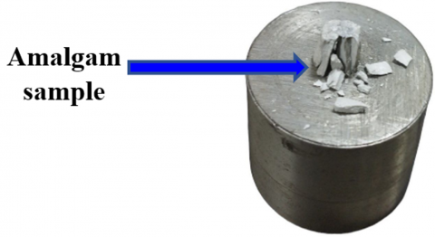
where, $\sigma$ (compressive strength) is in Mpa, P = the applied load (N) and A = the original area of the amalgam samples (mm2). Figure 3 shows the sample after the compression test.
Figure 3. The sample after the compression test
The density of sample was calculated according to the below Eq. (2) [13].
$\operatorname{Density} (\rho)=\frac{\operatorname{mass}(g)}{\text { volume }\left(\mathrm{mm}^3\right)}$ (2)
3.1 Physical properties
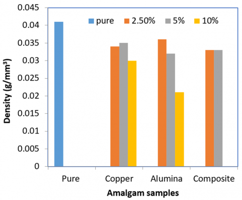
Figure 4 illustrates the density of amalgam samples after different weight percentages of copper (Cu) and alumina (Al2O3) have been added. The density of the pure amalgam sample was measured to be 0.041 g/mm3. Upon adding alumina in ratios of (2.5, 5, and 10 wt.%), the density of the samples decreases as the alumina ratio increases. Conversely, when copper is added to the pure amalgam in ratios of (2.5, 5, and 10 wt.%), the density increases with an increase in the copper ratio. When a composite of Cu+Al2O3 is added in different ratios by weight, the density gradually increases. This variation in density can be attributed to the difference in density between copper (⍴=7.59 g/cm3) and alumina (⍴=3.94 g/cm3).
Figure 4. Variation of densities of amalgam samples
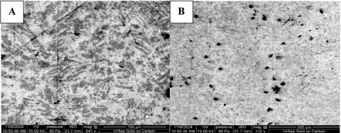
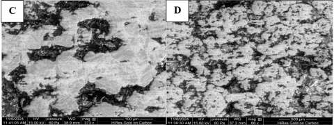
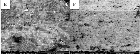
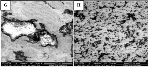
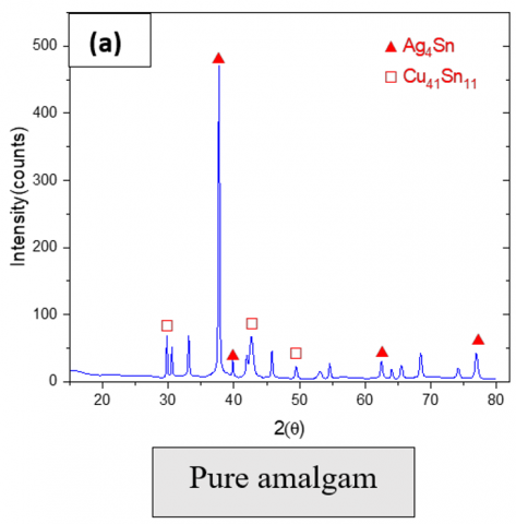
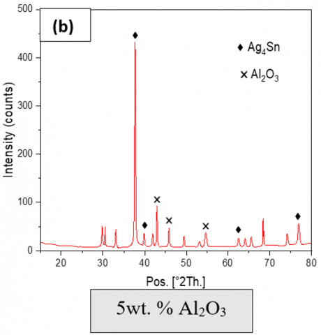
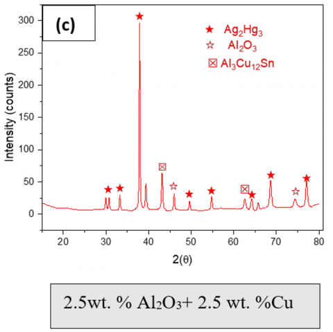
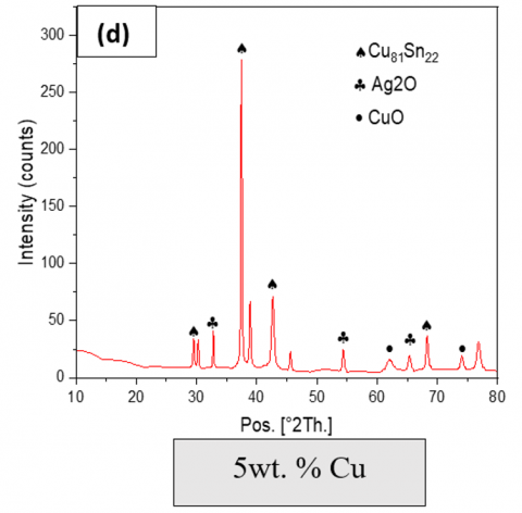
Figures 5 and 6 show SEM images and XRD results. Figure 5 (a and b) presents the microstructure of the pure amalgam (without additive). The white colour zones of the amalgam matrix show phases such as (Ag4Sn), while the gray zones refer to the second phase ƞ eta- prime (Cu6Sn5 and Cu41Sn11), and the dark gray zones refer to unreacted particles of (Cu-Ag-Sn). Figure 5 (c and d) shows the amalgam microstructure involving adding 5% copper nanoparticles. It can see regions in the white colour zones of the amalgam matrix refer to phases (γ1(Ag2 Hg3)) and the gray zones present the phase η (Cu6Sn5) and copper nanoparticle. Figure 5 (e and f) shows the amalgam's microstructure consisting of adding 5% alumina (Al2O3). It observes the alumina (Al2O3) as white zones in addition to the above-mentioned phases. Figure 5 (g and h) presents the microstructure of the amalgam alloy additive of 2.5 wt.% Al2O3+2.5 wt.% nano-copper. Also, can investigate the Al2O3 and nano-copper as white and black zones respectively in addition to the other phase as mentioned. The addition of nanoparticles leads to microstructure refinement and avoids creating a weaker phase (γ2) [14-16], resulting in a tougher material. Due to its fine particle size, and uniform dispersion of nano copper, nano copper can improve the bonding between mercury, tin, and silver particles, forming a more homogenous structure [17]. Particles of Al2O3 as a ceramic material, act as stress-scattering strengthening filler for the matrix and thus decrease the cracks propagation within the matrix. Also, the presence of alumina may reduce the degradation of surface and micro-leakage, which leads to limited mercury release during the time. The Al2O3 particles addition to amalgam aids enhance the amalgam matrix packing, decreasing porosity, and making the internal structure of amalgam more stable. This structural enhancement leads to minimizing mercury vapor and mercury ions dissolution [18].
Figure 5. SEM images (a&b) pure amalgam, (c&d) 5 wt.% Cu), (e&f) 5 wt.% Al2O3, and (g&h) 2.5 wt.% Al2O3+2.5 wt.% Cu
Figure 6. XRD test (a) Pure amalgam, (b) 5 wt.% Al2O3, (c) 2.5 wt.% Al2O3+2.5 wt.% Cu, and (d) 5 wt.% Cu)
One possible cause is inadequate density. Amalgam fillings typically consist of a mixture of mercury and alloy particles, such as silver, tin, and copper. If the process of densification is not carried out properly, air voids or trapped mercury may be present within the filler material, leading to the formation of pores [19]. Another factor that can contribute to the formation of pores is chemical interactions. The addition of copper to dental amalgam can impact its reactivity. Copper has the potential to react with mercury, forming copper amalgam compounds. These chemical reactions may contribute to the formation of pores if the copper content is not properly balanced or if the mixing and handling procedures are not followed correctly [20]. Trapped gases can also be a potential cause of pore formation in amalgam fillings. During the mixing or condensation process, gases like air or water vapor can become trapped within the amalgam block. As the filler material solidifies and hardens, these trapped gases can create voids or pores within the filling.
3.2 Mechanical properties
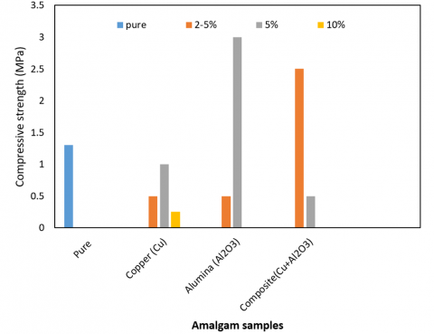
Figure 7 illustrates that the pure amalgam sample has a compressive strength of 0.0662 Mpa. It was observed that when alumina was added with different weight ratios the compressive strength gradually increased. While, when copper was added with different weight ratios the compressive strength increased with (2.5 and 5 wt.%) ratios but decreased at the 10 wt.% ratio. When a composite of copper and alumina is added in ratios of (2.5 and 5 wt.%) the compressive strength gradually decreases. The amalgam samples with alumina exhibit a significantly higher compressive strength of 0.1527 Mpa at a 5 wt% alumina ratio compared to the pure amalgam sample. This improvement in compressive strength can be attributed to the properties of alumina, such as its high hardness and rigidity [21]. Additionally, alumina particles are usually easily dispersed in the amalgam matrix, leading to uniform stress and strain distribution throughout the amalgam matrix. On the other hand, there is a decrease in the compressive strength of amalgam samples with a 10 wt.% copper ratio. This may be due to the ductility of copper compared with alumina, and copper particles tend to agglomerate and do not distribute stress in an effective form, which leads to localized concentrations of stress and potential repeated stress.
Figure 7. Variation of compressive strength of amalgam samples
The dental amalgam silver alloy was successfully reinforced through various investigations into its properties, including compressive strength, density, and microscopic evolution of the amalgam samples. The findings revealed that increasing the amount of pure nano-copper to 2.5%, 5% and 10% resulted in a decrease in strength and an increase in the appearance of grooves and tarnished, which can be attributed to the flexibility of copper. Similarly, similar effects were observed at ratios of 10% and 25% of pure alumina. Consequently, the optimal mechanical properties were achieved with 5% pure alumina and a composite of alumina and copper ratios, resulting in a doubling of the compressive strength. These findings suggest that incorporating small amounts of nano-alumina is a promising approach to enhancing the properties of this dental material. The study’s results indicate that the amalgam/composite [Cu 2.5%+alumina 2.5%] and amalgam/5% alumina combinations could be dental materials that offer improved characteristics for dental applications. The biocompatibility and corrosion resistance of these composites will be the future work in this field.
The Department of Biomedical Engineering/Karbala University is greatly acknowledged for the support rendered in making this work a success.
[1] Mehatlaf, A.A., Atiyah, A.A., Farid, S.B. (2021). An experimental study of porous hydroxyapatite scaffold bioactivity in biomedical applications. Engineering and Technology Journal, 39(6): 977-985. https://doi.org/10.30684/etj.v39i6.2059
[2] Durnan, J.R. (1971). Esthetic dental amalgam-composite resin restorations for posterior teeth. Journal of Prosthetic Dentistry, 25(2): 175-176. https://doi.org/10.1016/0022-3913(71)90104-1
[3] Mount, G.J., Hume, W.R., Ngo, H.C., Wolff, M.S. (2016). Preservation and Restoration of Tooth Structure. John Wiley & Sons. https://doi.org/10.1038/sj.bdj.4813126
[4] Uçar, Y., Brantley, W. (2017). Biocompatibility of dental amalgams. Biocompatibility of Dental Biomaterials. Woodhead Publishing, pp. 95-111. https://doi.org/10.1016/B978-0-08-100884-3.00007-2
[5] Hasheminezhad, A., Zebarjad, S.M., Sajjadi, S.A., Rahanjam, L. (2012). Effect of copper content on compressive strength and microstructure of dental amalgams. Engineering, 4(3): 155-159. http://doi.org/10.4236/eng.2012.43020
[6] Cochran, C.T., Van Hoose, J.R., McGill, P.B., Grugel, R.N. (2012). Improving the strength of amalgams by including steel fibers. Materials Science and Engineering: A, 545: 44-50. https://doi.org/10.1016/j.msea.2012.02.084
[7] Al Sarraj, Z.S., Atiyah, R.I. (2011). Preparation and characterization of high-copper restorative dental alloys corrected. Advanced Materials Research, 324: 69-72. https://doi.org/10.4028/www.scientific.net/AMR.324.6
[8] Khodaei, M., Amini, K., Mahdavian, P. (2018). Fabrication and evaluation of amalgam/nano hydroxyapatite composites for dental restoration. Materials Research Express, 5(10): 105403. https://doi.org/10.1088/2053-1591/aadb45
[9] Ortega-Arroyo, L., San-Martín-Martínez, E., Barceló, F.H., Vargas-Aparicio, J.H., Vengoechea-Gómez, F.A., Castaño, V.M. (2023). Nanosilver-reinforced AgSn alloys for dental applications: Mechanical behavior and hardness. Current Nanomaterials, 8(1): 77-81. https://doi.org/10.2174/2405461507666220309114149
[10] Moxon, R., Xu, Z., Tettey, F., Chris-Okoro, I., Kumar, D. (2024). Dental metal matrix composites: The effects of the addition of titanium nanoparticle particles on dental amalgam. Materials, 17(7): 1662. https://doi.org/10.3390/ma17071662
[11] Mehatlaf, A.A., Atiyah, A.A., Farid, S.B. (2022). Evaluation of mechanical and morphology properties of porous bioactive glass scaffolds. AIP Conference Proceedings, 2450(1): 020029. https://doi.org/10.1063/5.0094716
[12] Ali, M.S. (2024). Effect of nano-copper metal (NCM) particles on the mechanical properties and porosity of alumina ceramic composites (ACCS). Journal of the Australian Ceramic Society, 60(1): 323-330. https://doi.org/10.1007/s41779-023-00931-6
[13] Ali, M.S., Azmah Hanim, M.A., Tahir, S.M., Jaafar, C.N.A., Norkhairunnisa, M., Matori, K.A. (2017). The effect of nano-copper additives on the porosity, mechanical properties, and microstructure of alumina ceramics using commercial rice husk ash as a pore former. Journal of the Australian Ceramic Society, 53(2): 963-974. https://doi.org/10.1007/s41779-017-0112-0
[14] Johnson Jr, L.B. (1967). X-ray diffraction evidence for the presence of β (Ag Hg) in dental amalgam. Journal of Biomedical Materials Research, 1(2): 285-297. https://doi.org/10.1002/jbm.820010209
[15] Ghatee, M.H., Karimi, H., Shekoohi, K. (2015). Structural, mechanical and thermodynamical properties of silver amalgam filler: A Monte Carlo simulation study. Journal of Molecular Liquids, 211: 96-104. https://doi.org/10.1016/j.molliq.2015.06.062
[16] Bourgi, R., Doumandji, Z., Cuevas-Suárez, C.E., Ben Ammar, T., Laporte, C., Kharouf, N., Haikel, Y. (2025). Exploring the role of nanoparticles in dental materials: A comprehensive review. Coatings, 15(1): 33. https://doi.org/10.3390/coatings15010033
[17] Jandt, K.D., Watts, D.C. (2020). Nanotechnology in dentistry: Present and future perspectives on dental nanomaterials. Dental Materials, 36(11): 1365-1378. https://doi.org/10.1016/j.dental.2020.08.006
[18] Priyadarsini, S., Mukherjee, S., Mishra, M. (2018). Nanoparticles used in dentistry: A review. Journal of Oral Biology and Craniofacial Research, 8(1): 58-67. https://doi.org/10.1016/j.jobcr.2017.12.004
[19] Rathore, M., Singh, A., Pant, V.A. (2012). The dental amalgam toxicity fear: A myth or actuality. Toxicology International, 19(2): 81. https://doi.org/10.4103/0971-6580.97191
[20] Anusavice, K.J., Shen, C., Rawls, H.R. (2012). Phillips' Science of Dental Materials. Elsevier Health Sciences.
[21] Abdallah, R.M., Aref, N.S. (2021). Development of newly formulated Nanoalumina-Alkasite-based restorative material. International Journal of Dentistry, 2021(1): 9944909. https://doi.org/10.1155/2021/9944909