Atyaf Abdul Qahar Younis![]() | Laith Saleh Alhiti*
| Laith Saleh Alhiti*![]() | Ghassan A. Naeem
| Ghassan A. Naeem![]() | Bushra H. Musa
| Bushra H. Musa![]() | Huda R. Lateef
| Huda R. Lateef![]()
© 2025 The authors. This article is published by IIETA and is licensed under the CC BY 4.0 license (http://creativecommons.org/licenses/by/4.0/).
OPEN ACCESS
Gold nanoparticles have attracted significant attention in recent years due to their wide-ranging applications in catalysis, medicine, and electronics. While chemical synthesis methods can yield pure, homogeneous nanoparticles, they are costly and environmentally harmful. This approach also helps reduce environmental pollution caused by e-waste accumulation. Therefore, the utilization of e-waste can promote the concepts of sustainability and resource assessment. Green synthesis using plant extracts offers a cost-effective and environmentally friendly alternative. This study aimed to synthesize gold nanoparticles using cinnamon stick extract and investigate their optical and structural properties. Gold salts were extracted from electronic waste and reduced using cinnamon stick extract as a natural reducing agent. The reaction took place at 60 to 70℃, forming nanoparticles within minutes. The optical properties were investigated using UV-visible spectroscopy, the surface morphology was studied using atomic force microscopy (AFM), and the crystal structure was determined using X-ray diffraction (XRD). The UV-visible spectrum showed a distinct absorption peak at 564 nm, confirming the formation of gold nanoparticles. Atomic force microscopy (AFM) analysis revealed an average particle size of approximately 51 nm. X-ray diffraction (XRD) patterns revealed distinct diffraction peaks at the (111), (200), and (220) planes, indicating good crystallinity of the synthesized nanoparticles. The nanoparticles prepared by this method were uniform in shape and had a clear structure. These results demonstrate that cinnamon extract is an efficient, economical, and nontoxic reducing agent for the synthesis of gold nanoparticles. This green synthesis method provides a simple and environmentally friendly route with potential applications in nanomedicine, catalysis, and electronic devices.
gold nanoparticles, green synthesis, plant extract, biological applications
Nanoparticles are defined as particles with dimensions less than 100 nm, and they exhibit unique physical, chemical, and biological properties compared to their bulk counterparts [1]. Among these, noble metal nanoparticles such as gold (AuNPs) and silver (AgNPs) have attracted significant attention due to their optical, catalytic, and biomedical properties [2]. Gold nanoparticles, in particular, are known for their remarkable stability, surface plasmon resonance (SPR) behavior, and tunable size and shape, which make them highly valuable in biosensing, drug delivery, and nanomedicine [3]. The increasing global demand for nanoparticles has stimulated the search for sustainable and environmentally friendly synthesis approaches that avoid the drawbacks of conventional chemical and physical methods [4]. Previous studies have demonstrated that nanoparticles can be synthesized using physical and chemical methods, typically involving the reduction of metal salts in the presence of stabilizing agents. However, such methods often require toxic chemicals, generate hazardous byproducts, and raise concerns about environmental and biological safety [5]. To address these challenges, researchers have increasingly focused on biological or “green” synthesis approaches that employ microorganisms, enzymes, and especially plant extracts as reducing and stabilizing agents [6]. A wide range of plants, including tea, clove, coriander, rose, and olive, have been reported to mediate the synthesis of gold and silver nanoparticles [7]. Plant-derived metabolites such as polyphenols, terpenoids, proteins, and carbohydrates play a crucial role in the reduction and stabilization processes [8], imparting additional bioactivity and biocompatibility to the resulting nanoparticles [9].
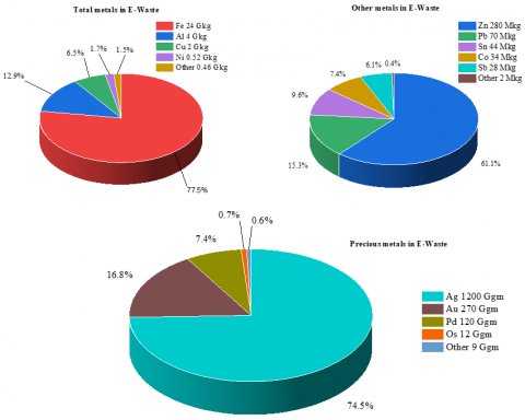
Despite the advances in green nanotechnology, there remain significant challenges in reducing costs, scaling up production, and utilizing waste-derived resources [10]. Electronic waste (e-waste), which has dramatically increased worldwide due to the rapid growth of the electronics industry, contains valuable metals including gold. However, conventional recovery methods are expensive and environmentally harmful [11]. Limited studies have investigated the possibility of integrating e-waste recycling with green nanoparticle synthesis using plant extracts as eco-friendly reducing agents [12]. Thus, there is a need to develop efficient, low-cost, and sustainable strategies that simultaneously address the problem of e-waste accumulation and enable the production of functional nanomaterials [13]. In 2022, the global total amount of e-waste was estimated to be approximately 31 billion kilograms, of which approximately 19 billion kilograms had been recycled and processed, as shown in Figure 1. This demonstrates the scale of global e-waste management and highlights its importance as a secondary source of precious metals, particularly gold. Given that the gold content of some electronic components is far higher than that of natural ores, recovering gold from e-waste not only contributes to resource sustainability but also provides a viable alternative to traditional mining [14].
Figure 1. Metals recovered from e-waste according to e-waste management [15]
The present study aims to investigate the synthesis of gold nanoparticles from electronic waste using cinnamon stick extract as a natural reducing and stabilizing agent. The research focuses on assessing the efficiency of this green approach under different reaction conditions, such as temperature and extract concentration, to control nanoparticle size and morphology. Furthermore, the study seeks to characterize the structural and optical properties of the synthesized nanoparticles using advanced analytical techniques, including UV–Vis spectroscopy, AFM, and XRD. This research is significant because it addresses two critical issues simultaneously: the environmental challenges associated with electronic waste and the demand for sustainable nanomaterial production. By utilizing electronic waste as a resource and employing cinnamon extract for green synthesis, this approach provides a cost-effective and eco-friendly alternative to conventional methods. The findings are expected to contribute to the advancement of green nanotechnology, promote waste valorization, and open new opportunities for applications of gold nanoparticles in medicine, sensing, and other nanotechnology-driven fields.
2.1 Gold deposition from electronic waste
The first method (mobile phone SIM): Mobile phone SIM cards were carefully collected and accurately weighed. 0.7 g of the SIM card chip was placed in a 1000 mL borosilicate glass beaker. To completely submerge the sample, concentrated hydrochloric acid (37%) was added, followed by a small amount of deionized water to ensure optimal mixing. Next, 50 mL of concentrated nitric acid (65-70%) was carefully added to form aqua regia. The mixture was gently stirred with a glass rod to ensure uniform interaction between the acid and the metal components in the SIM card chip. The reaction mixture was then exposed to direct sunlight, which served as a sustainable, clean heat source to promote the dissolution of the metal components. After approximately 48 hours, the solution color changed noticeably to dark green, indicating the formation of a gold-containing complex. A small amount of nitric acid (HNO₃) was then added to ensure complete dissolution of any remaining metal. The solution was then filtered through commercially available filter paper to remove any undissolved solid residue [16].
The resulting filtrate, containing dissolved gold ions, was treated with a freshly prepared solution of distilled sodium metabisulfite (Na₂S₂O₅). Additional distilled water was added to improve mixing and ensure uniform precipitation. This treatment resulted in a black, granular precipitate of metallic gold, which remained on the filter paper. The collected solid residue was carefully dried under ambient conditions, then weighed and analyzed to determine the gold content [17].
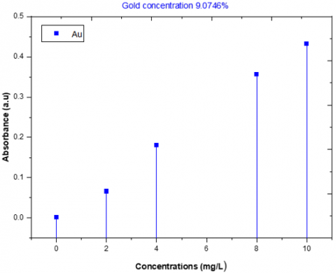
Gold (Au) content in samples is determined using atomic absorption spectrometry (AAS) at a wavelength of 242 nm using a gold-specific hollow cathode lamp. Standard solutions are prepared to generate a calibration curve, and samples are chemically treated and transferred into appropriate solutions [18]. By comparing the sample absorbance with the calibration curve, the gold concentration is determined and expressed as a percentage. Atomic absorption spectrometry (AAS) revealed the gold content of the mobile phone SIM sample, with a measured concentration of 9.0746% (Figure 2).
Figure 2. Gold percentage measured by atomic absorption spectrometry for the first method
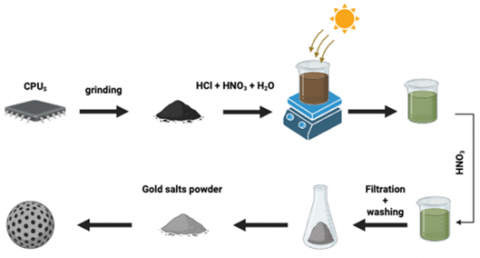
Figure 3. A diagram showing how to prepare gold salts from electronic waste
The second method (CPUS): To improve gold recovery, the precipitation process was repeated on freshly collected CPU samples, which are known to contain higher concentrations of precious metals. The CPU chips were cleaned, weighed, and treated under conditions similar to those used for SIM cards, including soaking in aqua regia and exposure to sunlight to promote dissolution, as shown in Figure 3 [19].
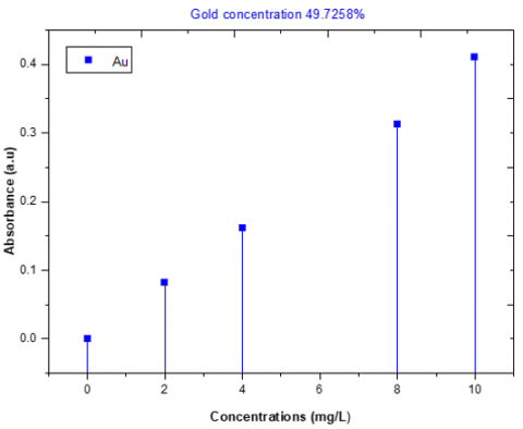
After filtration, the gold-containing filtrate was treated with sodium metabisulfite under controlled conditions to precipitate the metallic gold. The precipitate was collected, dried, and quantitatively analyzed. Atomic absorption spectroscopy (AAS) revealed a significantly higher gold content in the CPU sample, with a measured concentration of 49.72% (Figure 4).
These results clearly demonstrate that CPUs are a more efficient and higher-yielding source of gold than SIM cards. The improved recovery highlights the potential of CPU scrap as a practical and economically viable material for producing gold nanoparticles and other nanotechnology applications.
Figure 4. Gold percentage measured by atomic absorption spectrometer for the second method
2.2 Preparing herbal extract
Cinnamon stick samples were thoroughly washed, air-dried in a cool place, and then ground into a fine powder using a dedicated grinder. To prepare the extract, 5 g of cinnamon powder was mixed with 150 mL of deionized water in a 250 mL round-bottom flask. The mixture was placed on a magnetic stirrer with a reflux condenser and stirred continuously at 50℃ for 2 h. After extraction, the solution was filtered to remove solid residues, and the clear extract was collected for subsequent gold nanoparticle synthesis [20].
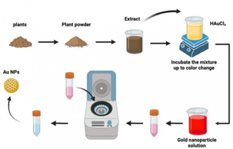
The gold used in this study was extracted from electronic waste. To prepare the gold salt solution, 0.1 g of the extracted gold was dissolved in a small amount of aqua regia (a mixture of concentrated nitric acid and hydrochloric acid) to obtain a gold (III) chloride solution (HAuCl₄). This solution was then diluted with deionized water to a final volume of 25 ml. To synthesize the nanoparticles, 150 ml of the previously prepared plant extract was added to a 250 ml beaker equipped with a magnetic stirrer. Subsequently, 3.6 ml of the HAuCl₄ solution was added to the extract, and the reaction mixture was heated to 60-70℃ and stirred for less than an hour. The formation of gold nanoparticles was confirmed by visually observing the reaction progress through color change [21], as shown in Figure 5.
Figure 5. A diagram showing the method of preparing gold nanoparticles using plant extracts
4.1 UV-vis spectroscopy
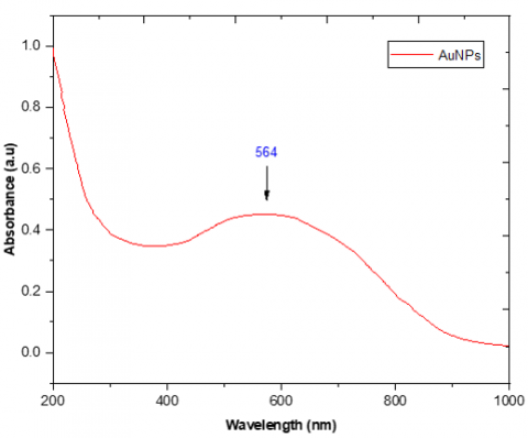
The successful formation of gold nanoparticles was verified by recording absorption spectra in the wavelength range of 400-800 nm using UV-vis spectroscopy [22]. As shown in Figure 6, the nanoparticles exhibited a clear surface plasmon resonance (SPR) band at 564 nm, which originates from the collective oscillation of free electrons in the conduction band. The position and sharpness of this SPR peak are characteristic of gold nanoparticles and are directly affected by the particle size, shape and degree of aggregation. The maximum absorption observed at approximately 564 nm indicates the presence of a strong plasmon response, confirming the presence of uniformly distributed nanoparticles with minimal aggregation. Since solid gold does not exhibit such obvious optical properties, the appearance of this absorption band in the visible light range further proves the formation of nanoparticles. In addition, the observed peak position is highly consistent with the values of previous studies [23]. This indicates that the biosynthesized nanoparticles exhibit optical properties comparable to those prepared by traditional chemical methods. These results highlight the effectiveness of plant extracts in reducing gold ions and stabilizing nanoparticles, thereby creating materials with desirable optical properties for potential applications in sensing, catalysis, and biomedicine.
Figure 6. UV-vis analysis of gold nanoparticles prepared using plant extracts
4.2 Atomic force microscopy (AFM)
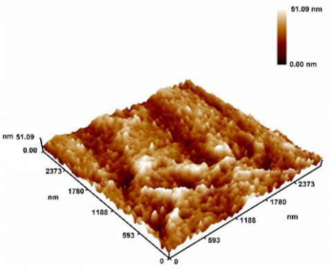
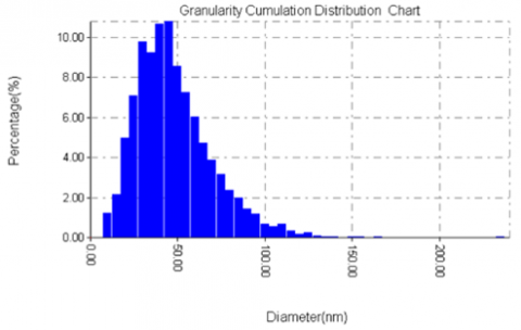
Atomic force microscopy (AFM) is considered one of the most powerful techniques for studying the surface morphology and topological features of nanostructured materials [24]. AFM generates three-dimensional images by scanning the sample surface, providing high-precision information about the particle shape, size, and distribution [25]. Figure 7 shows a three-dimensional AFM image of biosynthesized gold nanoparticles. Analysis shows that the particles are mainly spherical, relatively well dispersed, and have minimal aggregation. Figure 8 illustrates average particle size is approximately 51 nm, which is within the typical nanoscale range of gold nanoparticles [26].
Figure 7. AFM images of gold nanoparticles prepared using plant extracts
Figure 8. AFM images of gold nanoparticles prepared using plant extracts
Overall, AFM analysis not only confirms the spherical shape and nanoscale size of the synthesized gold nanoparticles but also supports the results of UV-visible spectroscopy, providing comprehensive evidence for successful synthesis. The combined application of AFM with spectroscopic and microscopic techniques underscores the reliability of the biosynthetic method and provides a deeper understanding of the nanoparticle structure and optical properties.
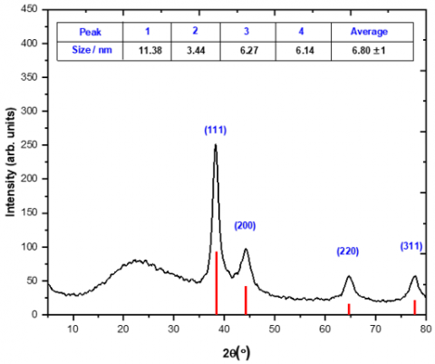
4.3 X-RAY diffraction (XRD)
The crystal structure and phase purity of gold nanoparticles (AuNPs) synthesized from plant extracts were investigated by X-ray diffraction (XRD). The diffraction pattern shown in Figure 9 shows characteristic Bragg reflections corresponding to the (111), (200), and (220) planes, confirming the face-centered cubic (FCC) structure of the gold crystals. The (111) reflection has the highest intensity, thus dominating the preferred orientation. This is consistent with JCPDS standard card No. 04-0784 and is consistent with previously published research results [27]. The average crystal size of the gold nanoparticles was calculated using the Scherrer equation:
$D=\frac{K \lambda}{\beta \cos \theta}$ (1)
where, D is the crystal size, K is the shape factor (0.9), λ is the X-ray wavelength, β is the half-maximum width (FWHM) of the diffraction peak, and θ is the Bragg angle [28].
Figure 9. XRD results of gold nanoparticles prepared using plant extracts
The crystal size estimated from the main peak (111) is in the nanometer range and agrees well with the particle size determined by atomic force microscopy (AFM) measurements (≈ 51 nm). This agreement between XRD and AFM confirms the reliability of the biosynthesis method and indicates that the nanoparticles have well-defined crystalline domains. In addition, the observed crystallite size correlates with the optical properties determined by UV-visible spectroscopy. A strong and sharp surface plasmon resonance (SPR) band at 564 nm supports the formation of nanoscale crystalline particles, as the SPR behavior is highly dependent on particle size and crystallinity. The consistency of XRD, AFM, and UV-vis data provides comprehensive evidence for the successful synthesis of morphologically uniform, stable, and crystalline gold nanoparticles. Importantly, no other peaks related to impurities were detected, except for some weak signals caused by residual phytochemicals in plant extracts, which act as reducing agents and stabilizers during the synthesis process [29]. The absence of obvious external reflections further confirms the purity of the biosynthesized gold nanoparticles. In summary, XRD analysis shows that the prepared gold nanoparticles have a highly crystalline FCC structure dominated by the (111) orientation, and the grain size determined by the Scherrer equation is consistent with the morphological and optical results. These findings emphasize the efficiency of the green synthesis method and the applicability of the nanoparticles for a variety of applications such as catalysis, optics, and biomedicine.
This study demonstrates the feasibility of utilizing e-waste as a valuable and environmentally friendly source for the extraction of gold and the subsequent synthesis of high-purity, highly stable nanoparticles. The method employed involves a series of carefully designed physical and chemical separation steps, optimized based on the composition of the selected e-waste. The results confirm that e-waste can be a cost-effective alternative to traditional chemical precursors and can produce gold nanoparticles with desirable physical and chemical properties. Furthermore, the biosynthesized nanoparticles exhibit promising properties, making them suitable for a wide range of applications, including optical devices, biomedical systems, and catalytic processes. Importantly, this approach not only facilitates the synthesis of functional nanomaterials but also helps alleviate the environmental issues associated with e-waste disposal. Overall, the results highlight a sustainable and economically viable strategy for developing nanomaterials through the environmentally friendly recycling of e-waste.
[1] Alhiti, L.S., Ali, I.H. (2025). Photodetector fabrication and characterization of gold-cerium oxide nanoparticles for next-generation high-efficiency devices. Journal of Physics: Conference Series, 2974(1): 012033. https://doi.org/10.1088/1742-6596/2974/1/012033
[2] Burlec, A.F., Corciova, A., Boev, M., Batir-Marin, D., et al. (2023). Current overview of metal nanoparticles’ synthesis, characterization, and biomedical applications, with a focus on silver and gold nanoparticles. Pharmaceuticals, 16(10): 1410. https://doi.org/10.3390/ph16101410
[3] Abbas, R., Luo, J., Qi, X., Naz, A., et al. (2024). Silver nanoparticles: Synthesis, structure, properties and applications. Nanomaterials, 14(17): 1425. https://doi.org/10.3390/nano14171425
[4] Altammar, K.A. (2023). A review on nanoparticles: Characteristics, synthesis, applications, and challenges. Frontiers in Microbiology, 14: 1155622. https://doi.org/10.3389/fmicb.2023.1155622
[5] Kumari, S., Raturi, S., Kulshrestha, S., Chauhan, K., et al. (2023). A comprehensive review on various techniques used for synthesizing nanoparticles. Journal of Materials Research and Technology, 27: 1739-1763. https://doi.org/10.1016/j.jmrt.2023.09.291
[6] Boruah, J.S., Devi, C., Hazarika, U., Reddy, P.V.B., Chowdhury, D., Barthakur, M., Kalita, P. (2021). Green synthesis of gold nanoparticles using an antiepileptic plant extract: In vitro biological and photo-catalytic activities. RSC Advances, 11(45): 28029-28041. https://doi.org/10.1039/D1RA02669K
[7] Pedroso‐Santana, S., Fleitas‐Salazar, N. (2023). The use of capping agents in the stabilization and functionalization of metallic nanoparticles for biomedical applications. Particle & Particle Systems Characterization, 40(2): 2200146. https://doi.org/10.1002/ppsc.202200146
[8] Alhiti, L.S., Hashim, E.H., Abdullah, F.H. (2025). Green methods for gold nanoparticle synthesis: Properties, characterization, and diverse applications–Review article. Humanities & Natural Sciences Journal, 6(3): 249-265. https://doi.org/10.53796/hnsj63/12
[9] Khan, N.A., López-Maldonado, E.A., Majumder, A., Singh, S., et al. (2023). A state-of-art-review on emerging contaminants: Environmental chemistry, health effect, and modern treatment methods. Chemosphere, 344: 140264. https://doi.org/10.1016/j.chemosphere.2023.140264
[10] Alsaiari, N.S., Alzahrani, F.M., Amari, A., Osman, H., Harharah, H.N., Elboughdiri, N., Tahoon, M.A. (2023). Plant and microbial approaches as green methods for the synthesis of nanomaterials: Synthesis, applications, and future perspectives. Molecules, 28(1): 463. https://doi.org/10.3390/molecules28010463
[11] Ahmed, F.K., Kalia, A., Ahmad, A., Alghuthaymi, M.A., Abd-Elsalam, K.A. (2023). Marine fungi and yeast: A green approach for production of bionanoparticles. Fungal Cell Factories for Sustainable Nanomaterials Productions and Agricultural Applications, 337-360. https://doi.org/10.1016/B978-0-323-99922-9.00016-7
[12] Vijayaram, S., Razafindralambo, H., Sun, Y.Z., Vasantharaj, S., Ghafarifarsani, H., Hoseinifar, S.H., Raeeszadeh, M. (2024). Applications of green synthesized metal nanoparticles—A review. Biological Trace Element Research, 202(1): 360-386. https://doi.org/10.1007/s12011-023-03645-9
[13] Huston, M., DeBella, M., DiBella, M., Gupta, A. (2021). Green synthesis of nanomaterials. Nanomaterials, 11(8): 2130. https://doi.org/10.3390/nano11082130
[14] Rao, M.D., Singh, K.K., Morrison, C.A., Love, J.B. (2020). Challenges and opportunities in the recovery of gold from electronic waste. RSC Advances, 10(8): 4300-4309. https://doi.org/10.1039/C9RA07607G
[15] Hagelüken, C., Lee-Shin, J.U., Carpentier, A., Heron, C. (2016). The EU circular economy and its relevance to metal recycling. Recycling, 1(2): 242-253. https://doi.org/10.3390/recycling1020242
[16] Saeed, Z., Pervaiz, M., Ejaz, A., Hussain, S., Shaheen, S., Shehzad, B., Younas, U. (2023). Garlic and ginger extracts mediated green synthesis of silver and gold nanoparticles: A review on recent advancements and prospective applications. Biocatalysis and Agricultural Biotechnology, 53: 102868. https://doi.org/10.1016/j.bcab.2023.102868
[17] Kinsman, L.M., Ngwenya, B.T., Morrison, C.A., Love, J.B. (2021). Tuneable separation of gold by selective precipitation using a simple and recyclable diamide. Nature Communications, 12(1): 6258. https://doi.org/10.1038/s41467-021-26563-7
[18] Giertyas, C.J., Silva, V.E., Oliveira, M.J.D., Freire, E.S., et al. (2022). Atomic absorption spectrometry as an alternative to determine the presence of gold nanoparticles on or in silica matrix. Journal of the Brazilian Chemical Society, 33: 406-412. https://doi.org/10.21577/0103-5053.20210158
[19] Hamelian, M., Varmira, K., Karmakar, B., Veisi, H. (2023). Catalytic reduction of 4-nitrophenol using green synthesized silver and gold nanoparticles over thyme plant extract. Catalysis Letters, 153(8): 2341-2351. https://doi.org/10.1007/s10562-022-04164-3
[20] Gowri, V.M., Kumar, P.S., Shankar, V.U., Thongmee, S., Rangasamy, G. (2024). Environmental analysis of nitrobenzene using newly synthesized anisotropic gold nanostructures: Reaction kinetics and electrocatalytic activity. Journal of Molecular Liquids, 408: 125303. https://doi.org/10.1016/j.molliq.2024.125303
[21] Semwal, V., Jensen, O.R., Bang, O., Janting, J. (2023). Investigation of performance parameters of spherical gold nanoparticles in localized surface plasmon resonance biosensing. Micromachines, 14(9): 1717. https://doi.org/10.3390/mi14091717
[22] Shanwaz, M.M., Shyam, P. (2023). Anti-bacterial effect and characteristics of gold nanoparticles (AuNps) formed with Vitex negundo plant extract. Applied Biochemistry and Biotechnology, 195(3): 1630-1643. https://doi.org/10.1007/s12010-022-04217-8
[23] Plaskova, A., Mlcek, J. (2023). New insights of the application of water or ethanol-water plant extract rich in active compounds in food. Frontiers in Nutrition, 10: 1118761. https://doi.org/10.3389/fnut.2023.1118761
[24] alhiti, L.S., Jawad, R.A., Abd Alwaahed, R.A., Sobhi, H.M. (2024). Study of the effect of thin layer thickness on the structural properties of copper phthalocyanine (CuPc) films prepared by vacuum thermal Evaporation Method. Al-Kitab Journal for Pure Sciences, 8(1): 81-91. https://doi.org/10.32441/kjps.08.01.p8
[25] Sharma, R., Suhendra, N.F., Jung, S.H., Lee, H.I. (2023). Gold recovery at ultra-high purity from electronic waste using selective polymeric film. Chemical Engineering Journal, 451: 138506. https://doi.org/10.1016/j.cej.2022.138506
[26] Cotty, S.R., Kim, N., Su, X. (2023). Electrochemically mediated recovery and purification of gold for sustainable mining and electronic waste recycling. ACS Sustainable Chemistry & Engineering, 11(9): 3975-3986. https://doi.org/10.1021/acssuschemeng.3c00227
[27] Pang, C., Lin, M., Wu, Y., Ruan, J. (2023). Enhanced Au bioleaching from waste central processing unit (CPU) slots in a bioreactor with 10 mA direct current. Resources, Conservation and Recycling, 198: 107171. https://doi.org/10.1016/j.resconrec.2023.107171
[28] Afdal, M., Kasim, A., Alimon, A.R., Abdullah, N. (2023). Investigation of the antioxidant activity of cinnamon bark extracted with different solvents. Jurnal Ilmiah Ilmu-Ilmu Peternakan, 26(1): 68-79. https://doi.org/10.22437/jiiip.v26i1.24368
[29] Osman, A.I., Zhang, Y., Farghali, M., Rashwan, A.K., Eltaweil, A.S., Abd El-Monaem, E.M., Mohamed, I.M.A., Badr, M.M., Ihara, I., Rooney, D.W., Yap, P.S. (2024). Synthesis of green nanoparticles for energy, biomedical, environmental, agricultural, and food applications: A review. Environmental Chemistry Letters, 22(2): 841-887. https://doi.org/10.1007/s10311-023-01682-3