Athar Al-Azzawi*![]() | Osman Nuri Uçan
| Osman Nuri Uçan![]()
© 2025 The authors. This article is published by IIETA and is licensed under the CC BY 4.0 license (http://creativecommons.org/licenses/by/4.0/).
OPEN ACCESS
Schizophrenia is a mental disorder condition that causes patients to become distracted from reality. Over time, the patient loses his cognitive and social abilities to communicate with the outside world. Due to machine learning's strong ability to analyze complicated brain data, it has become an increasingly important tool in recent years. This study considers the brain's neurologic signals in the resting state in two scenarios to classify schizophrenia disease by electroencephalography (EEG). The performed scenarios were to investigate the impact of selecting electrodes randomly (5 electrodes and 8 electrodes) and comparing it with applying the principal component analysis (PCA), utilizing four algorithms to extract features: Fast Fourier Transform (FFT), Approximate Entropy (ApEn), Log Energy Entropy (LogEn), and Shannon Entropy (ShnEn). We used publicly available datasets with 19 EEG channels consisting of two classes, which are schizophrenia and health control, using a one-second epoch window size. We applied a band-pass filter to decompose the EEG signals into five sub-bands. Also, the L2-normalization method has been applied to the derived features, which positively impacted the outcomes. The features were applied to three classifiers named K-nearest neighbor (KNN), support vector machine (SVM), and quadratic discriminant analysis (QDA). From all the scenarios, the five-electrode with random selection showed remarkable results of 99% using the SVM classifier in all evaluation metrics with LogEn+ Bandpass features.
schizophrenia, EEG, classification, SVM, KNN, QDA
The human nervous system is the main system that determines lifestyle and general behaviour, including decisions and emotional control. The brain may be exposed to various accidents or diseases that lead to abnormalities in the brain's neural structure. The severity of the abnormalities varies depending on many factors, such as the patient's age, the family’s medical history, or the brain injury's location, which leads to multiple differences occurring between one patient and another in the behaviour and diagnoses. Neurological diseases that affect the neural cells, for instance, epilepsy, schizophrenia (SZ), Parkinson’s, and Alzheimer's, effect different locations of the brain and have a specific effect on brain function that is discovered and diagnosed through symptoms, tests, and using different tools. One of the worst diseases that may completely destroy the neural system in the brain is schizophrenia.
Delusions, hallucinations, depression, and anxiety are just a few of the symptoms that come along with schizophrenia, which causes a severe behavioral change in character [1]. These symptoms can be clearly seen and distinguished between the ages of 16 and 30 and can be utilized to determine the patient's state, where timely detection is a crucial part of the patient's healing process [2, 3]. Besides, according to the official data of the World Health Organization, there are around 21 million patients, which is around 1% of people worldwide who are afflicted with this illness [4]. The traditional process of diagnosing schizophrenia is considered a complex process, according to some factors such as the psychiatrists' experience and the different case responses between one patient and another.
Indeed, the neuroimaging techniques field provided help in discovering various brain disorders like schizophrenia, which is considered a time-consuming process, encouraging scientific researchers to accomplish new medical aims.
Electroencephalography (EEG) devices have made tremendous strides in the diagnosis of nervous system disorders in recent years, including epilepsy [5], Alzheimer's disease [6-9], and schizophrenia [10, 11]. The non-invasive nature of EEG has led to its adoption as the ideal tool for recording and collecting the electrical activity of the brain, which in turn provides brain dimensions that contain huge amounts of data, which gives the capability to diagnose different brain diseases. Meanwhile, deep learning and machine learning (ML) are the most important methods in the medical domain that offer advanced processing and evaluation of diverse medical datasets, including brain signals [12].
ML used the feature extraction approach to retrieve the hidden information of signals from the data, where these features provide the possibility of obtaining clearer signals and identifying and measuring the most relevant data, which will be used with the frequency domain or the time domain [13, 14].
Many studies have addressed the subject of classifying schizophrenia based on EEG and analyzing the brain signals captured using the global 10-20 electrode system. The analysis of the relationship between these electrodes represents the nature of neural communication in the brain, which in turn leads to the detection of a person's mental health. According to the brain regions and the different functions and channels associated with them, the features in each channel of EEG are analyzed and extracted separately, and these features are subsequently linked and then classified.
Consequently, this study investigates the feasibility and importance of electrodes through the model's ability to classify schizophrenia without compromising classification accuracy by reducing electrodes and selecting electrodes randomly.
This approach supports the ability of electrodes for non-sophisticated devices as well as for devices with few electrodes, such as Emotiv Insights (5 channels), Muse EEG (4-6 channels), Neurosky Mindwave (1 channel), and InteraXon Muse S (4 channels). Clinically, random electrode reduction offers advantages by mitigating bias related to electrode type and placement. Furthermore, the proposed methodology simplifies traditionally employed techniques.
The manuscript is written in the following format: related work in Section 2, which included a comparison with the literature that has classified schizophrenia using different datasets and techniques. In Section 3, we illustrated the materials and methods: Firstly, we described the EEG dataset; secondly, we mentioned the preprocessing methods used; thirdly, we summarized the techniques of feature extraction; and finally, the normalization techniques applied to the dataset. In Section 4, we explained the classification through the description of machine learning classifiers; then in Section 5, we presented and discussed the results, and finally, the conclusion is clarified in Section 6.
Researchers in the last era increased their work that aimed to classify schizophrenia disorder utilizing a variety of methods, including MRI [15], eye tracking [16], facial features [17, 18], tracking handwriting [19], and schizophrenia EEG signal [20]. Various studies have been done on the use of several characteristics in machine learning to classify EEG data for the diagnosis of schizophrenia. They employed several ML models and achieved differing degrees of accuracy. Depending on the topic and the data utilized for classification, popular machine learning models in these investigations include SVM, RF, and Artificial Neural Networks (ANNs), with accuracy varying from 70% to over 90% [21].
One of these published papers that aimed to classify schizophrenia was done by Krishnan et al. [20], using datasets obtained from the Repository for Open Data (RepOD) with two classes: schizophrenia patients and healthy controls with 28 subjects. The EEG signals were recorded for 15 minutes, using 19 channels. The highest result achieved was by the SVM with Radial Basis Function, with an accuracy of up to 93%. Siuly et al. [22] used an EEG dataset including two groups: patients diagnosed with schizophrenia and healthy controls.
The signals underwent empirical mode decomposition, and the most significant features were then selected using the Kruskal-Wallis test. The SVM was then provided with all the attributes for classification. The highest percentage achieved was 93.21%. De Miras et al. [23] evaluated if machine learning methods may be helpful in the diagnosis of the condition. Additionally, they have created a pipeline for processing. Support vector machines (SVM), k-nearest neighbors (KNN), logistic regression (LR), decision trees (DT), and random forest (RF), were the five machine-learning techniques that they examined. SVM yielded the greatest results 89%.
Hartini and Rustam [24] proposed fuzzy kernel c-means using data from Northwestern University, including 171 schizophrenia and 221 non-schizophrenia samples. Using RBF and polynomial kernel functions, k-fold cross-validation was used for evaluation. Results showed that the RBF kernel with σ=0.01 and σ=1 performed better than the polynomial kernel with similar running time. Furthermore, among the five classification strategies employed by Khare et al. [25]-including SVM, KNN, DA, ensemble methods, and decision trees — SVM achieved the highest accuracy of 88%. They divide nonstationary EEG signals into Fourier spectrum modes, pull out linear and nonlinear time domain features, and use the Kruskal-Wallis test to choose highly discriminant features. This helps them put people into two groups: healthy and those with schizophrenia.
Hassan et al. [26] used a publicly available EEG signals dataset from Warsaw's Institute of Psychiatry and Neurology to automate the identification of schizophrenia using a channel selection mechanism based on a rigorous performance analysis of the Convolutional Neural Network. They used CNN in conjunction with other ML classifiers to train the classification model. Their highest findings reveal that LR and CNN yield 98% accuracy. Supakar et al. [27] proposed a DL model using RNN-LSTM to analyze the EEG signal data to diagnose schizophrenia. EEG signal data of 45 SZ patients and 39 healthy subjects. They had two scenarios: a complete feature set and a reduced feature set, which achieved an accuracy of 98% and 93%, respectively.
Moreover, Li et al. [28] introduced an innovative EEG data mapping technique using Vision Transformer (LeViT) as both a feature extractor and classifier for the early detection of schizophrenia. Their data was private, and they achieved 98%.
Table 1 presents and compares studies using various datasets, when all channels are used, with different techniques to classify EEG signals with SZ and healthy controls.
Previous studies have explored the researchers' utilization of various methods to categorize schizophrenia disorder and various datasets, and the outcomes were inconsistent and fell short of expectations. As a result, we examine and compare the possibility of improving the diagnostic precision of schizophrenia by using an electrode reduction technique with PCA as a traditional technique.
Table 1. Classifying schizophrenia utilizing various datasets
|
Ref. |
Chan. |
Dataset |
Methods |
Acc% |
Spe% |
Sen% |
|||||
|
SZ |
Free |
||||||||||
|
[29] |
64 |
49 |
32 |
RF |
81 |
NA |
NA |
||||
|
[30] |
16 |
37M, 10F |
14M, 11F |
SVM |
90 |
91 |
89 |
||||
|
[31] |
NA |
31M, 19F |
32M, 18F |
non-linear SVM |
73 |
56 |
62 |
||||
|
[32] |
16 |
39 |
45 |
MDC-CNN |
93 |
93 |
93 |
||||
|
[33] |
8 |
48 |
24 |
RF |
68 |
NA |
NA |
||||
|
[34] |
256 |
33M, 37F |
47M, 28F |
SVM |
82 |
81 |
82 |
||||
|
[35] |
64 |
36M, 18F |
3 M, 24F |
Hybrid DNN |
99 |
NA |
NA |
||||
|
[36] |
10 |
49 |
32 |
KNN |
99 |
90 |
95 |
||||
|
[37] |
64 |
49 |
32 |
CNN |
92 |
NA |
NA |
||||
|
[38] |
20 |
37M, 25F |
38M, 32F |
KNN |
96 |
98 |
95 |
||||
|
[39] |
NA |
158 |
76 |
Ensemble |
87 |
65 |
98 |
||||
|
[40] |
64 |
41M, 8F |
67M, 14F |
HDSS |
92 |
91 |
97 |
||||
|
[2] |
16 |
39 |
45 |
CNN |
98 |
NA |
NA |
||||
|
[41] |
64 |
13 |
11 |
SVM |
89 |
90 |
88 |
||||
|
[42] |
32 |
310 |
205 |
XGB |
94 |
NA |
NA |
||||
|
[43] |
16 |
39 |
45 |
AdaBoost |
99 |
100 |
98 |
||||
|
[44] |
64 |
49 |
32 |
DT |
99 |
95 |
95 |
||||
|
[23] |
31 |
9M, 20F |
13M, 7F |
SVM |
89 |
90 |
63 |
||||
|
[45] |
32 |
215M, 97F |
176M, 144F |
RBF |
93 |
NA |
NA |
||||
|
[46] |
19 |
626 |
516 |
KNN |
97 |
NA |
NA |
||||
|
[47] |
16 |
39 |
45 |
CNN |
99 |
99 |
100 |
||||
|
[48] |
64 |
36 |
22 |
CNN |
98 |
98 |
98 |
||||
Note: M refers to male and F refers to female, chan. refers to channel
3.1 Dataset
The EEG dataset used was accessible to the public [49]. Table 2 presents the details of the total signal used containing 28 subjects, 14 from each group: schizophrenia and healthy controls. The sampling frequency of the EEG dataset recording is 250Hz. The montage was executed with a conventional 10-20 system. Moreover, the dataset was assembled utilizing the subsequent 19 EEG channels: Fp1, Fp2, T3, C3, Cz, C4, T4, T5, P3, Pz, P4, T6, O1, and O2 [50].
Table 2. The used datasets details
|
Features |
Values |
|
|
SZ |
♂ |
7 |
|
♀ |
7 |
|
|
HC |
♂ |
7 |
|
♀ |
7 |
|
|
Mean age (SZ) |
28.1±3.7 years |
|
|
Mean age (HC) |
27.75±3.15 years |
|
|
SZ |
Mean age (♂) |
27.9±3.3 years |
|
Mean age (♀) |
28.3±4.1 years |
|
|
HC |
Mean age (♂) |
26.8±2.9 years |
|
Mean age (♀) |
28.7±3.4 years |
|
|
EEG segment |
15min |
|
|
No. of segments |
21702 |
|
|
No. of segments without artefacts |
30 |
|
3.2 Preprocessing
Preprocessing is a crucial step in ML due to EEG signals often containing a combination of noise resulting from several different factors, which in turn affects classification accuracy. There are several ways to reduce the noise and improve the EEG signals, for instance, by applying filters to enhance the signal quality.
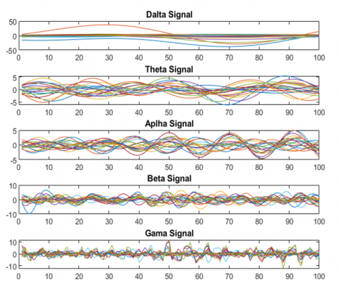
Thus, the bandpass filter was utilized to decompose the EEG data into five frequency sub-bands: beta rhythm (31-31Hz), alpha rhythm (10-14Hz), theta rhythm (5-9Hz), delta rhythm (0.1-4Hz), and gamma rhythm (32-100Hz).
The bandpass filter works on filtering out very high or low frequencies, as presented in Figure 1 with the 19 channels of the EEG signal after the band-pass filter was applied.
Figure 1. The impact of band-pass filtering on EEG signals
The brain's electrical activity generates billions of signals, so a multitude of electrodes are used to record these signals; adding more electrodes has a positive impact on the classification outcomes. It is important to note that there is no defined minimum or maximum number of electrodes, rules, or specific standards that should be used, as this varies depending on the kind of disorder being dealt with.
Hence, we examined our proposed models by the two electrode scenarios (8 and 5 electrodes); the electrodes were selected randomly to assess the suggested approach and determine whether or not it might yield encouraging outcomes. Additionally, we evaluated the computational efficiency of the classifier's final output, as well as estimated the training and speed time.
3.3 Feature extraction techniques
Different feature extraction techniques have been put out in the literature, such as entropy, approximate entropy, Shannon entropy, fuzzy entropy, fast Fourier transform, etc. Each one of these methods has a special mechanism for obtaining the feature from the signal. We identified the most commonly used methods with brain signals that helped identify subtle abnormalities associated with schizophrenia and combined these methods with our proposed approach to evaluate their effectiveness with varying constraints.
In this study, four extraction methods were calculated to extract the hidden features as presented in the following equations: Eq. (1) represents the mathematical formulation of Fast Fourier Transform (FFT), approximate entropy (ApEn) as shown in Eq. (2), log energy entropy (LogEn) by Eq. (3), and Shannon entropy (ShEn) as shown in Eq. (4).
FFT, LogEn, and ShEn were implemented with the band-pass filter; ApEn was applied in both cases with and without the band-pass filter. We selected these four techniques due to their established applications in EEG research and their accurate dealing with brain signals, which are explained in the following paragraphs.
FFT is one of the most important algorithms developed of all time, and the real reason for depending on FFT is its quick and effective method of denoising data. The FFT features have been implemented on the SZ EEG signals to convert the time domain to the frequency domain. Thus, it allows one to see the most prominent frequencies in the EEG signal and identify abnormalities or patterns that may appear in the brain wave frequencies [51].
$\begin{aligned} X(K)=\sum_{n=0}^{N-1} X[n] W_N^{k n} & =\sum_{n \text { even }} x(n) n w_N^{k n}+\sum_{n \text { odd }} x(n) w_N^{k n} \\ K & =0,1 \ldots \ldots, N-1,\end{aligned}$ (1)
$\begin{aligned} \operatorname{ApEn}(E, r, N)= & \frac{1}{(N-e+1)} \sum_{i-1}^{N-e+1} \log C_i^e(r) -\frac{1}{N-e} \sum_{i=1}^{N-e} \log C_i^{e+1}(r)\end{aligned}$ (2)
$\mathrm{ShEn}=\frac{H S h}{o g k}$ (3)
$\mathrm{E}=\sum_n \log \left(w_{i, j}^{n 2}\right)$ (4)
Entropy is the most frequently used feature to measure time-domain features. It is also widely used in disease detection by capturing any slight or subtle change in the brain signal, which assists when dealing with small data sizes. ApEn is defined as a measurement of the regularity or randomness of data in a time series and is used for short-length data due to its lower sensitivity to noise.
ShnEn is a time-domain complexity metric that does not rely on the signal spectrum and is similar to ApEn functionality. Since entropy may be used to ascertain the degree of randomness in the information, it is employed as the feature approach for schizophrenia due to ShEn decreasing with decreased neural complexity [52].
In signal processing, a common measure called LogEn, is used to extract relevant information. Since the frequency bands specific to schizophrenia have been identified, these bands can provide insights into the underlying neural systems. All these feature extraction methods are computed using a MATLAB routine.
3.4 Normalization
In general, normalization is the most common technique that deals with data in linear transformations. Its role is to rescale numerical features to a standard range to avoid larger values that may affect and bias the machine learning results. Although applying any normalization method is easy to implement, each method has strengths and weaknesses. Selecting the appropriate normalization method depends on some factors, such as the data and what is required from machine learning to obtain optimal results. In our work, we used L2 normalization to increase and enhance our accuracy results.
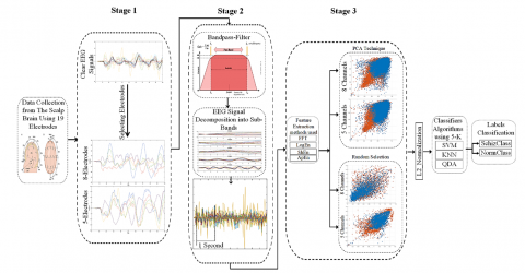
We used three machine learning (ML) classifiers, namely SVM, KNN, and QDA, together to classify the features computed by four different methods from the EEG signal using two scenarios for identifying a patient with schizophrenia (SZ). The main goal of our research is to achieve the highest possible accuracy regardless of the number and the location of the electrodes concerning different brain regions and prove that all channels are not necessary to achieve satisfactory results. Therefore, we implemented the 5 electrodes and 8 electrodes first by random selection, and then we implemented a traditional method, which is the PCA, as shown in Figure 2.
Figure 2. EEG signal data classification for schizophrenia with two classes: Healthy control and schizophrenia
4.1 SVM
In the field of machine learning, SVM is a popular choice for dealing with and processing various problems, most notably classification, as the strength of this approach lies in maximizing the margin separating the different classes, which leads to obtaining ideal accuracy in classification regardless of the type of data being dealt with. To lower classification error, certain unnecessary data are eliminated from the training data set at the ultra-optimal level, which affects the class borders. Multiple kernel tricks, such as polynomial, Gaussian, radial basis function (RBF), Laplace RBF, sigmoid, and Anove RBF, are used in SVM, making it a versatile tool.
4.2 K-nearest neighbors (KNN)
KNN is a non-parametric classification technique that finds the classifier's nearest neighbors by performing a distance check. Distance is calculated using the Euclidean Distance equation. During the training phase, the classifier check calculates the distance between the specific and the other data to categorize it. It gives it a unique label denoting its class, and KNN applies this to all of the data until all of the data is.
4.3 Quadratic discriminant analysis (QDA)
A machine learning and statistical classification classifier called QDA uses quadric surfaces to categorize two or more classes of disparate kinds of data. Compared to a linear classifier, this version is more suited. For every class, QDA specifically uses a Gaussian distribution.
By using the public database [49], we explore how bandwidth filtering and electrode reduction affect our ability to find schizophrenia-related EEG signals. We selected electrodes randomly, unlike previous studies that focused on identifying brain regions and selecting effective electrodes, which introduced monotony into the work and repeated the results. Most current studies now use all channels to obtain the highest possible classification accuracy to achieve the best computational efficiency in less time, which is often not the case when the electrodes are reduced. Therefore, our study investigates the effect of minimizing the data channel numbers in two different scenarios to achieve comparable results to studies that used all data channels.
Random channel selection is computationally inexpensive when compared to the traditional method (algorithms used that have the ability to rank channels or feature importance analysis), which is considered computationally expensive and helps to influence without prior knowledge of which are the most important channels.
Our experiments were based on 14 subjects for SZ and 14 subjects for healthy, so to maximize the data, we used one epoch window size for our two scenarios.
We examined the efficacy of four feature extraction methods: FFT, ApEn, LogEn, and ShnEn. Among the features, ApEn was applied twice: with and without the band-pass filter on the data. Then, the data was standardized using the L2-normalization approach and fed to three ML classifiers: SVM, KNN, and QDA. The original signals were recorded with 19 channels and a 250Hz frequency.
5.1 Experiment I: EEG classification based on five randomly selected electrodes
The brain's electrical activity generates billions of signals, so a multitude of electrodes are used to record these signals, and using more electrodes has a favourable impact on the classification outcomes. It is important to note that there is no defined minimum or maximum number of electrodes that should be used, as this varies depending on the kind of disorder being dealt with. As a result, there is no guideline, rule or specific standard for this. Therefore, we first tested the proposed models to evaluate our approach by reducing the number of electrodes to five, as the goal of choosing this number was to determine whether or not it could lead to encouraging results using a very limited electrode setup. And demonstrate the ability of the approach to extract and obtain information contained in a small set of EEG channels.
Classification was performed using the SVM, KNN, and QDA algorithms on the five electrodes for each class; the obtained confusion matrix values for the three classifiers are presented in Table 3, and the performance values are listed in Table 4.
Based on Table 4, the results show that when randomly selecting five electrodes, EEG signal classification with KNN with implementing LogEn outperformed the other two classifiers with results of 98%, 98%, and 99% for accuracy, sensitivity, and specificity, respectively.
Table 3. The confusion matrix obtained by the three classifiers with random 5-electrodes
|
Feature Name |
Classes Name |
SVM |
||
|
Predicted Class |
||||
|
FFT+Bandpass |
Actual class |
Sch |
1042 |
13703 |
|
Healthy |
12160 |
869 |
||
|
ApEn |
Sch |
4337 |
11496 |
|
|
Healthy |
10225 |
2804 |
||
|
ApEn+Bandpass |
Sch |
5093 |
10740 |
|
|
Healthy |
11247 |
1782 |
||
|
ShnEn+Bandpass |
Sch |
7176 |
8657 |
|
|
Healthy |
13181 |
963 |
||
|
LogEn+Bandpass |
Sch |
166 |
15666 |
|
|
Healthy |
12856 |
174 |
||
|
Feature Name |
Classes Name |
KNN |
||
|
Predicted Class |
||||
|
FFT+ Bandpass |
Actual Class |
Sch |
1402 |
13343 |
|
Healthy |
12184 |
845 |
||
|
ApEn |
Sch |
4095 |
11738 |
|
|
Healthy |
9782 |
3247 |
||
|
ApEn+Bandpass |
Sch |
8401 |
7432 |
|
|
Healthy |
12411 |
588 |
||
|
ShnEn+Bandpass |
Sch |
1297 |
14536 |
|
|
Healthy |
13312 |
832 |
||
|
LogEn+Bandpass |
Sch |
196 |
15636 |
|
|
Healthy |
12872 |
158 |
||
|
Feature Name |
Classes Name |
QDA |
||
|
Predicted Class |
||||
|
FFT+ Bandpass |
Actual Class |
Sch |
3580 |
11165 |
|
Healthy |
11804 |
1225 |
||
|
ApEn |
Sch |
7047 |
8786 |
|
|
Healthy |
10691 |
2338 |
||
|
ApEn+Bandpass |
Sch |
8072 |
7761 |
|
|
Healthy |
12667 |
362 |
||
|
ShnEn+Bandpass |
Sch |
9355 |
6478 |
|
|
Healthy |
11342 |
2802 |
||
|
LogEn+Bandpass |
Sch |
1910 |
13922 |
|
|
Healthy |
12781 |
249 |
||
Table 4. Classification performance results utilizing five random electrodes
|
Filter Condition |
Classifiers |
Feature Extraction Methods |
Evaluation Metrics |
||
|
Acc |
Sen |
Spe |
|||
|
With Bandpass |
SVM |
FFT |
93 |
92 |
94 |
|
KNN |
91 |
89 |
94 |
||
|
QDA |
82 |
76 |
90 |
||
|
SVM |
ApEn |
76 |
68 |
85 |
|
|
KNN |
68 |
59 |
92 |
||
|
QDA |
70 |
61 |
95 |
||
|
SVM |
ShEn |
72 |
64 |
89 |
|
|
KNN |
92 |
91 |
94 |
||
|
QDA |
59 |
54 |
69 |
||
|
SVM |
LogEn |
98 |
98 |
98 |
|
|
KNN |
98 |
98 |
99 |
||
|
QDA |
92 |
87 |
98 |
||
|
Without Bandpass |
SVM |
ApEn |
75 |
70 |
80 |
|
KNN |
74 |
70 |
78 |
||
|
QDA |
67 |
60 |
78 |
||
In contrast, SVM scored 98%, 98%, and 98%, and QDA scored 92%, 87%, and 98%, respectively, for accuracy, sensitivity, and specificity.
5.2 Experiment II: EEG classification based on eight randomly selected electrodes
In this scenario, we chose eight electrodes as an intermediate step between the first scenario of five electrodes and the previous studies mentioned in Section 2, which include more comprehensive electrodes. Here we verify the proposed approach to see if a small increase in the number of electrodes leads to a significant improvement in classification performance. Table 5 displays the confusion matrix obtained by using 8 electrodes, and Table 6 displays the performance values.
For this scenario reduction, the classification accuracy rates were 99%, 99%, and 95% for SVM, KNN, and QDA, respectively. The corresponding sensitivity and specificity of the SVM and KNN classifiers were identical at 99%, while QDA got 90% and 99%, respectively. Consequently, SVM outperformed the other classifiers.
Table 5. The confusion matrix was obtained by the three classifiers with random 8-electrodes
|
Feature Name |
Classes Name |
SVM |
||
|
Predicted Class |
||||
|
FFT+ Bandpass |
Actual class |
Sch |
504 |
15328 |
|
Healthy |
12543 |
486 |
||
|
ApEn |
Sch |
2753 |
13080 |
|
|
Healthy |
10997 |
2032 |
||
|
ApEn+Bandpass |
Sch |
1651 |
11379 |
|
|
Healthy |
11157 |
4676 |
||
|
ShnEn+Bandpass |
Sch |
4160 |
11673 |
|
|
Healthy |
12001 |
1028 |
||
|
LogEn+Bandpass |
Sch |
64 |
15768 |
|
|
Healthy |
12970 |
60 |
||
|
Feature Name |
Classes Name |
KNN |
||
|
Predicted Class |
||||
|
FFT+ Bandpass |
Actual Class |
Sch |
576 |
15256 |
|
Healthy |
12341 |
688 |
||
|
ApEn |
Sch |
2838 |
12995 |
|
|
Healthy |
10767 |
2262 |
||
|
ApEn+Bandpass |
Sch |
1053 |
11977 |
|
|
Healthy |
8763 |
7070 |
||
|
ShnEn+Bandpass |
Sch |
644 |
15189 |
|
|
Healthy |
12352 |
677 |
||
|
LogEn+Bandpass |
Sch |
96 |
15736 |
|
|
Healthy |
12979 |
51 |
||
|
Feature name |
Classes Name |
QDA |
||
|
Predicted Class |
||||
|
FFT+ Bandpass |
Actual Class |
Sch |
1511 |
14321 |
|
Healthy |
1192 |
1037 |
||
|
ApEn |
Sch |
6675 |
9158 |
|
|
Healthy |
11285 |
1744 |
||
|
ApEn+Bandpass |
Sch |
216 |
12814 |
|
|
Healthy |
7386 |
8447 |
||
|
ShnEn+Bandpass |
Sch |
11097 |
4736 |
|
|
Healthy |
12858 |
171 |
||
|
LogEn+Bandpass |
Sch |
1381 |
14451 |
|
|
Healthy |
12969 |
61 |
||
Table 6. Classification performance results utilizing eight random electrodes
|
Filter Condition |
Classifiers |
Feature Extraction Methods |
Evaluation Metrics |
||
|
Acc |
Sen |
Spe |
|||
|
With Bandpass |
SVM |
FFT |
96 |
96 |
96 |
|
KNN |
95 |
95 |
95 |
||
|
QDA |
91 |
88 |
93 |
||
|
SVM |
ApEn |
78 |
87 |
70 |
|
|
KNN |
71 |
89 |
62 |
||
|
QDA |
69 |
97 |
60 |
||
|
SVM |
ShEn |
82 |
74 |
91 |
|
|
KNN |
95 |
95 |
95 |
||
|
QDA |
61 |
53 |
96 |
||
|
SVM |
LogEn |
99 |
99 |
99 |
|
|
KNN |
99 |
99 |
99 |
||
|
QDA |
95 |
90 |
99 |
||
|
Without Bandpass |
SVM |
ApEn |
83 |
79 |
86 |
|
KNN |
82 |
79 |
85 |
||
|
QDA |
70 |
62 |
84 |
||
5.3 Experiment III: EEG classification based on PCA
Finally, we applied the principal component analysis (PCA); it preserves the most important features and reduces the dimensionality of the data by its formula illustrated in Eq. (5), but at the same time, these features may not be the most relevant or important for classifying the EEG data. This belongs to the PCA's lack of knowledge of the patterns that are important and required to function.
Tables 7 and 8 compare and demonstrate classifying EEG data for schizophrenia disorder by the confusion matrix for the two PCA scenarios, while Tables 9 and 10 show the performance results.
$\operatorname{cov}_{x, y}=\frac{\sum\left(x_i-\bar{x}\right)\left(y_i-\bar{y}\right)}{N-1}$ (5)
Given the earlier studies mentioned in section 2 and our study that investigated the limitations of random electrode selection and compared them to the PCA feature selection methods, this work stands out as a real demonstration of the validity of achieving impressive and unexpected results with fewer electrodes than expected, which demonstrates the existence of substantial discriminative information within this restricted grouping. This highlights the need to further explore the potential of EEG systems and the extent to which they can be extended.
Table 7. The confusion matrix was obtained using PCA with the 5-electrodes
|
Feature Name |
Classes Name |
SVM |
||
|
Predicted Class |
||||
|
FFT+ Bandpass |
Actual class |
Sch |
10929 |
2100 |
|
Healthy |
3061 |
11684 |
||
|
ApEn |
Sch |
7521 |
5508 |
|
|
Healthy |
4357 |
11476 |
||
|
ApEn+Bandpass |
Sch |
12744 |
285 |
|
|
Healthy |
7308 |
8525 |
||
|
ShnEn+Bandpass |
Sch |
7 |
14137 |
|
|
Healthy |
6 |
15827 |
||
|
LogEn+Bandpass |
Sch |
11447 |
1583 |
|
|
Healthy |
1978 |
13854 |
||
|
Feature Name |
Classes Name |
KNN |
||
|
Predicted Class |
||||
|
FFT+ Bandpass |
Actual Class |
Sch |
12274 |
755 |
|
Healthy |
1849 |
12896 |
||
|
ApEn |
Sch |
9782 |
3247 |
|
|
Healthy |
5357 |
10476 |
||
|
ApEn+Bandpass |
Sch |
11659 |
1370 |
|
|
Healthy |
7762 |
8071 |
||
|
ShnEn+Bandpass |
Sch |
8937 |
5207 |
|
|
Healthy |
5494 |
10339 |
||
|
LogEn+Bandpass |
Sch |
12689 |
341 |
|
|
Healthy |
448 |
15384 |
||
|
Feature name |
Classes Name |
QDA |
||
|
Predicted Class |
||||
|
FFT+ Bandpass |
Actual Class |
Sch |
11660 |
1369 |
|
Healthy |
6675 |
8070 |
||
|
ApEn |
Sch |
10587 |
2442 |
|
|
Healthy |
7190 |
8643 |
||
|
ApEn+Bandpass |
Sch |
12946 |
83 |
|
|
Healthy |
8950 |
6883 |
||
|
ShnEn+Bandpass |
Sch |
14106 |
38 |
|
|
Healthy |
15797 |
36 |
||
|
LogEn+Bandpass |
Sch |
11988 |
1042 |
|
|
Healthy |
4658 |
11174 |
||
Table 8. The confusion matrix was obtained using PCA with the 8-electrodes
|
Feature Name |
Classes Name |
SVM |
||
|
Predicted Class |
||||
|
FFT+ Bandpass |
Actual Class |
Sch |
10342 |
2687 |
|
Healthy |
1915 |
13917 |
||
|
ApEn |
Sch |
10269 |
2760 |
|
|
Healthy |
5510 |
10323 |
||
|
ApEn+Bandpass |
Sch |
8471 |
7361 |
|
|
Healthy |
128 |
12902 |
||
|
ShnEn+Bandpass |
Sch |
5212 |
7817 |
|
|
Healthy |
6332 |
9501 |
||
|
LogEn+Bandpass |
Sch |
10827 |
2203 |
|
|
Healthy |
5549 |
10283 |
||
|
Feature Name |
Classes Name |
KNN |
||
|
Predicted Class |
||||
|
FFT+ Bandpass |
Actual Class |
Sch |
11966 |
1063 |
|
Healthy |
427 |
15405 |
||
|
ApEn |
Sch |
10915 |
2114 |
|
|
Healthy |
3592 |
12241 |
||
|
ApEn+Bandpass |
Sch |
8632 |
7200 |
|
|
Healthy |
1199 |
11831 |
||
|
ShnEn+Bandpass |
Sch |
7500 |
5529 |
|
|
Healthy |
3201 |
12632 |
||
|
LogEn+Bandpass |
Sch |
12806 |
224 |
|
|
Healthy |
322 |
15510 |
||
|
Feature Name |
Classes Name |
QDA |
||
|
Predicted Class |
||||
|
FFT+ Bandpass |
Actual Class |
Sch |
11278 |
1751 |
|
Healthy |
4934 |
10898 |
||
|
ApEn |
Sch |
10916 |
2113 |
|
|
Healthy |
7482 |
8351 |
||
|
ApEn+Bandpass |
Sch |
7152 |
8680 |
|
|
Healthy |
171 |
12859 |
||
|
ShnEn+Bandpass |
Sch |
12942 |
87 |
|
|
Healthy |
15629 |
204 |
||
|
LogEn+Bandpass |
Sch |
12826 |
204 |
|
|
Healthy |
10258 |
5574 |
||
Table 9. Classification performance results with PCA for 5-electrodes
|
Filter Condition |
Classifiers |
Feature Extraction Methods |
Evaluation Metrics |
||
|
Acc |
Sen |
Spe |
|||
|
With Bandpass |
SVM |
FFT |
81 |
78 |
84 |
|
KNN |
90 |
86 |
94 |
||
|
QDA |
71 |
63 |
85 |
||
|
SVM |
ApEn |
73 |
63 |
96 |
|
|
KNN |
68 |
60 |
85 |
||
|
QDA |
68 |
59 |
98 |
||
|
SVM |
ShEn |
52 |
53 |
52 |
|
|
KNN |
64 |
61 |
66 |
||
|
QDA |
47 |
47 |
48 |
||
|
SVM |
LogEn |
87 |
85 |
89 |
|
|
KNN |
97 |
96 |
97 |
||
|
QDA |
80 |
72 |
91 |
||
|
Without Bandpass |
SVM |
ApEn |
65 |
63 |
67 |
|
KNN |
70 |
64 |
76 |
||
|
QDA |
66 |
59 |
77 |
||
Table 10. Classification performance results with PCA for 8-electrodes
|
Filter Condition |
Classifiers |
Feature Extraction Methods |
Evaluation Metrics |
||
|
Acc |
Sen |
Spe |
|||
|
With Bandpass |
SVM |
FFT |
84 |
84 |
83 |
|
KNN |
94 |
96 |
93 |
||
|
QDA |
76 |
69 |
86 |
||
|
SVM |
ApEn |
74 |
98 |
63 |
|
|
KNN |
70 |
87 |
62 |
||
|
QDA |
69 |
97 |
59 |
||
|
SVM |
ShEn |
50 |
45 |
54 |
|
|
KNN |
69 |
70 |
69 |
||
|
QDA |
45 |
45 |
70 |
||
|
SVM |
LogEn |
73 |
66 |
82 |
|
|
KNN |
98 |
97 |
98 |
||
|
QDA |
63 |
55 |
96 |
||
|
Without Bandpass |
SVM |
ApEn |
71 |
65 |
78 |
|
KNN |
80 |
75 |
85 |
||
|
QDA |
66 |
59 |
79 |
||
By comparing all the results obtained using 8 electrodes with the results using 5 electrodes in the two scenarios, our proposed methods result confirmed an acceptable balance between classification accuracy and the number of effective electrodes, taking into account the limited amount of data. We can set this random selection of electrodes in small numbers as a benchmark for comparison with more advanced electrode selection techniques. Where, the model based on PCA achieved a lower level of accuracy in identifying individuals with schizophrenia based on their EEG data, demonstrating the potential of our proposed approach as a valuable tool in the automated diagnosis of this mental disorder.
On the other hand, the datasets used are open-source data for all, and the number of subjects from whom data were collected was equal, which is an important factor in not biasing one group over another in our work. In addition, numerous previous studies have utilized the same dataset, as shown in Table 11.
To add reliability and achieve appropriate robustness to our approach, we calculated the prediction speed and training time, as computational efficiency is one of the indicators relied upon to determine the effectiveness of systems. Tables 12 and 13 show the prediction speed and training time for our approach when using 5 electrodes and 8 electrodes, while Tables 14 and 15 include the PCA prediction speed and training time.
These values are affected by several factors, such as the size of the medical dataset to be classified and the complexity of the model used. The model’s prediction speed was significantly slower using 5 electrodes, indicating faster classification and lower computational complexity. In the 8-electrode scenario, increasing the number of electrodes collected more features, enabled the model to make faster decisions. Thus, the 5-electrode model is faster in prediction but takes longer to train. This is a trade-off between performance speed and the actual time required by the system.
Table 11. Comparing the sensitivity (Sen), specificity (Spe), and accuracy (Acc) values of the studies using the same dataset, where Chan. Refer to channels
|
Study |
Year |
Chan. |
Preprocessing |
Time |
Features |
Cross-Validation |
Methods |
Acc% |
Sen% |
Spe% |
|
[53] |
2019 |
19 |
Butterworth filter |
25 S |
Nonlinear |
Unspecified |
SVM(RBF) |
92.91 |
NA |
NA |
|
[54] |
2019 |
19 |
Z-score |
25 S |
11-layer CNN |
10 |
SoftMax |
98.07 |
97.32 |
98.17 |
|
[55] |
2020 |
19 |
Independent Component Analysis |
Unspecified |
nonlinear features |
10 |
AdaBoost |
98.77 |
NA |
NA |
|
[56] |
2020 |
19 |
CWT |
Unspecified |
Pre-trained CNNs |
10 |
SVM |
98.60 |
99.65 |
96.92 |
|
[57] |
2020 |
19 |
Independent Component Analysis |
Unspecified |
Fast Fourier Transformation |
10 |
RF |
96.77 |
NA |
NA |
|
[20] |
2020 |
19 |
Multivariate Empirical Mode Decomposition |
2 S |
Several Entropies |
10 |
SVM(RBF) |
93 |
98 |
93.33 |
|
[58] |
2020 |
19 |
Band-pass filtered |
Unspecified |
Entropy |
Unspecified |
RF |
89.29 |
NA |
NA |
|
[59] |
2021 |
19 |
Z-score and L2 |
25 S |
13-layer |
5 |
Sigmoid |
99.25 |
NA |
NA |
|
[60] |
2021 |
19 |
Phase space dynamic |
255 S |
Graphical features |
10 |
KNN |
94.8 |
94.3 |
95.2 |
|
[61] |
2021 |
19 |
Iterative Filtering |
25 S |
Several features |
5&10 |
SVM (Cubic) |
98.9 |
99.1 |
98.8 |
|
[62] |
2021 |
19 |
Katz fractal dimension (KFD), approximate entropy (ApEn), and time-domain |
Unspecified |
Several Nonlinear features |
2 |
SVM |
99 |
99 |
NA |
|
[63] |
2021 |
19 |
Wavelet-based |
25 S |
Fourier transform |
10 |
KNN |
97.20 |
96.49 |
98.06 |
|
[36] |
2021 |
19 |
Collatz pattern |
15 S |
INCA-based |
10 |
KNN |
99 |
99.20 |
99.80 |
|
[64] |
2022 |
19 |
Transfer Entropy |
|
EfficientNetB0-LSTM |
10 |
BT |
96.26 |
95.48 |
97.02 |
|
[2] |
2022 |
19 |
CWT |
5 S |
Pre-trained CNNs |
Unspecified |
SoftMax |
99.5 |
NA |
NA |
|
[65] |
2022 |
19 |
Bandpass filtering |
Unspecified |
Entropy measures |
10 |
KNN |
93 |
NA |
NA |
|
[26] |
2023 |
19 |
2 nd order Butterworth filter |
20 S |
CNN+ML |
10 |
LR |
98 |
99 |
97 |
|
[14] |
2023 |
19 |
Wavelet Scattering Transform |
5 S & 1 S |
12 Statistical features |
10 |
DT |
97.98 |
98.2 |
97.72 |
|
[43] |
2023 |
19 |
Histogram of local variance (HLV) |
1 Min (60 S) |
Weighted local binary patterns (SLBP) |
10 |
AdaBoost |
99.36 |
98.8 |
100 |
|
[44] |
2023 |
19 |
Fourier transform |
4 S |
look ahead pattern |
Unspecified |
boosted trees |
96.12 |
95.20 |
96.99 |
|
[66] |
2024 |
19 |
Min–max normalization |
Unspecified |
Marine predator algorithm (MPA) |
10 |
DT |
99 |
NA |
NA |
|
[67] |
2024 |
6 |
Artifact subspace reconstruction (ASR) and fast, independent component analysis (Fast ICA) |
Unspecified |
Selected channels (Penalized sequential dictionary learning (PSDL)) |
Unspecified |
PSDL |
89.12 |
NA |
NA |
|
Ours |
2025 |
5 |
Bandpass filter |
1 S |
FFT, ApEn, LogEn, and ShEn |
10 |
SVM |
98 |
98 |
98 |
|
KNN |
98 |
98 |
99 |
|||||||
|
QDA |
92 |
87 |
98 |
|||||||
|
8 |
SVM |
99 |
99 |
99 |
||||||
|
KNN |
99 |
99 |
99 |
|||||||
|
QDA |
95 |
90 |
99 |
Table 12. The computational efficiency utilizing 5 electrodes
|
Filter Condition |
Classifiers |
Feature Extraction |
Prediction Speed (sec) |
Training Time (sec) |
|
With Bandpass |
SVM |
FFT |
21892 |
445.2 |
|
KNN |
1561.5 |
701.5 |
||
|
QDA |
198823.6 |
2.8 |
||
|
SVM |
ApEn |
5095.1 |
820.3 |
|
|
KNN |
2679.4 |
2679.4 |
||
|
QDA |
259110.1 |
2.6 |
||
|
SVM |
ShEn |
4463.7 |
2725.7 |
|
|
KNN |
2099.7 |
2791.5 |
||
|
QDA |
221274.4 |
14.1 |
||
|
SVM |
LogEn |
36298 |
27454.4 |
|
|
KNN |
1262.8 |
1120.5 |
||
|
QDA |
67659.2 |
23.1 |
||
|
Without Bandpass |
SVM |
ApEn |
4826.5 |
446.9 |
|
KNN |
15037.7 |
728.7 |
||
|
QDA |
315467.5 |
21.6 |
Table 13. The computational efficiency utilizing 8 electrodes
|
Filter Condition |
Classifiers |
Feature Extraction |
Prediction Speed (sec) |
Training Time (sec) |
|
With Bandpass |
SVM |
FFT |
39702.4 |
218.4 |
|
KNN |
936 |
673.9 |
||
|
QDA |
162248.9 |
17 |
||
|
SVM |
ApEn |
3550.2 |
1202.3 |
|
|
KNN |
915.3 |
1727.3 |
||
|
QDA |
97039.2 |
28.1 |
||
|
SVM |
ShEn |
7728.6 |
858.4 |
|
|
KNN |
2131.6 |
1789.2 |
||
|
QDA |
173752.5 |
3 |
||
|
SVM |
LogEn |
79433.4 |
197.3 |
|
|
KNN |
2924.3 |
383 |
||
|
QDA |
176420.4 |
4.44 |
||
|
Without Bandpass |
SVM |
ApEn |
6937.4 |
553.12 |
|
KNN |
14667.6 |
591.23 |
||
|
QDA |
272336.9 |
28.24 |
Table 14. PCA computational efficiency utilizing 5 electrodes
|
Filter Condition |
Classifiers |
Feature Extraction |
Prediction Speed (sec) |
Training Time (sec) |
|
With Bandpass |
SVM |
FFT |
8653.217 |
91.90725 |
|
KNN |
3795.987 |
77.97983 |
||
|
QDA |
47966.05 |
9.484103 |
||
|
SVM |
ApEn |
5288.737 |
137.3424 |
|
|
KNN |
3286.784 |
80.33712 |
||
|
QDA |
33422.95 |
12.59305 |
||
|
SVM |
ShEn |
4394.708 |
113.8068 |
|
|
KNN |
25778.1 |
93.64905 |
||
|
QDA |
63068.43 |
8.791682 |
||
|
SVM |
LogEn |
13111.01 |
79.90283 |
|
|
KNN |
4923.825 |
73.03309 |
||
|
QDA |
50694.2 |
11.28417 |
||
|
Without Bandpass |
SVM |
ApEn |
4538.36 |
121.9812 |
|
KNN |
9167.939 |
97.13916 |
||
|
QDA |
50401.99 |
11.33394 |
It is important to note that successful classification of machine learning-based data in MATLAB, such as neuroimaging, also depends on memory management, as dealing with such data is a challenge due to the large memory resources required for loading, preprocessing, feature extraction, and model training. Tables 16 and 17 show the memory utilization in megabytes for the random selection for each of the three ML models (SVM, KNN, and QDA) during the execution. In contrast, Tables 18 and 19 present the memory utilization values by PCA.
Upon calculating both M.A for all arrays and M.U by MATLAB for the model, the SVM model with random selection showed the highest memory usage when using five electrodes at 7356MB, while the KNN model with eight random electrodes required the most memory at 320MB. These results strengthen our conclusions and provide a more comprehensive understanding of the approaches for EEG-based schizophrenia classification.
Table 15. PCA computational efficiency utilizing 8 electrodes
|
Filter Condition |
Classifiers |
Feature Extraction |
Prediction Speed (sec) |
Training Time (sec) |
|
With Bandpass |
SVM |
FFT |
5216.988 |
150.1511 |
|
KNN |
1679.254 |
128.1679 |
||
|
QDA |
27115.5 |
16.29031 |
||
|
SVM |
ApEn |
2520.788 |
87.73264 |
|
|
KNN |
4312.89 |
171.1056 |
||
|
QDA |
45278.55 |
13.39469 |
||
|
SVM |
ShEn |
3378.736 |
143.4718 |
|
|
KNN |
15538.8 |
106.7791 |
||
|
QDA |
37936.46 |
12.65391 |
||
|
SVM |
LogEn |
9768.915 |
102.6538 |
|
|
KNN |
2490.387 |
97.01666 |
||
|
QDA |
37183.76 |
13.4246 |
||
|
Without Bandpass |
SVM |
ApEn |
5196.502 |
136.0106 |
|
KNN |
5977.845 |
102.8758 |
||
|
QDA |
46429.98 |
10.30246 |
Table 16. Summarizing the memory usage for the five electrodes, where M.A refers to memory availability and M.U refers to memory usage
|
Memory Usage (MB) |
Features Used |
5 Electrodes |
||
|
SVM |
KNN |
QDA |
||
|
M.A for all arrays |
FFT+Bandpass |
5284 |
3708 |
4389 |
|
M.U / MATLAB |
6979 |
6920 |
6899 |
|
|
M.A for all arrays |
ApEn |
4634 |
4326 |
3979 |
|
M.U / MATLAB |
6157 |
6251 |
6325 |
|
|
M.A for all arrays |
ApEn+Bandpass |
4914 |
3582 |
3902 |
|
M.U / MATLAB |
6804 |
6752 |
6749 |
|
|
M.A for all arrays |
ShEn+Bandpass |
8101 |
4965 |
5056 |
|
M.U / MATLAB |
7356 |
7269 |
7233 |
|
|
M.A for all arrays |
LogEn+Bandpass |
5249 |
5216 |
5513 |
|
M.U / MATLAB |
7144 |
7105 |
7094 |
|
Table 17. Summarizing the memory usage for the eight electrodes, where M.A refers to memory availability, and M.U refers to memory usage
|
Memory Usage (MB) |
Features Used |
8 Electrodes |
||
|
SVM |
KNN |
QDA |
||
|
M.A for all arrays |
FFT+Bandpass |
5541 |
5374 |
5490 |
|
M.U / MATLAB |
6888 |
6936 |
6957 |
|
|
M.A for all arrays |
ApEn |
6097 |
5991 |
6242 |
|
M.U / MATLAB |
6046 |
6281 |
6133 |
|
|
M.A for all arrays |
ApEn+Bandpass |
6196 |
6046 |
5857 |
|
M.U / MATLAB |
6652 |
6694 |
6696 |
|
|
M.A for all arrays |
ShEn+Bandpass |
5238 |
4820 |
5306 |
|
M.U / MATLAB |
7358 |
7409 |
7368 |
|
|
M.A for all arrays |
LogEn+Bandpass |
5352 |
5268 |
5188 |
|
M.U / MATLAB |
7134 |
7160 |
7172 |
|
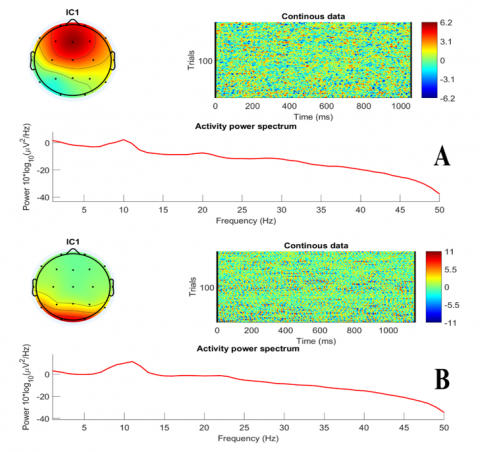
Lastly, the visual examination aids clinicians in identifying the disorder's signal and location inside the brain. Figure 3 displays the power spectral density, highlighting the disparity between the signals of a patient with schizophrenia and those of a healthy individual. The healthy signals are mostly uniform and stable, whereas the patient signals display irregularities and unusual rhythms. Furthermore, a topographical picture delineates the electrode's position, enabling physicians to ascertain the etiology of the neurological issue more precisely and identify the indicators of schizophrenia.
Figure 3. Visualisation method for power spectrum density and topography image, whereas A represents the Schizophrenia person and B represents the patient
Above all, the five random electrodes using KNN with LogEn features outperformed the performances of the two other classifiers (SVM and QDA). Whereas SVM with LogEn achieved the best performance compared with KNN and QDA in the eight-electrode scenario. To conclude, the two random scenarios, based on the findings, indicate that superior outcomes may be achieved with a reduced number of electrodes.
In this study, some limitations are listed below:
(1) The dataset used was publicly available and not privately collected for this study.
(2) The one-epoch window size technique was an advantage that allowed us to cover and provide a much bigger amount of the data, with an overlap was 50%, which was fed into the machine-learning models.
Table 18. PCA memory usage with the five electrodes, where M.A refers to memory availability, and M.U refers to memory usage
|
Memory Usage (MB) |
Features Used |
5 Electrodes |
||
|
SVM |
KNN |
QDA |
||
|
M.A for all arrays |
FFT+ Bandpass |
4409 |
4473 |
4173 |
|
M.U / MATLAB |
6139 |
6143 |
6135 |
|
|
M.A for all arrays |
ApEn |
4711 |
4744 |
4470 |
|
M.U / MATLAB |
5795 |
5876 |
5919 |
|
|
M.A for all arrays |
ApEn+Bandpass |
3881 |
3877 |
4075 |
|
M.U / MATLAB |
6321 |
6320 |
6327 |
|
|
M.A for all arrays |
ShEn+Bandpass |
4227 |
4088 |
4346 |
|
M.U / MATLAB |
6489 |
6497 |
6491 |
|
|
M.A for all arrays |
LogEn+Bandpass |
4298 |
4266 |
4179 |
|
M.U / MATLAB |
6423 |
6413 |
6426 |
|
(3) The model processed the data and diagnosed schizophrenia, not its three severity stages (early, active, or residual).
(4) The study results may lack the ability to generalize to patients with schizophrenia for several reasons that may be due to some characteristics of the data, such as age and gender.
Table 19. PCA memory usage with the eight electrodes, where M.A refers to memory availability and M.U refers to memory usage
|
Memory Usage (MB) |
Features Used |
8 Electrodes |
||
|
SVM |
KNN |
QDA |
||
|
M.A for all arrays |
FFT+Bandpass |
4773 |
4796 |
4931 |
|
M.U / MATLAB |
6717 |
6712 |
6722 |
|
|
M.A for all arrays |
ApEn |
4002 |
4009 |
4065 |
|
M.U / MATLAB |
6505 |
6499 |
6515 |
|
|
M.A for all arrays |
ApEn+Bandpass |
5059 |
4834 |
3829 |
|
M.U / MATLAB |
6666 |
6661 |
6679 |
|
|
M.A for all arrays |
ShEn+Bandpass |
5597 |
5270 |
4755 |
|
M.U / MATLAB |
6777 |
6777 |
6787 |
|
|
M.A for all arrays |
LogEn+ Bandpass |
4773 |
4660 |
4874 |
|
M.U / MATLAB |
6762 |
6752 |
6754 |
|
The person's life and behavior are influenced by the changing electrical activity of the brain, which can be observed through an electroencephalogram (EEG). It can be asserted that a healthy brain functions more actively compared to a brain affected by schizophrenia. In this study, we proposed a method to classify schizophrenia using an EEG signal dataset containing 28 subjects: 14 individuals suffering from schizophrenia and 14 healthy controls. Due to the variations in the EEG signal, we applied a band-pass filter to decompose the EEG signal into five sub-bands. Next, we implemented four feature extraction methodologies. We applied the first three methods (FFT, ApEn, LogEn, and ShnEn) to the band-pass filter, and then we used ApEn again without band-pass filters to compare the impact of the filter on the results.
Normalization was applied to all features to ensure they were on the same scale, using the L2 normalization technique. Consequently, we fed the features into the SVM, KNN, and QDA classifiers.
The hypothesis in this work demonstrates the ability to use a smaller number of electrodes, a randomly selected subset, that can achieve classification accuracy similar to all data electrodes. We provide support to EEG-based diagnostic tools that are less sophisticated and have fewer electrode numbers, such as Emotiv Insight, Muse EEG, Neurosky Mindwave, and InteraXon Muse S.
Our proposed approach was simple and effective with the most imposed constraints. First, we reduced the number of channels to 5 electrodes, which means we decreased by (5/19=73.68%). Then, we used 8 electrodes to 8, which means increased by (8/19=57.89%), which in turn increased the accuracy by 1% using the random channel selection. Thus, both experiments showed remarkable classification performance despite reducing the number of electrodes.
The results show that our approach provides a clear and significant improvement in accuracy compared to the PCA conventional methods. In addition, recent advances indicate that using EEG with fewer electrodes could completely transform the usability and affordability of the technology. This technique simplifies electrode preparation, which not only saves time and reduces complexity but also improves mobility, allowing EEG to be used in a variety of contexts outside of clinical laboratories.
According to the study limitation mentioned above, future work may include:
(1) Combining EEG data with other neuroimaging methods (such as fMRI and PET) may provide a more comprehensive understanding of brain changes caused by schizophrenia.
(2) Implement techniques to enhance the dataset, improve model robustness, and mitigate overfitting.
(3) Utilize the Graph Neural Networks (GNNs) for analyzing brain networks.
[1] Zhu, Y., Nakatani, H., Yassin, W., Maikusa, N., Okada, N., Kunimatsu, A., Abe, O., Kuwabara, H., Yamasue, H., Kasai, K., Okanoya, K., Koike, S. (2022). Application of a machine learning algorithm for structural brain images in chronic schizophrenia to earlier clinical stages of psychosis and autism spectrum disorder: A multiprotocol imaging dataset study. Schizophrenia Bulletin, 48(3): 563-574. https://doi.org/10.1093/schbul/sbac030
[2] Aslan, Z., Akin, M. (2022). A deep learning approach in automated detection of schizophrenia using scalogram images of EEG signals. Physical and Engineering Sciences in Medicine, 45(1): 83-96. https://doi.org/10.1007/s13246-021-01083-2
[3] Jauhar, S., Johnstone, M., McKenna, P.J. (2022). Schizophrenia. The Lancet, 399(10323): 473-486. https://doi.org/10.1016/s0140-6736(21)01730-x
[4] Sadeghi, D., Shoeibi, A., Ghassemi, N., Moridian, P., Khadem, A., Alizadehsani, R., Teshnehlab, M., Gorriz, J.M., Khozeimeh, F., Zhang, Y.D., Nahavandi, S., Acharya, U.R. (2022). An overview of artificial intelligence techniques for diagnosis of Schizophrenia based on magnetic resonance imaging modalities: Methods, challenges, and future works. Computers in Biology and Medicine, 146: 105554. https://doi.org/10.1016/j.compbiomed.2022.105554
[5] Al-jumaili, S., Ibrahim, A.A., Duru, A.D. (2022). Classification of epileptic seizure features from scalp electrical measurements using KNN and SVM based on Fourier Transform. In AIP Conference Proceedings. AIP Publishing. LLC., 2499: 1. https://doi.org/10.1063/5.0105034
[6] Al-Jumaili, S., Al-Azzawi, A., Uçan, O.N., Duru, A.D. (2023). Classification of the level of Alzheimer's disease using anatomical magnetic resonance images based on a novel deep learning structure. In Diagnosis of Neurological Disorders Based on Deep Learning Techniques, pp. 29-46.
[7] Kim, S.K., Kim, H., Kim, S.H., Kim, J.B., Kim, L. (2024). Electroencephalography-based classification of Alzheimer’s disease spectrum during computer-based cognitive testing. Scientific Reports, 14(1): 5252. https://doi.org/10.1038/s41598-024-55656-8
[8] Nour, M., Senturk, U., Polat, K. (2024). A novel hybrid model in the diagnosis and classification of Alzheimer's disease using EEG signals: Deep ensemble learning (DEL) approach. Biomedical Signal Processing and Control, 89: 105751. https://doi.org/10.1016/j.bspc.2023.105751
[9] Ferdowsi, M., Liu, H., Kwan, B.H., Goh, C.H. (2024). Automated detection of Alzheimer's disease using EEG signal processing and machine learning. In Artificial Intelligence Enabled Signal Processing based Models for Neural Information Processing, pp. 118-135.
[10] Khare, S.K., Bajaj, V., Acharya, U.R. (2023). SchizoNET: A robust and accurate Margenau-Hill time-Frequency distribution based deep neural network model for schizophrenia detection using EEG signals. Physiological Measurement, 44(3): 035005. https://doi.org/10.1088/1361-6579/acbc06
[11] Alazzawı, A., Aljumaili, S., Duru, A.D., Uçan, O.N., Bayat, O., Coelho, P.J., Pires, I.M. (2024). Schizophrenia diagnosis based on diverse epoch size resting-state EEG using machine learning. PeerJ Computer Science, 10: e2170. https://doi.org/10.7717/peerj-cs.2170
[12] Al-Azzawi, A., Al-Jumaili, S., Duru, A.D., Duru, D.G., Osman, N.U. (2023). Evaluation of deep transfer learning methodologies on the COVID-19 radiographic chest images. Traitement du Signal, 40(2): 407. https://doi.org/10.18280/ts.400201
[13] Aksöz, A., Akyüz, D., Bayır, F., Yıldız, N.C., Orhanbulucu, F., Latifoğlu, F. (2022). Analysis and classification of schizophrenia using event related potential signals. Computer Science, 32-36. https://doi.org/10.53070/bbd.1173093
[14] Gosala, B., Kapgate, P.D., Jain, P., Chaurasia, R.N., Gupta, M. (2023). Wavelet transforms for feature engineering in EEG data processing: An application on Schizophrenia. Biomedical Signal Processing and Control, 85: 104811. https://doi.org/10.1016/j.bspc.2023.104811
[15] Tyagi, A., Singh, V.P., Gore, M.M. (2022). Machine learning approaches for the detection of schizophrenia using structural MRI. In International Conference on Advanced Network Technologies and Intelligent Computing, Varanasi, India, pp. 423-439. https://doi.org/10.1007/978-3-031-28183-9_30
[16] Lim, J.Z., Mountstephens, J., Teo, J. (2022). Eye-Tracking feature extraction for biometric machine learning. Frontiers in Neurorobotics, 15: 796895. https://doi.org/10.3389/fnbot.2021.796895
[17] Wang, J., Wang, M. (2021). Review of the emotional feature extraction and classification using EEG signals. Cognitive Robotics, 1: 29-40. https://doi.org/10.1016/j.cogr.2021.04.001
[18] Rahman, M.M., Sarkar, A.K., Hossain, M.A., Hossain, M.S., Islam, M.R., Hossain, M.B., Quinn, J.M.W., Moni, M.A. (2021). Recognition of human emotions using EEG signals: A review. Computers in Biology and Medicine, 136: 104696. https://doi.org/10.1016/j.compbiomed.2021.104696
[19] Rashid, M., Sulaiman, N., PP Abdul Majeed, A., Musa, R.M., Ab. Nasir, A.F., Bari, B.S., Khatun, S. (2020). Current status, challenges, and possible solutions of EEG-based brain-computer interface: A comprehensive review. Frontiers in Neurorobotics, 14: 25. https://doi.org/10.3389/fnbot.2020.00025
[20] Krishnan, P.T., Raj, A.N.J., Balasubramanian, P., Chen, Y. (2020). Schizophrenia detection using Multivariate Empirical mode decomposition and entropy measures from multichannel EEG signal. Biocybernetics and Biomedical Engineering, 40(3): 1124-1139. https://doi.org/10.1016/j.bbe.2020.05.008
[21] Lai, J.W., Ang, C.K.E., Acharya, U.R., Cheong, K.H. (2021). Schizophrenia: A survey of artificial intelligence techniques applied to detection and classification. International Journal of Environmental Research and Public Health, 18(11): 6099. https://doi.org/10.3390/ijerph18116099
[22] Siuly, S., Khare, S.K., Bajaj, V., Wang, H., Zhang, Y. (2020). A computerized method for automatic detection of schizophrenia using EEG signals. IEEE Transactions on Neural Systems and Rehabilitation Engineering, 28(11): 2390-2400. https://doi.org/10.1109/TNSRE.2020.3022715
[23] De Miras, J.R., Ibáñez-Molina, A.J., Soriano, M.F., Iglesias-Parro, S. (2023). Schizophrenia classification using machine learning on resting state EEG signal. Biomedical Signal Processing and Control, 79: 104233. https://doi.org/10.1016/j.bspc.2022.104233
[24] Hartini, S., Rustam, Z. (2021). Schizophrenia classification using fuzzy kernel c-means. In Journal of Physics: Conference Series, 1752(1): 012039. https://doi.org/10.1088/1742-6596/1752/1/012039
[25] Khare, S.K., Bajaj, V., Siuly, S., Sinha, G.R. (2020). Classification of schizophrenia patients through empirical wavelet transformation using electroencephalogram signals. In Modelling and Analysis of Active Biopotential Signals in Healthcare, Bristol, UK: IOP Publishing, 1: 1-26.
[26] Hassan, F., Hussain, S.F., Qaisar, S.M. (2023). Fusion of multivariate EEG signals for schizophrenia detection using CNN and machine learning techniques. Information Fusion, 92: 466-478. https://doi.org/10.1016/j.inffus.2022.12.019
[27] Supakar, R., Satvaya, P., Chakrabarti, P. (2022). A deep learning based model using RNN-LSTM for the detection of schizophrenia from EEG data. Computers in Biology and Medicine, 151: 106225. https://doi.org/10.1016/j.compbiomed.2022.106225
[28] Li, B., Wang, J., Guo, Z., Li, Y. (2023). Automatic detection of schizophrenia based on spatial-temporal feature mapping and LeViT with EEG signals. Expert Systems with Applications, 224: 119969. https://doi.org/10.1016/j.eswa.2023.119969
[29] Zhang, L. (2019). EEG signals classification using machine learning for the identification and diagnosis of schizophrenia. In 2019 41st Annual International Conference of The IEEE Engineering in Medicine and Biology Society (EMBC), Berlin, Germany, pp. 4521-4524. https://doi.org/10.1109/EMBC.2019.8857946
[30] Li, F., Wang, J., Liao, Y., Yi, C., Jiang, Y., Si, Y., Peng, W., Yao, D., Zhang, Y., Dong, W., Xu, P. (2019). Differentiation of schizophrenia by combining the spatial EEG brain network patterns of rest and task P300. IEEE Transactions on Neural Systems and Rehabilitation Engineering, 27(4): 594-602. https://doi.org/10.1109/TNSRE.2019.2900725
[31] Winterburn, J.L., Voineskos, A.N., Devenyi, G.A., Plitman, E., de la Fuente-Sandoval, C., Bhagwat, N., Graff-Guerrero, A., Knight, J., Chakravarty, M.M. (2019). Can we accurately classify schizophrenia patients from healthy controls using magnetic resonance imaging and machine learning? A multi-method and multi-dataset study. Schizophrenia Research, 214: 3-10. https://doi.org/10.1016/j.schres.2017.11.038
[32] Phang, C.R., Ting, C.M., Noman, F., Ombao, H. (2019). Classification of EEG-Based brain connectivity networks in schizophrenia using a multi-Domain connectome convolutional neural network. arXiv preprint arXiv: 1903.08858. https://doi.org/10.1109/JBHI.2019.2941222
[33] Jo, Y.T., Joo, S.W., Shon, S.H., Kim, H., Kim, Y., Lee, J. (2020). Diagnosing schizophrenia with network analysis and a machine learning method. International Journal of Methods in Psychiatric Research, 29(1): e1818. https://doi.org/10.1002/mpr.1818
[34] Baradits, M., Bitter, I., Czobor, P. (2020). Multivariate patterns of EEG microstate parameters and their role in the discrimination of patients with schizophrenia from healthy controls. Psychiatry Research, 288: 112938. https://doi.org/10.1016/j.psychres.2020.112938
[35] Sun, J., Cao, R., Zhou, M., Hussain, W., Wang, B., Xue, J., Xiang, J. (2021). A hybrid deep neural network for classification of schizophrenia using EEG Data. Scientific Reports, 11(1): 4706. https://doi.org/10.1038/s41598-021-83350-6
[36] Baygin, M., Yaman, O., Tuncer, T., Dogan, S., Barua, P.D., Acharya, U.R. (2021). Automated accurate schizophrenia detection system using Collatz pattern technique with EEG signals. Biomedical Signal Processing and Control, 70: 102936. https://doi.org/10.1016/j.bspc.2021.102936
[37] Guo, Z., Wu, L., Li, Y., Li, B. (2021). Deep neural network classification of EEG data in schizophrenia. In 2021 IEEE 10th Data Driven Control and Learning Systems Conference (DDCLS), Suzhou, China, pp. 1322-1327. https://doi.org/10.1109/DDCLS52934.2021.9455509
[38] Ciprian, C., Masychev, K., Ravan, M., Manimaran, A., Deshmukh, A. (2021). Diagnosing schizophrenia using effective connectivity of resting-state EEG data. Algorithms, 14(5): 139. https://doi.org/10.3390/a14050139
[39] Chilla, G.S., Yeow, L.Y., Chew, Q.H., Sim, K., Prakash, K.B. (2022). Machine learning classification of schizophrenia patients and healthy controls using diverse neuroanatomical markers and Ensemble methods. Scientific Reports, 12(1): 2755. https://doi.org/10.1038/s41598-022-06651-4
[40] Khare, S.K., Bajaj, V. (2022). A hybrid decision support system for automatic detection of Schizophrenia using EEG signals. Computers in Biology and Medicine, 141: 105028. https://doi.org/10.1016/j.compbiomed.2021.105028
[41] Zandbagleh, A., Mirzakuchaki, S., Daliri, M.R., Premkumar, P., Sanei, S. (2022). Classification of low and high schizotypy levels via evaluation of brain connectivity. International Journal of Neural Systems, 32(4): 2250013. https://doi.org/10.1142/S0129065722500137
[42] Soria, C., Arroyo, Y., Torres, A.M., Redondo, M.Á., Basar, C., Mateo, J. (2023). Method for classifying schizophrenia patients based on machine learning. Journal of Clinical Medicine, 12(13): 4375. https://doi.org/10.3390/jcm12134375
[43] Kumar, T.S., Rajesh, K.N., Maheswari, S., Kanhangad, V., Acharya, U.R. (2023). Automated schizophrenia detection using local descriptors with EEG signals. Engineering Applications of Artificial Intelligence, 117: 105602. https://doi.org/10.1016/j.engappai.2022.105602
[44] Agarwal, M., Singhal, A. (2023). Fusion of pattern-based and statistical features for Schizophrenia detection from EEG signals. Medical Engineering & Physics, 112: 103949. https://doi.org/10.1016/j.medengphy.2023.103949
[45] Bretones, C.S., Parra, C.R., Cascón, J., Borja, A.L., Sotos, J.M. (2023). Automatic identification of schizophrenia employing EEG records analyzed with deep learning algorithms. Schizophrenia Research, 261: 36-46. https://doi.org/10.1016/j.schres.2023.09.010
[46] WeiKoh, J.E., Rajinikanth, V., Vicnesh, J., Pham, T.H., Oh, S.L., Yeong, C.H., Sankaranarayanan, M., Kamath, A., Bairy, G.M., Barua, P.D., Cheong, K.H. (2024). Application of local configuration pattern for automated detection of schizophrenia with electroencephalogram signals. Expert Systems, 41(5): e12957. https://doi.org/10.1111/exsy.12957
[47] Hussain, M., Alsalooli, N.A., Almaghrabi, N., Qazi, E.U.H. (2024). Schizophrenia detection on EEG signals using an ensemble of a lightweight convolutional neural network. Applied Sciences, 14(12): 5048. https://doi.org/10.3390/app14125048
[48] Srinivasan, S., Johnson, S.D. (2024). A novel approach to schizophrenia detection: Optimized preprocessing and deep learning analysis of multichannel EEG data. Expert Systems with Applications, 246: 122937. https://doi.org/10.1016/j.eswa.2023.122937
[49] Olejarczyk, E., Wojciech, J. (2017). EEG in schizophrenia. IBIB PAN-Department of Methods of Brain Imaging and Functional Research of Nervous System. https://repod.icm.edu.pl/dataset.xhtml?persistentId=doi:10.18150/repod.0107441.
[50] Sazgar, M., Young, M.G., Sazgar, M., Young, M.G. (2019). Overview of EEG, electrode placement, and montages. In Absolute Epilepsy and EEG Rotation Review: Essentials for Trainees. Springer, Cham, pp. 117-125. https://doi.org/10.1007/978-3-030-03511-2_5
[51] Sedik, A., Marey, M., Mostafa, H. (2023). WFT-Fati-Dec: Enhanced fatigue detection AI system based on wavelet denoising and Fourier Transform. Applied Sciences, 13(5): 2785. https://doi.org/10.3390/app13052785
[52] Prasanna, J., George, S.T., Subathra, M.S.P. (2023). Classification of gait signals for detection of neurodegenerative diseases using log energy entropy and ANN classifier. In Computational Techniques in Neuroscience, pp. 101-112.
[53] Jahmunah, V., Oh, S.L., Rajinikanth, V., Ciaccio, E.J., Cheong, K.H., Arunkumar, N., Acharya, U.R. (2019). Automated detection of schizophrenia using nonlinear signal processing methods. Artificial Intelligence in Medicine, 100: 101698. https://doi.org/10.1016/j.artmed.2019.07.006
[54] Oh, S.L., Vicnesh, J., Ciaccio, E.J., Yuvaraj, R., Acharya, U.R. (2019). Deep convolutional neural network model for automated diagnosis of schizophrenia using EEG signals. Applied Sciences, 9(14): 2870. https://doi.org/10.3390/app9142870
[55] Prabhakar, S.K., Rajaguru, H., Lee, S.W. (2020). A framework for schizophrenia EEG signal classification with nature inspired optimization algorithms. IEEE Access, 8: 39875-39897. https://doi.org/10.1109/ACCESS.2020.2975848
[56] Shalbaf, A., Bagherzadeh, S., Maghsoudi, A. (2020). Transfer learning with deep convolutional neural network for automated detection of schizophrenia from EEG signals. Physical and Engineering Sciences in Medicine, 43: 1229-1239. https://doi.org/10.1007/s13246-020-00925-9
[57] Buettner, R., Beil, D., Scholtz, S., Djemai, A. (2020). Development of a machine learning based algorithm to accurately detect schizophrenia based on one-minute EEG recordings. In Proceedings of the 53rd Hawaii International Conference on System Sciences, Maui, Hawaii, USA, pp. 3216-3225. https://hdl.handle.net/10125/64135.
[58] Racz, F.S., Stylianou, O., Mukli, P., Eke, A. (2020). Multifractal and entropy-based analysis of delta band neural activity reveals altered functional connectivity dynamics in schizophrenia. Frontiers in Systems Neuroscience, 14: 49. https://doi.org/10.3389/fnsys.2020.00049
[59] Shoeibi, A., Sadeghi, D., Moridian, P., Ghassemi, N., Heras, J., Alizadehsani, R., Khadem, A., Kong, Y., Nahavandi, S., Zhang, Y.D., Gorriz, J.M. (2021). Automatic diagnosis of schizophrenia in EEG signals using CNN-LSTM models. Frontiers in Neuroinformatics, 15: 777977. https://doi.org/10.3389/fninf.2021.777977
[60] Akbari, H., Ghofrani, S., Zakalvand, P., Sadiq, M.T. (2021). Schizophrenia recognition based on the phase space dynamic of EEG signals and graphical features. Biomedical Signal Processing and Control, 69: 102917. https://doi.org/10.1016/j.bspc.2021.102917
[61] Das, K., Pachori, R.B. (2021). Schizophrenia detection technique using multivariate iterative filtering and multichannel EEG signals. Biomedical Signal Processing and Control, 67: 102525. https://doi.org/10.1016/j.bspc.2021.102525
[62] Nikhil Chandran, A., Sreekumar, K., Subha, D.P. (2020). EEG-based automated detection of schizophrenia using long short-term memory (LSTM) network. In Advances in Machine Learning and Computational Intelligence: Proceedings of ICMLCI 2019. Singapore: Springer Singapore, pp. 229-236. https://doi.org/10.1007/978-981-15-5243-4_19
[63] Sharma, M., Acharya, U.R. (2021). Automated detection of schizophrenia using optimal wavelet-based l 1 norm features extracted from single-channel EEG. Cognitive Neurodynamics, 15(4): 661-674. https://doi.org/10.1007/s11571-020-09655-w
[64] Bagherzadeh, S., Shahabi, M.S., Shalbaf, A. (2022). Detection of schizophrenia using hybrid of deep learning and brain effective connectivity image from electroencephalogram signal. Computers in Biology and Medicine, 146: 105570. https://doi.org/10.1016/j.compbiomed.2022.105570
[65] Goshvarpour, A., Goshvarpour, A. (2022). Schizophrenia diagnosis by weighting the entropy measures of the selected EEG channel. Journal of Medical and Biological Engineering, 42(6): 898-908. https://doi.org/10.1007/s40846-022-00762-z
[66] Garip, Z., Ekinci, E., Serbest, K., Eken, S. (2024). Chaotic marine predator optimization algorithm for feature selection in schizophrenia classification using EEG signals. Cluster Computing, 27(8): 11277-11297. https://doi.org/10.1007/s10586-024-04511-6
[67] Haider, U., Hanif, M., Rashid, A., Qaisar, S.M., Subasi, A. (2024). EEG-based schizophrenia classification using penalized sequential dictionary learning in the context of mobile healthcare. Biomedical Signal Processing and Control, 90: 105856. https://doi.org/10.1016/j.bspc.2023.105856