Ruifang Bi*![]() | Hualin Shi
| Hualin Shi![]()
© 2025 The authors. This article is published by IIETA and is licensed under the CC BY 4.0 license (http://creativecommons.org/licenses/by/4.0/).
OPEN ACCESS
The rapid advancement of Artificial Intelligence (AI) has led to the displacement of traditional fossil pattern recognition methods in paleontological studies, particularly through the application of image processing technologies. This study focuses on the fossilized whorls of ancient organisms from the Yangquan region, employing state-of-the-art AI-driven techniques to identify and extract distinctive features from these fossils for automated pattern recognition. Existing paleontological databases of whorl fossils were reviewed, and a deep learning model was developed using convolutional neural networks (CNNs) to facilitate the extraction and classification of fossil whorl patterns. The model incorporates multi-level feature abstraction through various image preprocessing techniques to enhance both the accuracy and robustness of the recognition process. A transfer learning strategy based on CNNs was introduced, allowing for rapid adaptation to new fossil patterns despite limited sample sizes. Furthermore, an improved feature extraction algorithm leveraging Scale-Invariant Feature Transform (SIFT) for feature point matching was implemented, significantly accelerating the speed and accuracy of the feature extraction process. In the experimental phase, over 300 images of fossilized whorls were utilized for model training and validation, achieving a recognition accuracy exceeding 95%, which represents an improvement of nearly 30% over traditional manual methods. The generalization ability of the model was also evaluated, confirming its stability and reliability across diverse fossil data sets. This research underscores the transformative potential of AI-based image processing technologies in the extraction and analysis of paleontological patterns, offering new tools for the study of Yangquan fossils while also contributing to broader applications in cultural heritage preservation and scientific education. This work provides a solid foundation for the further integration of AI methodologies into paleontological research and presents valuable insights and technical references for future studies in related fields.
paleontological whorl fossils, AI image processing, CNN, feature point extraction, pattern recognition
The rapid advancement of AI has demonstrated its significant potential across various domains, particularly in the field of image processing, where it has become a pivotal driver of innovation. AI technologies, especially those based on deep learning, are increasingly applied in paleontology, where they offer promising solutions for the precise identification and classification of fossil patterns. The accurate interpretation of fossilized patterns is essential for understanding geological history and the evolution of life on Earth, making it a central objective in paleontological research.
The Yangquan region, located in northern China, is one of the country’s primary coal-producing areas, but it is also renowned for its rich paleontological fossil resources. The region has attracted considerable scholarly attention due to its diverse and well-preserved fossil records. Among the various types of fossils found in the area, the whorl fossils from the Carboniferous and Permian periods hold particular scientific and economic significance. These fossils represent a key element of the region's paleontological heritage. However, the identification and analysis of these fossils using traditional methods have been inefficient and are often influenced by human error, limiting the potential for large-scale, high-accuracy studies.
To address these challenges, AI-based image processing techniques have been increasingly integrated into the study of paleontological fossils. In this work, CNNs, automatic feature recognition algorithms, and transfer learning strategies have been employed to enhance the recognition and feature extraction of Yangquan paleontological whorl fossils [1]. By leveraging these advanced AI technologies, the accuracy and robustness of pattern recognition have been significantly improved, while the speed of feature extraction has been enhanced [2]. Deep learning models have been constructed and trained on a dataset of over 300 fossil images, resulting in an exceptional recognition accuracy exceeding 95%, nearly 30% higher than traditional manual methods.
A critical aspect of this study is the incorporation of the SIFT algorithm, which has substantially improved the extraction of feature points [3]. This has not only increased the efficiency of fossil pattern recognition but also made the process more accurate, enabling the reliable classification of fossilized whorls. The application of transfer learning has further contributed to the model’s ability to quickly adapt to new fossil patterns, ensuring accurate recognition even with limited sample data.
This research provides a powerful tool for the scientific study of paleontological fossils in the Yangquan region and offers significant implications for the protection of cultural heritage. Furthermore, the methods introduced have the potential to transform educational approaches in paleontology and related fields. The generalization capability and stability of the developed AI system have been rigorously tested, and the results confirm its robustness and reliability in real-world applications [4]. These findings establish a solid foundation for the continued application of AI technologies in paleontological research, paving the way for future advancements in both fossil analysis and broader scientific inquiries into geological history and evolutionary biology.
With the rapid advancements in information technology, particularly in the domains of big data and computational power, image processing technologies have undergone unprecedented transformations [5]. In the field of AI, and more specifically in image recognition, the application of deep learning—particularly CNNs—has significantly advanced the field by shifting the focus from surface-level features to deeper semantic understanding [6]. These technologies are increasingly demonstrating their unique value within the domain of paleontology, with the recognition of Yangquan Paleontological Fossil Patterns serving as a key empirical case. As crucial evidence of life’s evolutionary history, the intricate and diverse patterns found in paleontological fossils present significant challenges for researchers. Traditional recognition methods were not only time-consuming and subjective but also prone to human error. The integration of modern AI-driven image processing technologies, however, has substantially enhanced both the efficiency and robustness of the recognition process, greatly reducing the potential for human bias and error. As a result, these advancements have allowed for a more accurate and comprehensive understanding of the complex evolutionary narratives embedded within fossil records.
2.1 AI image processing in fossil pattern recognition
The application of AI-driven image processing technologies encompasses various aspects, including image denoising, enhancement, classification, feature extraction, and recognition. Through the integrated application of these techniques, the full potential of image processing is continuously explored and leveraged.
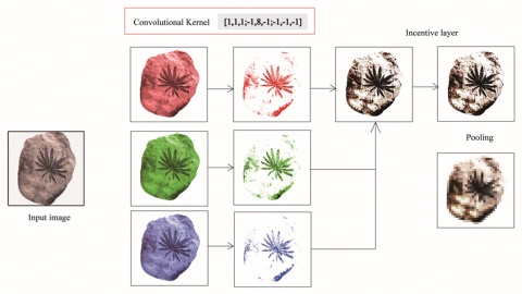
In the context of pattern recognition for fossilized whorls of ancient organisms in the Yangquan region, as illustrated in Figure 1, efficient image preprocessing methods are employed to enhance the quality of input images, mitigate the impact of noise during the training process, and allow the model to focus on more relevant and informative features. Additionally, by utilizing multi-level feature abstraction and the capacity to recognize complex patterns, along with the integration of modern feature matching techniques such as SIFT and multi-scale analysis methods, both the accuracy and efficiency of recognition are significantly improved [7]. Furthermore, the transfer learning capabilities of CNNs are harnessed to rapidly and effectively learn new patterns of paleontological whorl fossils based on limited sample data, thereby facilitating the recognition and classification of fossil patterns in the Yangquan region. The ongoing advancements in image processing technologies have resulted in both visual and technological innovations within the field of paleontology, and the research into fossilized whorl patterns in Yangquan stands as an exemplary case of the fusion of cutting-edge technology with the study of ancient science.
Figure 1. Process diagram of pattern recognition of Yangquan paleontological fossils
2.2 Application of AI in fossil pattern recognition
The rapid advancements in AI, particularly within the domain of image recognition, have enabled unprecedented speed and accuracy in numerous scientific studies. AI technologies are increasingly being employed to analyze image data at deeper levels, achieving precise and efficient automatic recognition and classification. This is especially true in the field of paleontological research, where AI’s image recognition capabilities offer new methodologies for the identification and classification of paleontological fossils, significantly accelerating progress in related disciplines. In the study of the abundant paleontological fossil resources in the Yangquan region, traditional manual identification methods, limited by their efficiency and accuracy, fail to meet the growing demands of contemporary scientific research. The integration of AI image processing technology has not only addressed these limitations but has also provided a robust technical framework and methodological foundation for ongoing research [8].
In the recognition of paleontological fossil images from the Yangquan area, advanced technologies such as deep neural networks, feature learning, and pattern matching enable AI systems to accurately extract relevant information from complex image data, including lines, patterns, shapes, and colors—critical features for analyzing fossil patterns. By combining classifiers such as CNNs and Support Vector Machines (SVMs), AI technology not only enhances the speed of pattern extraction but also substantially improves the accuracy and reliability of recognition [9].
Through multi-level feature abstraction and synthesis, a highly efficient and intelligent recognition model has been developed. An improved least squares ellipse fitting method, in conjunction with the Faster Regional Convolutional Neural Network (Faster RCNN) algorithm, has enabled the model to achieve precise and robust pattern recognition across diverse and complex backgrounds. When trained and tested on an actual dataset of paleontological whorl fossils, the AI model demonstrated a recall rate exceeding 95% and a recognition accuracy of 99%, significantly outperforming traditional methods. Furthermore, AI image recognition technology exhibits excellent adaptability. By employing strategies such as transfer learning and data augmentation, the system can quickly adapt and accurately recognize new fossil patterns, even when faced with sparse data. This highlights the powerful capabilities of AI in handling diverse and evolving image datasets [10].
3.1 Image features of fossilized whorls
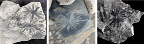
Whorl leaf fossils represent a unique type of fossil from the Carboniferous and Permian periods. These ancient plant fossils exhibit distinctive morphological features, often displaying an herbaceous or shrub-like appearance. Such characteristics can be attributed to the warm and humid environment in which the original plants thrived, typically near rivers or swamps, during the late Carboniferous to Permian periods. The fossilized leaves of these plants are usually arranged in a whorled pattern, with the number of leaves per whorl varying, though typically around 20. The leaves are predominantly lanceolate in shape, measuring approximately 2-3 centimeters in length. The arrangement of the leaves tends to follow a radial pattern, with the leaves tightly clustered and slightly fused at the base. In certain species, such as those with long elliptical blades, the leaf morphology is characterized by horizontally elongated, vertically shortened forms, with nearly parallel upper and lower sides, resulting in a pronounced elliptical contour.
The stems of these fossilized whorls exhibit segmented and branching features, often leaving discernible lines or traces on the fossils. These patterns reflect the growth structure and morphology of the original plants. The color and texture of fossilized whorls can vary depending on the preservation conditions and the specific fossil species. Some fossils retain well-defined contours and intricate details, with clear leaf veins and textures, while others may appear indistinct or damaged due to age and poor preservation conditions. In terms of coloration, fossilized whorls can range from gray to black, brown, or other hues, depending on their preservation environment and the diagenetic processes they underwent, as summarized in Table 1.
Fossilized whorls are also colloquially known as the "flowers of the coal sea" among fossil enthusiasts, owing to their delicate leaf morphology, which bears a resemblance to flowers.
Table 1. Image features of fossilized whorls
|
Image Features |
Image of Fossilized Whorls |
Inductive Analysis |
|
Form |
Typically, in a whorled shape, the leaves are arranged in a regular, radial pattern, closely packed together and slightly fused at the base. |
|
|
Colour |
Fossil colours may vary, typically appearing in shades of grey, black, or brown, depending on the preservation environment and diagenetic processes. |
|
|
Texture |
Some fossil leaves exhibit clear contours and vein patterns, while others show blurred or damaged textures. |
Table 2. Classification and induction of whorl fossils
|
Fossil Classification |
Image of Fossilized Whorls |
Inductive Analysis |
|
Star wheel leaf |
The stem is segmented, with opposite branches and whorled leaves. The leaves are loosely arranged, occasionally showing subtle defects on the upper surface. The leaf shape is linear or needle-like, with a blunt tip and a connected base. The midrib is unclear. |
|
|
Dongfang wheel leaf |
The leaves are arranged radially, with shorter blades positioned beneath the whorl. They are linear to linear-lanceolate in shape, featuring a broad midrib, a pointed apex, and a slightly connected base. The widest part of the leaf is typically located in the middle. |
3.2 Classification and identification of wheel leaf fossils
More common fossils in the genus of whorls include star whorls and oriental whorls. The morphological characteristics of star shaped leaves are that the final branches are thicker, with 15-20 leaves per round. The arrangement of leaves is loose, sometimes with inconspicuous upper leaf defects. The longest leaf length can reach 3 cm, and the leaf shape is linear or needle shaped. The top of the leaf end is blunt, and the base of the leaf base is connected; the midrib is not prominent. The stem of the star shaped leaf is segmented, branches are opposite, and leaves are whorled. Generally, each round has 20 lanceolate leaves of 2-3 centimeters. The terminal secondary branches of the oriental whorls are slender. The final branch width is less than 1.5 mm, slightly narrower than the leaf width. Each round usually has about 20 blades arranged radially, with shorter blades below the impeller. The leaf is linear to linear lanceolate, generally 3 cm long, with a wide midrib, pointed tip, and slightly connected base. The widest part is often located in the middle of the leaf, as shown in Table 2.
In the classification and recognition process, this study introduces a pattern extraction technique based on deep learning models. Unlike traditional algorithms that rely solely on basic features such as image grayscale mean and texture, this approach leverages CNN to automatically and effectively abstract higher-level features, such as contours, shapes, and texture contrasts, from ancient biological leaf fossil patterns. The adaptive network hierarchy can learn rich local and global information from complex images, improving the accuracy of localization and classification performance in the recognition process. In practice, considering the dependence of image recognition on data volume, data augmentation strategies such as rotation, scaling, cropping, etc. were adopted to generate diverse training datasets to ensure the effectiveness and generalization ability of model training. At the same time, in order to accelerate the feature extraction process, an improved basic grayscale matrix (I-BGLAM) technique was introduced for preliminary feature extraction, preserving the basic information of the image and reducing the computational load during model training. To address the issue of CNN being susceptible to background interference and noise, an attention mechanism is introduced to enhance the model's response to key features of patterns, thereby improving the accuracy of feature extraction and the reliability of recognition. In addition, this article also combines the advantages of multi feature fusion, deeply integrating different aspects of feature representation such as color, texture, shape, etc., and proposes a comprehensive feature vector, which enables the entire model to demonstrate excellent recognition performance on various paleontological leaf fossil patterns, and significantly improves the recognition accuracy on the test set. For subtle differences in the patterns of paleontological whorl fossils, such as feature differences caused by different fossil ages or preservation conditions, the model can more accurately capture these details through fine feature layering and relationship modeling. Not only can it significantly improve the recognition efficiency of fossil patterns, but it also provides solid technical support for the subsequent classification and research of paleontological whorl fossils [11].
4.1 Collection of wheel leaf fossils
As early as 2014, lobe fossils were found in waste rock piles of some coal mining mines in Yangquan. In 2016, the staff of the Yangquan Paleontological Fossil Protection Association and Yangquan Planning and Natural Resources Bureau collected them in the open-pit coal mining pit in the Yangquan mining area, and then they were found to be rich in paleontological fossils in several engineering construction areas and collected them.
The fossil images of Verticillata used in this paper mostly come from collecting fossils in the Yangquan area. A total of 200 fossils were collected, and 30 high-resolution images were sourced from China's Geological Museum. An additional 70 images were added from the public Paleobiology Database. All images are standardized to ensure the consistency of resolution and format. The data set consists of 280 images of fossil lobes, covering different species. Among them, complete fossil images account for 60%, and partial fossil images account for 40%. Shooting angles include front, side, and top view angles to ensure the comprehensiveness of features. In addition, the backgrounds of the fossil images in the data set are varied, with rocks, soil, and lab backgrounds, which can help the model work better in more complex settings. Fossils span from the Carboniferous to the Permian, reflecting the characteristics of different geological periods.
4.2 Preprocessing of wheel leaf fossil images
In the data preprocessing stage, we adjust all images to uniform resolution (350×300 pixels) and convert them into BMP format to preserve high-quality details. Through background separation technology, the interference of soil and rock in the image is removed. In addition, we have enhanced the data set, including rotation, scaling, and flipping, and processed the collected original data to enhance the training effect and further expand the data set scale.
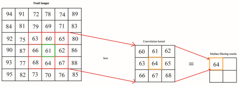
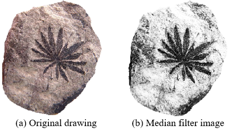
When capturing and transmitting images, due to the influence of the environment and equipment, the image information inevitably contains noise, and it is necessary to smooth the image. The median filtering method has a significant effect on eliminating this type of noise and can effectively preserve image edge information. Sort the grayscale values of all pixels within the pixel and its neighboring window, and take the median value as the new grayscale value for that pixel [12]. The principle is shown in Figure 2. Taking a 3×3 window as an example, the original pixel point in the figure is 61, and the grayscale values in adjacent windows are arranged in ascending order, with the sequence being [60, 61, 62, 63, 64, 65, 66, 67, 68]. The median value 64 is replaced with the original value 61 as the new grayscale value, so that the surrounding grayscale values are close to the actual value, thereby eliminating noise points, as shown in Figure 3.
Figure 2. Principle of binary filtering
Figure 3. Image denoising
Image segmentation is an important step in image preprocessing, extracting fossil image regions from the image. In practical applications, the use of the image local segmentation ROI style algorithm is widely adopted due to its fast calculation speed and low computational complexity. Assuming the image function is f(x,y), its grayscale value is compared with the set threshold T to generate a new image function through the following method:
g(x,y)={1, if f(x,y)>T0, if f(x,y)≤T
In this formula, the pixel value f(x,y) is compared with a threshold T. If the grayscale value is higher than the threshold, the pixel is marked as an object (with a value of 1); if it is below the threshold, it is marked as background (value 0). In this way, through grayscale threshold segmentation, the image is divided into two parts, usually foreground and background. However, using a fixed grayscale threshold can be affected by factors such as uneven image lighting, resulting in unsatisfactory segmentation results. To solve this problem, the image can be divided into multiple sub images, and the grayscale values of neighboring pixels of a certain pixel point in each sub image can be weighted and averaged to obtain the threshold of the local region [13].
Figure 4. Local image segmentation
By using the above methods, the impact of uneven lighting can be effectively avoided, thereby achieving effective segmentation of images. Especially for images with different brightness and backgrounds, such as fossil images, using local thresholding can significantly improve segmentation performance. The effect of the segmented fossil image is shown in Figure 4. This method can effectively address the issue of lighting changes in images, thereby improving the accuracy of image segmentation.
4.3 Pattern extraction and feature analysis
The multi-level CNN structure used effectively extracts the texture features and color differences of the surface of paleontological whorl fossils. In order to improve the accuracy of the recognition model, an HSV (hue, saturation, brightness) model for the color of paleontological leaf fossils was also added. The RGB image corresponds to the hardware output, while the HSV image is more in line with the intuitive vision of the human eye. Therefore, when processing leaf fossil images, the RGB image is first converted to an HSV image, and the image is processed in the HSV color space. First, the R, G, and B values need to be converted between 0 and 1. Let the largest component of the R, G, and B components of the image be Xmax and the smallest component be Xmin, then the conversion relationship between the RGB model and the HSV model is:
R=R/255G=G/255 B=B/255 (1)
H={60∗(G−B)/(X−min (2)
S=\left\{\begin{array}{cc}\frac{X-\min (R, G, B)}{X}, \text { if } & X \neq 0 \\ 0, & \text { else }\end{array}\right\} (3)
\mathrm{V}=\max (\mathrm{R}, \mathrm{G}, \mathrm{B}) (4)
Using the conversion relationship from the RGB model to the HSV model, the image of the fossilized wheel leaf was transformed, and the HSV model image is shown in Figure 5.
Figure 5. Original image of wheel leaf fossil and HSV model image
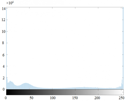
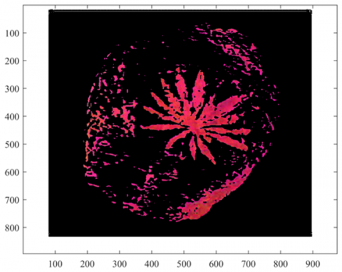
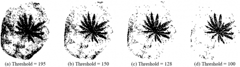
In the HSV model, binarization was performed on the dataset of fossilized wheel leaf images [14]. Based on the HSV color reference range in Table 3, the approximate range of feature thresholds for HSV model wheel leaf fossil images is preliminarily determined to be (H=35~77, S=43~255, V=46~255); By observing the histogram of the HSV model (see Figures 6-7) and continuously refining and adjusting it, the binary threshold range of the HSV model was finally determined to be (H=35-60, S=162-214, V=48-125). The features of the wheel leaf fossil image are shown in Figure 8. By statistically analyzing the color characteristics of fossil images of paleontological whorls, the color tone variation patterns of different paleontological whorls were finely distinguished.
Table 3. HSV color reference range
|
|
Hmax |
Hmix |
Smax |
Smix |
Vmax |
Vmix |
||
|
Black |
180 |
0 |
255 |
0 |
46 |
0 |
||
|
White |
180 |
0 |
30 |
0 |
255 |
221 |
||
|
Grey |
180 |
0 |
43 |
0 |
220 |
46 |
||
|
Red |
10 |
180 |
0 |
156 |
255 |
43 |
255 |
46 |
|
Orange |
25 |
11 |
255 |
43 |
255 |
46 |
||
|
Yellow |
34 |
26 |
255 |
43 |
255 |
46 |
||
|
Green |
77 |
35 |
255 |
43 |
255 |
46 |
||
|
Cyan |
99 |
78 |
255 |
43 |
255 |
46 |
||
|
Blue |
124 |
100 |
255 |
43 |
255 |
46 |
||
|
Purple |
155 |
125 |
255 |
43 |
255 |
46 |
||
(1) Original histogram
(2) HSV model histogram
Figure 6. Image histogram
Figure 7. Image segmentation map of HSV model
Figure 8. Binary different threshold maps
Figure 9. Partial wheel leaf fossil image dataset
Further combining texture based gray level co-occurrence matrix, entropy, contrast and other parameters, by analyzing the spatial relationship between pixels and the statistical distribution of gray levels in the image, the co-occurrence relationship between different gray levels in the image can be captured. It can provide information about the grayscale direction, spacing, and magnitude of changes in the image, and calculate feature values through it to describe various texture features in the image, thereby improving the model's ability to recognize the texture of paleontological whorl fossils.
4.4 Experimental model design and methods
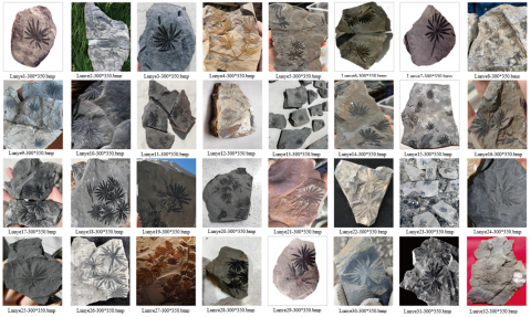
In order to effectively extract and identify the patterns of paleontological whorl fossils in the Yangquan area, an image processing technique combining gray level co-occurrence matrix (GLCM) and CNN was adopted. Firstly, over 300 images of paleontological whorl fossils were collected from the fossil repository in the Yangquan area, and the images were uniformly output as 350 pixels by 300 pixels using Matlab, as shown in Figure 9.
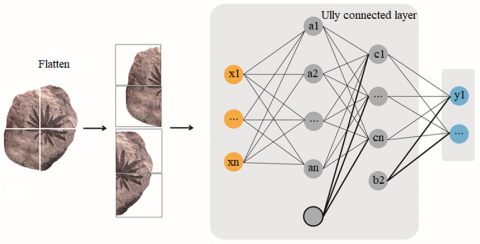
Divide the training set and testing set in a 9:1 ratio, with 270 training sets and 30 testing sets. These images need to undergo strict preprocessing before processing to ensure the quality and consistency of the input data. Preprocessing includes steps such as grayscale conversion, noise removal, and contrast enhancement to ensure the accuracy of feature extraction. Subsequently, GLCM was used to extract texture features from the preprocessed image. Based on the texture parameters obtained from the gray level co-occurrence matrix, such as energy, contrast, uniformity, entropy, etc., representative feature vectors were constructed. These complex texture features provide an important basis for identifying patterns of fossilized whorls in ancient organisms. Subsequently, a CNN based deep learning model was designed, which includes multiple convolutional layers, pooling layers, and fully connected layers, as shown in Figure 10. ReLU was used as the activation function to ensure that the model can effectively learn and extract advanced features from images [15].
In order to speed up the model training and improve the recognition accuracy, the transfer learning strategy is adopted, and the pre-trained network weights are used as the starting point to train the model, which shortens the training time and improves the recognition rate [16]. In the experiment, firstly, the SIFT algorithm is used to extract local feature points and descriptors of images, and preliminary feature matching is carried out to screen out possible similar image subsets. Then, using CNN's powerful feature learning ability, these candidate images are more finely extracted and classified to judge the similarity of images and improve the accuracy and efficiency of retrieval. Aiming at the image data set of paleontological lobe fossils, the experiment adopts cross-validation to ensure the generalization ability of model training. All experiments are carried out on the same training set and test set, which ensures the objectivity and accuracy of the evaluation results. Figure 11 shows the network architecture diagram of this paper. The features of the input image are extracted through multiple convolution layers and pooling layers, and finally the classification results are output through the fully connected layer and SoftMax layer.
By comparing the baseline model with the model using advanced feature extraction techniques, the results show that the latter can significantly improve recognition accuracy, reaching over 95%, which is significantly better than manual pattern matching and traditional machine learning methods. The AI image processing technology presented in this article provides a powerful tool for extracting and applying fossil patterns of ancient organisms in the Yangquan area, opening up new paths for paleontological research and cultural heritage protection [17].
In order to verify the effectiveness of the network architecture, this paper uses ResNet-50 as the basic model. We compare the effects of different convolution kernel sizes of 3×3 and the activation function ReLU, and fine-tune them to adapt to the recognition task of paleontological lobe fossil patterns. The experimental results show that the classification accuracy of the network using a 3×3 convolution kernel and ReLU activation function is above 95% on the test set, which is obviously superior to manual pattern matching and traditional machine learning methods. The AI image processing technology in this paper provides a powerful tool for the extraction and application of paleontological fossil patterns in the Yangquan area, and opens up a new path for paleontological research and cultural heritage protection.
Figure 10. Model architecture
Figure 11. Model training diagram
4.5 Analysis of experimental results
In this paper, the pattern recognition and feature extraction of paleontological lobe fossils in the Yangquan area are discussed in depth. Through the large-scale data training of more than 300 fossil pictures of paleontology, the experiment uses advanced AI image processing technology and CNNs, which significantly improve the accuracy and efficiency of pattern recognition. In the specific experimental operation, the CNN model used in this paper is optimized by a specific algorithm, and in the process of multi-layer feature extraction and abstraction, supplemented by appropriate image enhancement and regularization skills, which ensures the network's deep learning and recognition ability for the complex texture of paleontological lobe fossil patterns. The experiments on the verification set show that the recognition accuracy of the optimized model for the fossil patterns of Yangquan paleontology is over 95%, which is much higher than the traditional manual recognition efficiency, and it shows excellent transfer learning ability and generalization performance in the new pattern recognition task. Further analyzing the experimental results, it is found that the model is particularly obvious in learning the unique texture features of paleontological lobe fossils, such as patterns with clear texture and strong color contrast, and the visual results show the high sensitivity of the model in feature recognition. Through SIFT feature matching technology, the model realizes more accurate and stable extraction of key feature points, which greatly improves the speed and accuracy of feature matching. In the face of pattern change and noise disturbance, the model can still maintain high robustness and recognition rate.
In addition, AI technology has important practical application value in pattern extraction and classification recognition of paleontological fossils. Traditional methods rely on manual observation and measurement, which is inefficient and subjective, while AI technology can automatically and efficiently process a large number of fossil image data, which significantly improves the research efficiency. AI models can complete the classification and feature extraction of a fossil image in a few seconds, while traditional methods may take hours or even days. Fossil features extracted by the deep learning model are more accurate and can identify subtle differences that are difficult for human eyes to detect. As can be seen from Figure 12, the F1-score value of the SIFT and CNN algorithms is 0.099 higher than that of the SIFT algorithm, but the performance of the traditional image processing method is lower than that of the deep learning method. Compared with the image contour extraction algorithm based on the ResNet-50 model, the improved image feature point extraction algorithm based on the SIFT algorithm improves the F1-score value by 0.028 and has better performance. The automated process of AI technology ensures the repeatability of research results and reduces human error. AI technology can process massive fossil image data, which provides unprecedented data support for paleontology research.
Figure 12. Graph of artificial and AI technology algorithm
The analysis of paleontological fossil patterns from the Yangquan region has demonstrated the efficacy of AI image processing technologies in the precise identification and efficient extraction of features. The deep learning model, which employs CNNs, not only facilitates systematic image preprocessing and feature learning based on a comprehensive ancient fossil pattern database but also significantly enhances the model’s capability to learn and recognize novel patterns through the application of transfer learning techniques.
In terms of feature extraction, the integration of an enhanced SIFT algorithm has led to a marked improvement in the matching speed and accuracy of extracted details. The algorithm effectively strengthens the model's ability to detect subtle characteristics of fossil patterns, thereby improving the practical application of AI-driven recognition systems in paleontological research.
Experimental results have shown that the developed AI model achieves a recognition accuracy exceeding 95% when applied to a dataset of over 300 paleontological fossil patterns from the Yangquan region. This performance not only substantially reduces the time required for manual identification but also mitigates the dependence on specialized knowledge and minimizes the potential for human error in subjective judgment. These results underscore the significant potential and advantages of AI image processing technologies in advancing paleontological research, offering an efficient, reliable tool for future studies.
Furthermore, the model’s robustness has been validated through its performance across diverse sample types and environmental conditions, establishing its stability and generalizability in real-world applications. This paves the way for the model’s broader adoption in tasks beyond pattern recognition, including other aspects of paleontological fossil identification. As such, the research contributes to both local advancements in paleontology and the global preservation and study of fossilized life forms.
In addition to its impact on paleontology, the successful application of AI image processing technologies has opened new avenues for interdisciplinary research in fields such as cultural heritage preservation, biodiversity conservation, and geological education. The potential for further interdisciplinary collaboration has been significantly enhanced, showcasing the wider societal contributions of technological innovations.
In summary, the research has made substantial strides in the recognition, analysis, and extraction of paleontological fossil patterns, laying a solid foundation for future scientific inquiry. The outcomes not only mark a critical advancement in paleontological research but also demonstrate the broader applicability of AI technologies, offering promise for future technological innovations in the field.
This paper was supported by 2023 Shanxi Provincial Higher Education Reform and Innovation Project (Grant No.: J20231472); 2024 Shanxi Provincial Culture, Tourism, Creative Industry Development research Project (Grant No.: SXSKWC2023064); 2024 Innovation and Entrepreneurship Training Program for College Students of Shanxi Education Department (Grant No.: X202414527013); Shanxi Federation of Social Sciences 2024-2025 Key Project (Grant No.: SSKLZDKT2024-205).
[1] Li, Y., Jiang, L., Li, X., Feng, W. (2023). Non-contact clothing anthropometry based on two-dimensional image contour detection and feature point recognition. Industria Textila, 74(1): 67-73. https://doi.org/10.35530/IT.074.01.202279
[2] Lu, X., Zhang, Y. (2021). An improved canny detection method for detecting human flexibility. In Intelligent IoT Systems in Personalized Health Care, pp. 207-234. https://doi.org/10.1016/B978-0-12-821187-8.00008-3
[3] Gupta, K.K., Vijay, R., Pahadiya, P. (2022). Detection of abnormality in breast thermograms using Canny edge detection algorithm for thermography images. International Journal of Medical Engineering and Informatics, 14(1): 31-42. https://doi.org/10.1504/IJMEI.2022.119308
[4] Kieu, S.T.H., Bade, A., Hijazi, M.H.A. (2022). Modified canny edge detection technique for identifying endpoints. Journal of Physics: Conference Series, 2314(1): 012023. https://doi.org/10.1088/1742-6596/2314/1/012023
[5] Yudhana, A., Fahmi, M. (2023). SVM-CNN hybrid classification for waste image using morphology and HSV color model image processing. Traitement du Signal, 40(4): 1763-1769. https://doi.org/10.18280/ts.400446
[6] Hu, S., Duan, H., Zhao, J., Zhao, H. (2023). A rust extraction and evaluation method for navigation buoys based on improved U-net and hue, saturation, and value. Sensors, 23(21): 8670. https://doi.org/10.3390/s23218670
[7] Lv, Q., Zhang, S., Wang, Y. (2022). Deep learning model of image classification using machine learning. Advances in Multimedia, 2022(1): 3351256. https://doi.org/10.1155/2022/3351256
[8] Taye, M.M. (2023). Understanding of machine learning with deep learning: Architectures, workflow, applications and future directions. Computers, 12(5): 91. https://doi.org/10.3390/computers12050091
[9] Amirian, S., Rasheed, K., Taha, T.R., Arabnia, H.R. (2020). Automatic image and video caption generation with deep learning: A concise review and algorithmic overlap. IEEE Access, 8: 218386-218400. https://doi.org/10.1109/ACCESS.2020.3042484
[10] Chu, G., Peng, Y., Luo, X. (2023). ALGD-ORB: An improved image feature extraction algorithm with adaptive threshold and local gray difference. Plos One, 18(10): e0293111. https://doi.org/10.1371/journal.pone.0293111
[11] Liu, X., Zhao, X., Xia, Z., Feng, Q., Yu, P., Weng, J. (2023). Secure outsourced SIFT: Accurate and efficient privacy-preserving image SIFT feature extraction. IEEE Transactions on Image Processing, 32: 4635-4648. https://doi.org/10.1109/TIP.2023.3295741
[12] Alfaro-Ponce, M., Chairez, I. (2020). Continuous and recurrent pattern dynamic neural networks recognition of electrophysiological signals. Biomedical Signal Processing and Control, 57: 101783.
[13] Verbitskiy, S., Berikov, V., Vyshegorodtsev, V. (2022). Eranns: Efficient residual audio neural networks for audio pattern recognition. Pattern Recognition Letters, 161: 38-44. https://doi.org/10.1016/j.patrec.2022.07.012
[14] Prashanth, D.S., Mehta, R.V.K., Sharma, N. (2020). Classification of handwritten Devanagari number–an analysis of pattern recognition tool using neural network and CNN. Procedia Computer Science, 167: 2445-2457.
[15] Maghari, A.M., Al-Najjar, I.A., Al-Laqtah, S.J., Abu-Naser, S.S. (2020). Books’ rating prediction using just neural network. International Journal of Engineering and Information Systems, 4(10): 17-22.
[16] Khan, A.H., Hussain, M., Malik, M.K. (2021). Arrhythmia classification techniques using deep neural network. Complexity, 2021(1): 9919588. https://doi.org/10.1155/2021/9919588
[17] Gong, H., Gong, L., Ma, T., Sun, Z., Li, L. (2023). AHY-SLAM: Toward faster and more accurate visual SLAM in dynamic scenes using homogenized feature extraction and object detection method. Sensors, 23(9): 4241. https://doi.org/10.3390/s23094241