Sethulekshmi Rugmini*![]() | Justus Arul Linsely
| Justus Arul Linsely![]()
© 2023 IIETA. This article is published by IIETA and is licensed under the CC BY 4.0 license (http://creativecommons.org/licenses/by/4.0/).
OPEN ACCESS
Melanoma, a prevalent invasive skin cancer, is treatable in its early stages with timely diagnosis and prompt intervention. However, accurately diagnosing lesions through visual inspection or based on their characteristics remains challenging for physicians. Medical imaging techniques play a crucial role in the rapid and precise prognosis of skin lesions. Our research focuses on analyzing and classifying early-stage melanoma using machine learning techniques. In this paper, we propose a metaheuristic algorithm, Differential Evolution optimized Artificial Neural Network (DEO-ANN), for melanoma diagnosis. Color features are assessed from the Region of Interest (ROI) of the lesion using RGB and opponent color space to enhance classification accuracy. Classification is performed using an artificial neural network trained by a differential evolution algorithm. Simulated output demonstrates that the trained DEO-ANN classifier achieves an Area Under Curve (AUC) of 0.98966 with an accuracy of 94.9% on an ISIC dataset.
color features, computer aided diagnosis, differential evolution algorithm, melanoma, receiver operating characteristic curve
Melanoma, the deadliest form of skin cancer, poses a significant threat to global health [1]. The incidence of melanoma has risen dramatically over the past few decades, outpacing all other cancer types. Alarmingly, melanoma has also become increasingly prevalent among younger populations. Melanoma originates in melanocytes and progresses rapidly, often leading to metastasis and a mere 30% survival rate. Factors such as uncontrollable growth of melanocytes and exposure to ultraviolet (UV) rays contribute to melanoma development. Recent research suggests that sun exposure during childhood may damage melanocyte DNA and predispose individuals to melanoma later in life. This could explain melanoma occurring in areas not directly exposed to sunlight. Additionally, excessive sun exposure contributes to melanoma found on arms, face, and neck. Public education on melanoma symptoms, warning signs, and risk factors is crucial for prevention. Early detection and treatment can cure melanoma, minimize treatment-associated pain, disfigurement, and discomfort, reduce surgical intervention and therapy side effects, and decrease overall medical costs. Thus, early diagnosis of melanoma is of utmost importance.
Manual detection remains challenging due to varying skin textures. As a result, dermoscopy techniques have been developed to improve accuracy. However, even with these techniques, detection accuracy depends on the expertise of specialists, with success rates averaging around 85% [2]. To enhance prognosis efficiency, researchers have turned to computer-aided diagnosis (CAD) methods [3, 4]. Our study proposes an innovative machine learning-based diagnostic system to increase melanoma survivability.
The ABCD (Asymmetry, Border, Color, Diameter) criteria are widely used by specialists to diagnose melanoma [5]. Several studies have proposed CAD systems based on these criteria to detect malignancy [6-8]. Among the various visual attributes, color is particularly important in melanoma diagnosis. Researchers have utilized color spaces to manipulate image visual content [9] and applied histogram equalization to filter unwanted noise while maintaining image intensity [10]. Pixel saturation values have been employed for histogram segmentation processes [11].
Advancements in neural networks have led to the development of convolutional neural network (CNN) systems for malignancy identification [12-14]. Support vector machine (SVM) classifiers have been employed to extract features from ABCD parameters for malignancy detection [15]. Deep CNN (DCNN) architectures have been explored for melanoma diagnosis, incorporating regularization approaches [16], Gray Level Co-occurrence Matrix (GLCM) feature extraction [17], and the importance of color features in malignancy detection [18].
Ensemble schemes have been integrated with CNN models to improve malignancy prognosis [19]. Spatial detail extraction has been incorporated into DCNN frameworks to enhance melanoma diagnosis [20, 21]. Wavelet analysis and logical operators have been employed for melanoma detection [22]. Researchers have also investigated the use of k-nearest neighbor (kNN) and SVM classifiers for distinguishing melanoma from healthy skin [23, 24], as well as optimization algorithms in combination with CNN for malignancy diagnosis [25, 26]. Intensity-based features have been used in conjunction with SVM classifiers for malignant lesion identification [27]. The importance of MATLAB in melanoma identification has also been explored [28].
Additional research on machine learning methodologies for melanoma diagnosis and classification can contribute to early disease detection and reduce the risk of human error [29]. In this paper, we propose a CAD system based on DEO-ANN for early melanoma diagnosis using ISIC dataset images. Our significant contributions include:
Experimental results highlight the importance of color features and optimization algorithms in melanoma diagnosis. Our proposed DEO-ANN CAD system demonstrates high accuracy, facilitating efficient melanoma diagnosis. The remainder of this paper is organized as follows: Section 2 describes the methodology and optimization algorithm; Section 3 presents training and experimental results; and Section 4 concludes the paper.
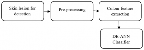
In our methodology, MATLAB platform together with its Neural Network and Image Processing tool boxes are used for the diagnosis of malignant Melanoma. In this paper various steps are used for the diagnosis of the skin lesion to obtain the highest accuracy in its results. The different phases of the skin lesion detection are shown in the Figure 1.
For this we used color feature extraction technique and implemented a system with DEO-ANN which also reduces the computational costs. ROI of the lesion is marked and colour features are extracted from the segmented region. In the final step, optimized classifier is used to detect malignancy.
Figure 1. Diagnosis system block diagram
2.1 Preprocessing
Different cameras or dermoscopes will have different lighting and resolution characteristics. Pre-processing is used for improving the lesion images by supressing unwanted elements or enhancing relevant features. ROI of the lesion is taken and further processing of the image is done in the later stages.
2.2 Feature extraction
Skin lesion colour variation can be studied by assessing chromatic values of red, green and blue colours. In our DEO-ANN classifier, four colour attributes taken out from each pixel of the ROI are used for malignancy identification. Normalized Red and Green values mentioned in Eq. (1) and Eq. (2) are calculated from the RGB colour space:
$r_N=\frac{R}{(R+G+B)}$ (1)
$g_{N=} \frac{G}{(R+G+B)}$ (2)
Increased colour perception is obtained by using channel (Op1) and G-R channel (Op2), the two components from the opponent colour space [30] shown in Eq. (3) and Eq. (4):
$\mathrm{Op} 1(\mathrm{R}, \mathrm{G}, \mathrm{B})=\frac{R-G}{2}$ (3)
$\mathrm{Op} 2(\mathrm{R}, \mathrm{G}, \mathrm{B})=\frac{2 B-R-G}{2}$ (4)
2.3 DEO-ANN classification
The objective of this study is to determine an optimized DEO-ANN for creating a novel prediction system for the identification of melanoma with minimal errors. The ANN methodology is based on a biological neural network imitating the human brain, in which neurons are interrelated to create complex prototypes. Through training, the ANN identifies the bond between the data fed into the system. The ANN model has better inbuilt capabilities to deal with noisy data and outliers for the accurate prognosis of output [31, 32].
ANN strictly depends on Learning rate parameter (Lr), Momentum constant parameter (Mc) and number of hidden neurons and layers. If we fail to set Lr properly, it can increase training time indefinitely and end in oscillations. Back propagation, a gradient descent algorithm can get confined into deceiving local minima making it more complex. So, Momentum constant parameter is used to hasten the convergence. The critical task is to perceive the optimal number of hidden layers. If not done properly the ANN can be more complex and time consuming. In order to enhance the classifier accuracy, initial parameters were tuned by DEO algorithm. So, in the proposed system, to evolve as fully connected optimised neural network, DEO algorithm is applied to optimize the learning rate, hidden layer neurons and momentum factor. When unusual patterns are present in the dataset, the change in learning direction can be avoided by optimizing learning rate. Based on the system the number of input /output neurons are defined.
Figure 2. DE algorithm block diagram
DEO is the most versatile computing algorithm with exceptional convergence for global optimization. DEO can operate in non-linear functions which needs a few control specifications [33]. The block diagram for the DEO algorithm is given in Figure 2.
Np indicates population size, Dn represents the dimension of the vectors which is equal to the entire control parameters and the optimization procedure call for 3 actions: Mutation, Crossover and Selection [34].
DEO algorithm begins with a primary candidate solution / vector population with upper and lower bounds and primary attribute values are arbitrarily chosen at each interval.
The candidate solution / vector is given in Eq. (5):
$Y_{i, g n}=\left(Y_{1, i, g n,}, Y_{2, i, g n}, Y_{3, i, g n}, Y_{4, i, g n}, \ldots \ldots Y_{D n, i, g n}\right)$ (5)
where, i=1, 2, 3, 4, …, Np and gn is the generation number.
Each candidate solution/parameter is having lower and upper bounds as shown in Eq. (6) and Eq. (7):
$Y_j^l=\left(Y_1^l, Y_2^l, Y_3^l, \ldots . Y_{D n}^l\right)$ (6)
$Y_j^u=\left(Y_1^u, Y_2^u, Y_3^u, \ldots \ldots Y_{D n}^u\right)$ (7)
Initialize the jth parameter in the ith population as in Eq. (8):
$Y_{j, i, 0}=Y_j^l+R N_{i, j}[0,1] \times\left(Y_j^u-Y_j^l\right)$ (8)
where, RNi,j[0,1] is the random number.
These N parameter vectors undergo the above-mentioned optimization steps until required criteria is reached. Target vectors are the candidate solution of the present population.
Mutation process begins to initiate a new candidate solution at each iteration, by adding the weighted difference between arbitrarily taken vectors to generate mutant vector, a tertiary vector.
Select three vectors randomly Yrn1,gn, Yrn2,gn and Yrn3,gnfrom the parameter vector given in Eq. (5) in generation gn where i,rn1,rn2,rn3 are mutually exclusive random numbers from {1,2, 3,…, Np}.
The donor vector / mutant vector is given in Eq. (9):
$V_{i . g n}=Y_{r n 1, g n}+F m\left(Y_{r n 2, g n}-Y_{r n 3, g n}\right)$ (9)
Fm, mutation factor is a constant integer from [0, 2].
In crossover process, another vector Ui,gn called Trial vector is obtained from the attributes of target (Yi,gn) and mutant (Vi,gn)vectors as shown in Eq. (10) and Eq. (11):
$U_{i, g n}=V_{j, i, g n}$ if $R N_{j, i} \leq C_o R$ or $j=I_{R N}$ (10)
$U_{i, g n}=Y_{j, i, g n}$ if $R N_{j, i} \leq C_o R$ or $j \neq I_{R N}$ (11)
CoR, a control parameter which is crossover rate constant, Cor ∈ [0,1], random number, IRN is [1, 2, 3, …., Dn] with Yi,gn≠Vi,gn and $R N_{j, i} \in U[0,1]$.
Comparing the target and the trial vectors, the algorithm retains the fittest candidate solution, that yields the best objective value for the next iteration by selection process as shown by Eq. (12) and Eq. (13):
$Y_{i, g n+1}=U_{i, g n+1}$ if $f\left(U_{i, g n+1}\right) \leq f\left(Y_{i, g n}\right)$ (12)
$Y_{i, g n+1}=Y_{i, g n+1}$ if $f\left(U_{i, g n+1}\right)>f\left(Y_{i, g n}\right)$ (13)
where, the function f(.) is to be minimized. This selection activity is replicated till given termination criteria obtained. The DEO algorithm pseudo code is shown below: Assume gn as the Generation, Np as the Population size, CoR as the Crossover rate constant.
Algorithm 1
Initialize
$g n=0, C_o R \in[1,0], N_p \geq 4$
Set
upper and lower bounds for each parameter
$Y_j^l \leq Y_{j, i 1}<Y_j^u$
Set intial values of each j individuals randomly in the interval [Yjl,Yju]
for each j value in the Np population at gn=0,
Initialise using Eq. (8)
end
while termination criteria is not reached
do
for i=1, 2, ..., Np,
Select i, rn1, rn2, rn3 as mutually exclusive random numbers from {1, 2, 3, …, Np}.
Generate mutant vector as in Eq. (9).
select $I_R \in(1,2, \ldots, N p)$
Generate trial vector using Eq. (10) and Eq. (11)
end
Evaluate trial vector using Eq. (12) and Eq. (13).
Increment gn=gn+1
end while
Our proposed methodology DEO-ANN is applied to images collected from the ISIC data base. The momentum constant (MC) and learning rate (LR) is ranging from 0 to 1 and the hidden neurons (NH) are from 31 to 200.The population size of candidate solution is N, equal to 5 with a maximum of 100 generations and 500 training epochs is used for training the images. Minimum Mean Square Error (MSE), the best fitness score is obtained during each generation is stored. The CoR, cross over constant and Fm, mutation factor value is set to 0.5 and 1.2 respectively. By our suggested method, an optimized neural network is obtained with NH=132, LR=0.01254 and MC=0.9583. Using this DEO-ANN method, for developing entirely connected neural network with sigmoid function, NH, LR and MC are optimized. Optimal MC helps to accelerate the convergence and the optimal LR avoids the learning directions in the presence of unusual training patterns.
The output and input layer neurons are fixed according to the problem defined and the hidden layer number is made to one for the entire configuration. The optimization process range is given by the arrays RAmin = {LRmin, NHmin, MCmin} and RAmax = {LRmax, NHmax, MCmax}. Activation function, f is the sum of bias and weighted inputs and is given in Eq. (14) and Eq. (15):
$X_k^p=f\left(S_k^p\right)$ (14)
$S_k^p=\sum_i W_{i, k} X_i^p+b_k$ (15)
kth neuron output is Xkp with pattern p, weight from the ith neuron is Wi,k, kth neuron bias value is bk in hidden layer which is expressed by the activation function, hyperbolic tan sigmoid as in Eq. (16):
$\tanh =\frac{e^x-e^{-x}}{e^x+x^{-x}}$ (16)
Output layer utilizes identity function. For optimal training MSE is the fitness function as in Eq. (17):
$M S E_{D E O-A N N}=\sum_{p \in T} \sum_{k=1}^{N_0}\left(t_k^p-X_k^{p, o}\right)^2$ (17)
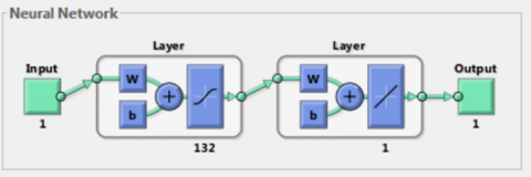
Training set with pattern p, desired output is tkp and actual output Xkp,o is from kth neuron in the output layer o. DEO-ANN algorithm automatically advance to best solution using the fitness function. The ANN structure and classification results are shown in Figure 3 and Figure 4 respectively. The high discriminative power of the proposed methodology yields 94.9% accuracy. The performance of the classifier is increased by choosing optimal network parameters by the proposed differential evolution optimization algorithm. Thus, the accuracy gives the efficiency of the proposed DEO-ANN classifier for the timely and accurate malignancy detection. Performance evaluation of the suggested model is the prime function of machine learning work flow. The different performance metrics and techniques for evaluating the classifier performance are: Accuracy, ROC curve, Sensitivity, Specificity, Precision, Recall and Youden’s index [35]. Accuracy gives the exact predictions of the detection system, expressed as in Eq. (18):
Accuracy $=\frac{(\mathrm{tp}+\mathrm{tn})}{(\mathrm{tp}+\mathrm{fp}+\mathrm{tn}+\mathrm{fn})}$ (18)
Figure 3. Artificial neural network structure
Figure 4. a) Image from ISIC database b) ROI c) Malignant lesion image
ROC curve is used to plot the proposed model accuracy using fPR and tPR, false positive rate and true positive rates respectively [36].
tPR, calculated using Eq. (19) is the percentage of melanoma patients identified as positive.
fPR is the percentage of non-melanoma patients identified as positive is shown in Eq. (20):
$t_{P R}=\frac{t p}{t p+f n}$ (19)
$f_{P R}=\frac{f p}{f p+t n}$ (20)
tp is melanoma patients, tn is benign patients, fp is incorrectly predicted melanoma patients and fn is incorrectly predicted non melanoma patients.
Sensitivity shown in Eq. (21), is the ratio of truly melanoma patients to actual melanoma patients:
Sensitivity $=\frac{t p}{t p+f n}$ (21)
Specificity given in Eq. (22) is the ratio of truly benign patients to total benign patients:
Specificity $=\frac{t n}{t n+f p}$ (22)
Precision in Eq. (23) is the ratio of truly melanoma patients to total predicted melanoma patients:
Precision $=\frac{t p}{t p+f p}$ (23)
Recall in Eq. (24) is the ratio of truly melanoma patients to actual melanoma patients:
Recall $=\frac{t p}{t p+f n}$ (24)
Fmeasure in Eq. (25) shows how precise and robust the classifier is:
$F_{\text {measure }}=\frac{2 * \text { Precision } * \text { Recall }}{\text { Precision }+ \text { Recall }}$ (25)
Youden’s index (J) in Eq. (26) indicates the performance at a given cut-off:
$J=$ Sensitivity + Specificity -1 (26)
Misclassification Rate in Eq. (27) gives what fraction of classifier predictions are incorrect:
Misclassification Rate $=1-$ Accuracy (27)
From the ROC curve shown in Figure 5, area under curve (AUC) of the proposed DEO-ANN system is calculated as 0.98966. Rose plot for the CAD system is shown in Figure 6.
The complete performance of the proposed system is given by Confusion Matrix as shown in Figure 7. From the confusion matrix, the accuracy of the proposed system is obtained as 94.9% with sensitivity of 95% and Specificity of 94.9%.
Figure 5. ROC curve for DEO-ANN classifier
Figure 6. ROSE plot for DEO-ANN classifier
Figure 7. Confusion matrix for DEO-ANN classifier
The performance study of the suggested technique is shown in the Table 1.
The comparison with previous state of art results is shown in Table 2.
Our results show that optimization technique can enhance the classification accuracy.
Table 1. Performance analysis of the DEO-ANN system
|
Evaluation Metrics |
Performance Measure of ISIC Database of DEO-ANN |
|
Sensitivity (%) |
95.0 |
|
Specificity (%) |
94.9 |
|
Accuracy (%) |
94.9 |
|
Youden’s index |
0.8983 |
|
F-Measure |
0.966 |
|
Misclassification rate |
0.051 |
|
AUC |
0.98966 |
Table 2. Comparison with previous work
|
Methodology |
Accuracy |
|
SVM (Babu et al.) [37] |
76.0% |
|
CNN (Gouda et al.) [38] |
83.1% |
|
Resnet50 (Gouda et al.) [38] |
83.6% |
|
Resnet50-Inception (Gouda et al.) [38] |
84.1% |
|
Inception V3(Gouda et al.) [38] |
85.7% |
|
SVM (Murugan et al.) [24] |
89.5% |
|
KNN (Murugan et al.) [24] |
86.0% |
|
CNN (Yilmaz et al.) [39] |
93.09% |
|
CNN (Gaikwad et al.) [40] |
93.9% |
|
Proposed system |
94.9% |
In our paper, a computer aided detection system is developed for identifying malignancy using Artificial Neural Network optimized by differential evolution algorithm. Through our work, we looked into the ability of machine learning algorithms in early diagnosis of melanoma. The skin lesions are taken from ISIC database. Color features like normalized rg and opponent colour features are taken from the ROI to identify the lesion types. ROC is used for performance evaluation of DEO-ANN classifier and AUC is obtained as 0.98966 with an accuracy of 94.9%. The sensitivity of the system is 95% with specificity level of 94.9%. These performance metrics show that the suggested method is a powerful tool for the detection of Melanoma. Our results show that implementing optimized neural network architectures for the detection of melanoma can achieve better classification accuracy and these models can be used in assiting the dermatologist by implementing in dermocopy systems or smart phones.
[1] Khan, N.H., Mir, M., Qian, L., Baloch, M., Khan, M.F.A., Rehman, A., Ngowi, E.E., Wu, D.D., Ji, X. (2022). Skin cancer biology and barriers to treatment: Recent applications of polymeric micro/nanostructures. Journal of Advanced Research, 36: 223-247. https://doi.org/10.1016/j.jare.2021.06.014
[2] Dermoscopy. https://www.dermnetnz.org/topics/dermoscopy, accessed on Oct 15, 2022.
[3] Jana, E., Subban, R., Saraswathi, S. (2017). Research on skin cancer cell detection using image processing. In 2017 IEEE International Conference on Computational Intelligence and Computing Research (ICCIC), pp. 1-8. https://doi.org/10.1109/ICCIC.2017.8524554
[4] Saba, T. (2020). Recent advancement in cancer detection using machine learning: Systematic survey of decades, comparisons and challenges. Journal of Infection and PublicHealth, 13: 1274-1289. https://doi.org/10.1016/j.jiph.2020.06.033
[5] Senan, E.M., Jadhav, M.E. (2021). Analysis of dermoscopy images by using ABCD rule for early detection of skin cancer. Global Transitions Proceedings, 2(1): 1-7. https://doi.org/10.1016/j.gltp.2021.01.001
[6] Lattoofi, N.F., Al-Sharuee, I.F., Kamil, M.Y., Obaid, A.H., Mahidi, A.A., Omar, A.A. (2019). Melanoma skin cancer detection based on ABCD rule. In 2019 First International Conference of Computer and Applied Sciences (CAS), pp. 154-157. https://doi.org/10.1109/CAS47993.2019.9075465
[7] Premaladha, J., Ravichandran, K.S. (2014). Asymmetry analysis of Malignant Melanoma using image processing, A survey. Journal on Artificial Intelligence, 2: 45-53. https://doi.org/10.3923/jai.2014.45.53
[8] Hoshyar, A.N., Al-Jumaily, A., Hoshyar, A.N. (2014). The beneficial techniques in preprocessing step of skin cancer detection system comparing. Procedia Computer Science, 42: 25-31. https://doi.org/10.1016/j.procs.2014.11.029
[9] Menotti, D., Najman, L., Facon, J., de A Albuquerque, A. (2012). Fast hue-preserving histogram equalization methods for color image contrast enhancement. International Journal of Computer Science & Information Technology, 4(5): 243.
[10] Bagade, S.S., Shandilya, V.K. (2011). Use of histogram equalization in image processing for image enhancement. International Journal of Software Engineering Research & Practices, 1(2): 6-10.
[11] Sural, S., Qian, G., Pramanik, S. (2002). Segmentation and histogram generation using the HSV color space for image retrieval. In Proceedings. International Conference on Image Processing, pp. II-II. https://doi.org/10.1109/ICIP.2002.1040019
[12] Nasr-Esfahani, E., Samavi, S., Karimi, N., Soroushmehr, S.M.R., Jafari, M.H., Ward, K., Najarian, K. (2016). Melanoma detection by analysis of clinical images using convolutional neural network. In 2016 38th Annual International Conference of the IEEE Engineering in Medicine and Biology Society (EMBC), pp. 1373-1376. https://doi.org/10.1109/EMBC.2016.7590963
[13] Dubal, P., Bhatt, S., Joglekar, C., Patil, S. (2017). Skin cancer detection and classification. In 2017 6th International Conference on Electrical Engineering and Informatics (ICEEI), pp. 1-6. https://doi.org/10.1109/ICEEI.2017.8312419
[14] Wei, L., Ding, K., Hu, H. (2020). Automatic skin cancer detection in dermoscopy images based on ensemble lightweight deep learning network. IEEE Access, 8: 99633-99647. https://doi.org/10.1109/ACCESS.2020.2997710
[15] Alquran, H., Qasmieh, I.A., Alqudah, A.M., Alhammouri, S., Alawneh, E., Abughazaleh, A., Hasayen, F. (2017). The melanoma skin cancer detection and classification using support vector machine. In 2017 IEEE Jordan Conference on Applied Electrical Engineering and Computing Technologies (AEECT), pp. 1-5. https://doi.org/10.1109/AEECT.2017.8257738
[16] Hossin, M.A., Rupom, F.F., Mahi, H.R., Sarker, A., Ahsan, F., Warech, S. (2020). Melanoma skin cancer detection using deep learning and advanced regularizer. In 2020 International Conference on Advanced Computer Science and Information Systems (ICACSIS), pp. 89-94. https://doi.org/10.1109/ICACSIS51025.2020.9263118
[17] Thaajwer, M.A., Ishanka, U.P. (2020). Melanoma skin cancer detection using image processing and machine learning techniques. In 2020 2nd International Conference on Advancements in Computing (ICAC), pp. 363-368. https://doi.org/10.1109/ICAC51239.2020.9357309
[18] Pathan, S., Aggarwal, V., Prabhu, K.G., Siddalingaswamy, P.C. (2019). Melanoma detection in dermoscopic images using color features. Biomedical and Pharmacology Journal, 12(1): 107-115. https://doi.org/10.13005/bpj/1619
[19] Chen, D., Ziyuan, Z., Ji, H., Huang, Y. (2020). Melanoma classification using deep convolutional neural networks with ensemble scheme. In 2020 2nd International Conference on Information Technology and Computer Application (ITCA), pp. 363-366. https://doi.org/10.1109/ITCA52113.2020.00082
[20] Ashraf, R., Kiran, I., Mahmood, T., Butt, A.U.R., Razzaq, N., Farooq, Z. (2020). An efficient technique for skin cancer classification using deep learning. In 2020 IEEE 23rd International Multitopic Conference (INMIC), pp. 1-5. https://doi.org/10.1109/INMIC50486.2020.9318164
[21] Ali, M.S., Miah, M.S., Haque, J., Rahman, M.M., Islam, M.K. (2021). An enhanced technique of skin cancer classification using deep convolutional neural network with transfer learning models. Machine Learning with Applications, 5: 100036. https://doi.org/10.1016/j.mlwa.2021.100036
[22] Fassihi, N., Shanbehzadeh, J., Sarrafzadeh, H., Ghasemi, E. (2011). Melanoma diagnosis by the use of wavelet analysis based on morphological operators. International MultiConference of Engineers and Computer Scientists, IMECS 2011, Hong Kong, pp. 193-196. https://hdl.handle.net/10652/3051
[23] Jain, S., Pise, N. (2015). Computer aided melanoma skin cancer detection using image processing. Procedia Computer Science, 48: 735-740. https://doi.org/10.1016/j.procs.2015.04.209
[24] Murugan, A., Nair, S.A.H., Kumar, K.S. (2019). Detection of skin cancer using SVM, random forest and kNN classifiers. Journal of Medical Systems, 43: 1-9. https://doi.org/10.1007/s10916-019-1400-8
[25] Zhang, L., Gao, H.J., Zhang, J., Badami, B. (2019). Optimization of the convolutional neural networks for automatic detection of skin cancer. Open Medicine, 15(1): 27-37. https://doi.org/10.1515/med-2020-0006
[26] Arif, M., Philip, F.M., Ajesh, F., Izdrui, D., Craciun, M.D., Geman, O. (2022). Automated detection of nonmelanoma skin cancer based on deep convolutional neural network. Journal of Healthcare Engineering, 2022: 6952304. https://doi.org/10.1155/2022/6952304
[27] Masood, A., Al-Jumaily, A. (2015). Differential evolution based advised SVM for histopathalogical image analysis for skin cancer detection. In 2015 37th Annual International Conference of the IEEE Engineering in Medicine and Biology Society (EMBC), pp. 781-784. https://doi.org/10.1109/EMBC.2015.7318478
[28] Lazaridis, P., Zaharis, Z.D., Kampitaki, D.G. (2006). Automated malignant melanoma detection using MATLAB. Proceedings of the 5th WSEAS Int. Conf. on Data networks, Communications & Computers, Bucharest, Romania.
[29] Das, K., Cockerell, C.J., Patil, A., Pietkiewicz, P., Giulini, M., Grabbe, S., Goldust, M. (2021). Machine learning and its application in skin cancer. International Journal of Environmental Research and Public Health, 18(24): 13409. https://doi.org/10.3390/ijerph182413409
[30] Gevers, T., Stokman, H. (2004). Robust histogram construction from color invariants for object recognition. IEEE Transactions on Pattern Analysis and Machine Intelligence, 26(1): 113-118. https://doi.org/10.1109/TPAMI.2004.1261083
[31] Gupta, A. (2019). Implementation of ANN classifier for skin cancer detection. International Journal of Recent Technology and Engineering (IJRTE), 8(4): 12214-12217. https://doi.org/10.35940/ijrte.D8260.118419
[32] Abiodun, O.I., Jantan, A., Omolara, A.E., Dada, K.V., Mohamed, N.A., Arshad, H. (2018). State-of-the-art in artificial neural network applications: A survey. Heliyon, 4(11): e00938. https://doi.org/10.1016/j.heliyon.2018.e00938
[33] Ahmad, M.F., Isa, N.A.M., Lim, W.H., Ang, K.M. (2022). Differential evolution: A recent review based on state-of-the-art works. Alexandria Engineering Journal, 61(5): 3831-3872. https://doi.org/10.1016/j.aej.2021.09.013
[34] Sahu, V.S.D.M., Samal, P., Panigrahi, C.K. (2020). Application of differential evolution algorithm to optimal control problem with state variable constraints. In 2020 IEEE 17th India Council International Conference (INDICON), pp. 1-7. https://doi.org/10.1109/INDICON49873.2020.9342273
[35] Tharwat, A. (2018). Classification assessment methods. Applied Computing and Informatics, 17(1): 168-192. https://doi.org/10.1016/j.aci.2018.08.003
[36] Fawcett, T. (2006). An introduction to ROC analysis. Pattern Recognition Letters, 27(8): 861-874. https://doi.org/10.1016/j.patrec.2005.10.010
[37] Babu, G.N.K., Peter, V.J. (2021). Skin cancer detection using support vector machine with histogram of oriented gradients features. ICTACT Journal on Soft Computing, 11(02): 2301-2305. https://doi.org/10.21917/ijsc.2021.032
[38] Gouda, W., Sama, N.U., Al-Waakid, G., Humayun, M., Jhanjhi, N.Z. (2022). Detection of skin cancer based on skin lesion images using deep learning. In Healthcare, 10(7): 1183. https://doi.org/10.3390/ healthcare1007118
[39] Yilmaz, E., Trocan, M. (2021). A modified version of GoogLeNet for melanoma diagnosis. Journal of Information and Telecommunication, 5(3): 395-405. https://doi.org/10.1080/24751839.2021.1893495
[40] Gaikwad, M., Gaikwad, P., Jagtap, P., Kadam, S., Patil, R.R. (2020). Melanoma cancer detection using deep learning. International Journal of Scientific Research in Science, Engineering and Technology (IJSRSET), 7(3): 394-400. https://doi.org/10.32628/IJSRSET