Sabreen Ghanem Al-Fatlawi![]() | Ibtihal Riyadh Najeeb
| Ibtihal Riyadh Najeeb![]() | Narimann Neamah Hussein
| Narimann Neamah Hussein![]() | Kahtan A. Mohammed*
| Kahtan A. Mohammed*![]()
© 2025 The authors. This article is published by IIETA and is licensed under the CC BY 4.0 license (http://creativecommons.org/licenses/by/4.0/).
OPEN ACCESS
The study demonstrates the production of nanomagnesium oxide prepared using the hydrothermal method. As well as studying the optical, morphological and structural properties of magnesium oxide. As well as studying the effect of concentration on inhibiting negative Gram bacteria Escherichia coli (E. coli) activity. According to the structural parameters analyzed with FESEM, all preparations with varying amounts of MgO had an erratic crystal structure. According to the optical properties, changes in material concentration resulted in an increase in the absorption value (2.011, 2.11, 2.25 a.u). energy gaps are determined to be between 1.7-2.03 eV since this intersection represents the energy gap of the permitted direct digital transitions. MgO nanoparticles were agglomerated with irregular morphologies and displayed a spherical perimeter. The MgO nanoparticles that were produced using hydrothermal synthesis have a morphology that is characterized by a particle size of sixty nanometers. A crystalline -structure of a produced MgO NPs was examined. Major powerful peaks with 2θ values of 39.52, 36, 33.72, 33.705, 33.64, 33.63, 32.49, 20.61, 19 and 18.53 degree. A data indicated that an inhibitory effect of MgO nanoparticles is depending on (concentration). A highest concentration of 2 g magnesium oxide was the most inhibitory to Gram-negative bacteria E. coli.
MgO, nanoparticles, inhibition, bacteria, hydrothermal, optical properties, morphological investigation, Escherichia coli (E. coli)
Recently, materials with notably new and improved characteristics that are biological, chemical, and physical have been a focus on a global technological revolution driven by nanosciences and nanotechnology. In this sense, the size, structure, and surface characteristics of nanoparticles make them known as antibacterial agents. Consequently, Through the application of nanotechnology, it is possible to enhance the effectiveness of inorganic antibacterial agents [1-3], research has focused on metal oxide nanoparticles like zinc oxide, magnesium oxide, and calcium oxide, which serve as these agents. There are various techniques for producing nanoparticles from liquid solutions, including the hydrothermal method [1]. Among these methodologies, hydrothermal synthesis distinguishes itself as a superb technique for modifying a sample’s morphology by tweaking the creation parameters. Research has demonstrated that various forms can significantly boost the unique traits required for particular applications [4]. In this approach, water-based precursors are subjected to elevated temperatures and pressures within a chamber and an autoclave reactor. For hydrothermal synthesis to produce a singular crystal, it is crucial that the minerals are able to dissolve in hot water under high pressure [5, 6]. This technique provides the advantage of producing crystalline phases that remain unstable near their melting points, facilitating precise compositional regulation and the generation of large, high-grade crystals [7]. The hydrothermal production method ranks among the most frequently employed for crafting nanostructured materials. This approach involves a mixed chemical reaction within liquid solutions at temperatures surpassing room temperature (exceeding 100℃) and pressures above 1 bar, typically realized using an autoclave system [8]. Magnesium is found in more significant amounts within the Earth’s crust, rendering it more accessible than other metals. Nanosized MgO materials [9], generated through hydrothermal means, are utilized to create MgO nanostructures capable of forming crystalline phases [10]. Metal oxide nanoparticles comprise a vital category of nanomaterials extensively used across diverse sectors, including biosensing technology, tissue engineering, food packaging, biomedicine, antibacterial applications, and environmental sciences, owing to their distinctive physical and chemical properties. Magnesium oxide (MgO) is highlighted as an essential inorganic compound with a wide band-gap, applied in numerous fields such as antibacterial functions, heavy fuel oil additives, catalysis, catalyst supports, toxic waste remediation, refractory materials, adsorbents, superconductors, lithium-ion batteries [11], and as substrates in superconducting and ferroelectric thin films. Among these metal oxide nanoparticles, MgO nanoparticles have garnered considerable interest due to their noteworthy biocompatibility, non-toxic nature, remarkable stability in unpredictable environments, and extensive range of applications, especially in biomedicine. At the nanoscale, MgO is a significant inorganic compound characterized by unique qualities. Various methodologies, including hydrothermal, aerogel, and direct chemical transformation, have been employed to fabricate MgO nanoparticles [12]. The hydrothermal process is widely acknowledged as one of the simplest methods for producing MgO nanoparticles. Generally, a basic solution and magnesium compounds are designed using water. The base solution is then mixed with the magnesium compound solution by adjusting the molecular ratio of Mg2+/OH-. Ultimately, the resultant precipitate is washed and subjected to calcination in an oven. Several factors, including magnesium sources, types of solvents, and reaction conditions, significantly influence the dimensions and morphology of MgO nanoparticles. The development of MgO nanostructures and their effects on various bacterial species have been thoroughly investigated. In 2023, Abinaya and Kavitha [9] conducted an experimental study where they synthesized MgO nanoparticles using four different chemical methods: sol-gel, green synthesis, microwave, and hydrothermal techniques [13-15]. The produced magnesium oxide (MgO) nanoparticles were characterized using FT-IR, XRD, HRSEM, and EDAX techniques. The functional groups within the synthesized nanoparticles were identified through FT-IR analysis. XRD and HRSEM were utilized to examine the structural and morphological features. The presence of O and Mg in the prepared samples was confirmed through EDAX. The antibacterial efficacy of the synthesized magnesium oxide nanoparticles was tested against Staphylococcus aureus, Escherichia coli (E. coli), Enterococcus faecalis, and Klebsiella pneumoniae at varying concentrations. Both Gram-positive and Gram-negative bacteria demonstrated susceptibility to the remarkable antibacterial properties [16]. In a significant study carried out in 2025, Jayakumar et al. [17] focused on magnesium oxide nanoparticles (MgO NPs). They generated and characterized the nanoparticles employing transmission electron microscopy to analyze their size, distribution, shape, and grain size, along with ultraviolet-visible (UV-Vis) spectrophotometry to evaluate their optical properties. Antibacterial action was assessed using agar well diffusion at different volumes (50–150 µL). The minimum inhibitory concentration (MIC) was evaluated through the broth microdilution method. Colony-forming units were counted during time-kill tests at 15-minute intervals over a period of 90 minutes. A one-way analysis of variance combined with Tukey's post hoc test (P < 0.05) was implemented for statistical analysis [17]. In 2024, Vidaarth et al. [18] conducted a research project on MgO nanoparticles aimed at determining whether magnesium oxide (MgO) nanoparticles could effectively eliminate the dyes congo red and malachite green from textile wastewater. The nanoparticles were synthesized using seed extracts from Moringa oleifera (MO) and Strychnous potatorum (SP), both separately and in combination. Various characteristics of the MgO nanoparticles were examined, such as structural integrity, grain size, shape, surface dimensions, void fraction, energy gap, and surface electric potential. A phytochemical component in the seed extract significantly enhanced the surface properties of the MgO nanoparticles, functioning as surfactants and reducing agents during the environmentally friendly synthesis process. The antibacterial potency of all magnesium oxide nanoparticles (MgO NPs) was assessed against the pathogens E. coli and Staphylococcus aureus at different concentrations (25 mg ml−1, 50 mg ml−1, and 100 mg ml−1). The nanoparticles derived from the SP-MO combined extract, designated as MgO-M NPs, showed inhibition zones of 17 and 22 millimeters, attaining efficacy rates of 96% and 91% against E. coli and S. aureus, respectively. Furthermore, the DPPH scavenging activity of MgO-M NPs signified robust antioxidant capabilities, with an effectiveness of 91%. A variety of MgO nanoparticles were subjected to congo red and malachite green for a 120-minute exposure to evaluate their photocatalytic degradation potential under sunlight. Notably, MgO-M nanoparticles, which displayed an exceptional surface potential (-43 mv), demonstrated superior photocatalytic performance for negatively charged malachite green dyes (98.5%) compared to positively charged congo red dyes (95.8%). The stability of the structural phase and functional attributes of the MgO nanoparticles throughout the dye degradation process was examined utilizing XRD and FTIR techniques [18]. The potency of magnesium oxide nanoparticles as antimicrobial agents was investigated through various diagnostic approaches, with the broth dilution method being one of the most common. In this methodology, bacterial cultures and dilutions of MgO nanoparticles are arranged in sequence. Thereafter, these dilutions are cultured on agar media under optimal conditions, followed by colony counting. Numerous investigations have indicated that the antibacterial efficacy of magnesium oxide nanoparticles is size-dependent, suggesting that smaller particles may improve antibacterial action. The findings of the study establish a correlation between the size of nano-MgO particles and their bactericidal effectiveness. As the particle size diminished, especially for sizes within the range of 45 to 70 nanometers, the antibacterial efficiency of nano-MgO in eradicating bacteria progressively amplified. However, the bactericidal efficiency below approximately 45 nm was found to be significantly more dependent on size compared to larger sizes. Despite their diminutive size, magnesium oxide nanoparticles effectively hinder E. coli. The cytoplasm of bacteria treated with magnesium oxide nanoparticles exhibited small, electron-dense spots. Investigation of E. coli revealed a low-density region at the cell's core, suggesting the presence of magnesium oxide nanoparticles within the cellular framework [11].
There is a high probability that the shaped like rods actively anaerobic, Gram-negative coliform bacterium E. coli can be found in the lower intestine of warm-blooded creatures. Together with other facultative anaerobes, the majority of E. coli strains comprise approximately 0.1% of the normal gut microbiota [19-23]. Aim of study focused on preparation of MgO in hydrothermal method and examine the main physicochemical properties like optical, structural and morphological properties and study the ability of using the prepared nanoparticles as an agent to kill the bacteria.
Hydrothermally dissolving MgCl2 (1,1.5, and 2 g) MgO nanostructures were created in 100 milliliters of distilled water and 0.1M NaOH. MgH-NPs were confirmed by white suspension particles with a pH of 8. The solution was moved to a Teflon-lined stainless steel slave and maintained at 250℃ for 10 hours at 180 bar following 45 minutes of magnetic stir-frying at 1500 rpm. The pH got close to 7 after being cleaned with ethanol and water. The washing solution was calculated at 200℃ for eight hours and centrifuged at 3000 rpm. From January to March 2025, 100 patients at Al-Sadr Medical City in Al-Najaf Governorate had bacterial species collected and purified. to test MgO's ability to inhibit E. coli. E. coli strains are known. underwent subculturing in nutrient broth, followed by incubation at 37 degrees Celsius for 18 to 24 hours. The resulting bacterial stock solution was then utilized for subsequent investigations. Use a minimum inhibitory concentration (MIC) method, the antibacterial activity of magnesium oxide nanoparticles (MgO NPs) was evaluated against the Gram-negative pathogen E. coli in a microtiter polystyrene plate that contained 96 wells made of flat-bottomed material. In order to create the freshly created bacterial subcultures, the bacteria were first inoculated into a 10 ml test tube that was filled with nutrient broth. After that, the bacteria were allowed to sit for between 18 and 24 hours before the experiment began. For each MgO NPs concentration, 100 µl was coupled with 100 µl of bacterial growth. This was done after the concentration of the bacterial growth was adjusted to 1.5*108 cells/ml and the McFarland turbidity was set at 0.5.
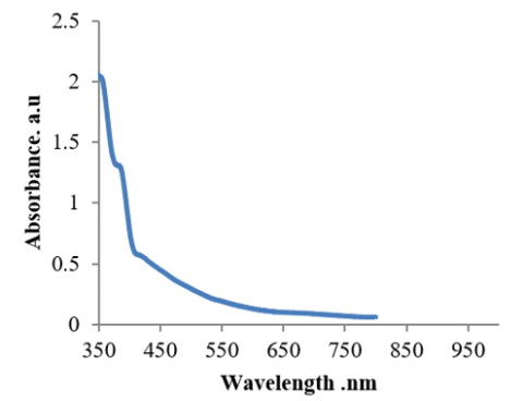
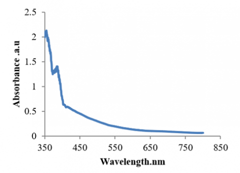
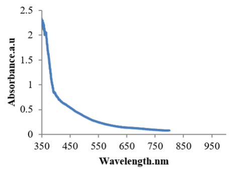
Figures 1, 2, and 3 illustrate the relationship between absorbance and wavelength for MgO nanoparticles produced at different concentrations (2.011,2.11,2.25 a.u). The image illustrates how absorbance increases with wavelength and concentration for all produced MgO nanoparticles.
Figure 1. Absorbance of MgO nanoparticles in 1 g
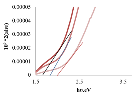
The energy gap of MgO nanoparticles is displayed in Figure 4. The connection between photon energy (hυ) plotted on the x-axis and (α hυ)² represented on the y-axis is illustrated in the image. By extending the curve tangentially until it strikes the photon energy axis at (αhυ)2=0, the energy gap of the permitted direct transitions is computed. The step sizes of the energy gaps are determined to be between 1.7- 2.03 eV since this intersection represents the energy gap of the permitted direct digital transitions.
Figure 2. Absorbance of MgO nanoparticles in 1.5 g
Figure 3. Absorbance of MgO nanoparticles in 2 g
Figure 4. Energy gap of MgO nanoparticles in 1,1.5, 2 g morphological investigation
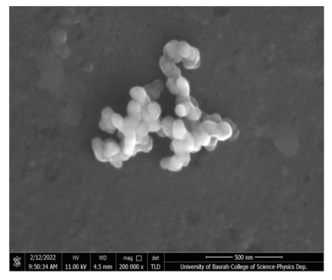
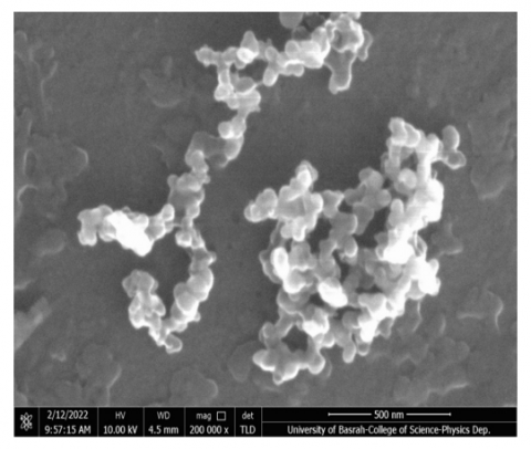
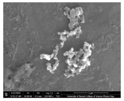
Scanning electron microscopy of MgO nanoparticles using field emission.
The compounds' crystal surfaces were imaged using the scanning electron microscopy technique, which showed a clear variance in surface homogeneity and crystal structures. At a magnification power of K and a cross-sectional distance of 200 nm, the scanning electron microscopy technique was employed. The size, shape, and distribution of the particles, together with their surface properties and aggregation, all have a significant effect on the compounds' characteristics and efficacy.
Scanning electron microscopy equipment was used to image the material's crystal surfaces because it showed a clear variance in crystal structure and surface uniformity. The surface morphological characteristics of each of the different concentrations of the chemical were investigated because the kind and form of the surface greatly affects its attributes and efficacy. As shown in Figures 5, 6, and 7, the morphology of MgO NPs includes particles with a size of 60 nm. After careful inspection, it was discovered that the NPs had a spherical shape. These micrographs also showed agglomerated and porous formations.
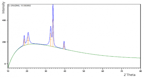
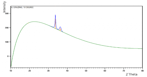
X-ray diffraction of magnesium oxide nanoparticles.
Using XRD examination, the crystalline assembly of the formed MgO NPs was examined. Major powerful peaks with 2θ values of 39.52, 36,33.72, 33.705, 33.64,33.63, 32.49, 20.61, 19 and 18.53 degree were visible in the XRD spectra. Figures 8, 9 and 10, along with Table 1, display the X-ray diffraction patterns of these magnesium oxide nanoparticles.
Figure 5. FESEM of MgO nanoparticles in 1 g
Figure 6. FESEM of MgO nanoparticles in 1.5 g
Figure 7. FESEM of MgO nanoparticles in 2 g
Figure 8. X-ray diffractions pattern of MgO nanoparticles in 1 g
Figure 9. X-ray diffractions pattern of MgO nanoparticles in 1.5 g
Figure 10. X-ray pattern of MgO nanoparticles in 2 g
Table 1. X-ray diffraction of magnesium oxide nanoparticles
|
2θ (Degree) |
Intensity (Counts) |
FWHM Left (Degree) |
D-Spacing [Å] |
Relative. Intensity. [%] |
|
33.63 |
77 |
0.79 |
2.66254 |
100.00 |
|
33.72 |
39 |
0.79 |
2.66254 |
50.00 |
|
18.53 |
58 |
0.54 |
4.78415 |
23.04 |
|
20.61 |
74 |
1.08 |
4.30516 |
29.32 |
|
32.49 |
119 |
0.88 |
2.75353 |
47.05 |
|
33.705 |
254 |
0.44 |
2.65706 |
100.00 |
|
39.52 |
51 |
0.50 |
2.27836 |
20.03 |
|
19 |
0 |
0 |
4.62059 |
0.00 |
|
33.64 |
67 |
0 |
2.66203 |
100.00 |
|
36 |
18 |
1 |
2.47542 |
27.52 |
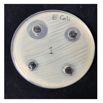
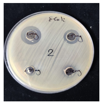
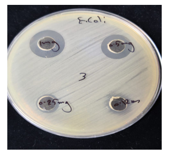
Magnesium oxide nanoparticles (MgO NPs) demonstrated a notable ability to inhibit the growth of E. coli, a Gram-negative bacterium, when tested for antibacterial activity using MIC analysis in a microtiter polystyrene plate containing 96 of these particles. This inhibition's intricate mechanism includes interactions with the cell wall. The thin, multilayered cell wall of Gram-negative bacteria contains an outer layer of lipopolysaccharide (LPS), which provides them with extra resistance. This outer layer can interact with MgO NPs, changing its structure and making it more permeable. Reactive oxygen species and magnesium ions are able to enter the bacterial cell through this disruption. f Reactive oxygen species (ROS) production Superoxide and hydroxyl ions are reactive oxygen species produced when water and magnesium oxide at the nanoscale interact. The bacterial cell's proteins, DNA, and cell membranes are all harmed by these reactive oxygen species. These reactive oxygen species can more easily reach the interior components of the cell when the cell membrane is damaged. The results of the inhibitory zone diameters on E. coli are shown in Figures 11, 12, and 13. Table 2 shows the inhibitor of Gram-negative bacteria of MgO nanoparticles.
Figure 11. Inhibitor of Gram-negative bacteria E. coli in MgO nanoparticles in 1 g
Figure 12. Inhibitor of Gram-negative bacteria E. coli in MgO nanoparticles in 1.5 g
Figure 13. Inhibitor of Gram-negative bacteria E. coli in MgO nanoparticles in 2 g
Table 2. Inhibitor of Gram-negative bacteria of MgO nanoparticles
|
Weight of MgO |
Inhibition Zone (mm) |
Percentage of Inhibition Zone (%) |
|
1g |
12 |
81.2 |
|
1. 5g |
14 |
76.4 |
|
2g |
15 |
85.1 |
MgO-nanostructures have been effectively created with the use of a hydrothermal process. UV, XRD, and FESEM methods were used to analyze the produced MgO nanostructures. E. coli and other harmful bacterial strain was used to test an antibacterial- properties. Additionally, the research demonstrated that MgO NPs' inhibitory function is concentration-dependent. A highest concentration of 2 g magnesium oxide was the most inhibitory to Gram-negative bacteria E. coli. Because of the special qualities of nano magnesium oxide, including its huge surface area and capacity to interact with biomaterials. It has a wide range of uses, including antibacterial ones. Because of its potent antibacterial qualities, nano magnesium oxide works well against a variety of bacteria, including E. coli, which is resistant to antibiotics. Therefore, it is hypothesized that it might be applied in the creation of medicinal ointments and bandages to treat burns, wounds, and skin infections. Even though nanomagnesium oxide has several advantages, its safety must be taken into account. To fully comprehend the long-term impacts of exposure to nanomagnesium oxide, more investigation is still required.
[1] Sundrarajan, M., Suresh, J., Gandhi, R.R. (2012). A comparative study on antibacterial properties of MgO nanoparticles prepared under different calcination temperature. Digest Journal of Nanomaterials and Biostructures, 7(3): 983-989.
[2] Wani, A.H., Shah, M.A. (2012). A unique and profound effect of MgO and ZnO nanoparticles on some plant pathogenic fungi. Journal of Applied Pharmaceutical Science, 2(3): 40-44.
[3] Hornak, J. (2021). Synthesis, properties, and selected technical applications of magnesium oxide nanoparticles: A review. International Journal of Molecular Sciences, 22(23): 12752. https://doi.org/10.3390/ijms222312752
[4] Kumar, P., Kumar, R. (2021). Synthesis process of functionalized ZnO nanostructure for additive manufacturing: A state-of-the-art review. Additive Manufacturing with Functionalized Nanomaterials, 2021: 135-153. https://doi.org/10.1016/B978-0-12-823152-4.00002-8
[5] Vendruscolo, V., Fritzen, D.L., de Mattos, E.A., Rodrigues, L.C.V. (2023). Light storage perovskites: Synthesis, mechanisms, and applications. Perovskite Ceramics, pp. 517-546. https://doi.org/10.1016/B978-0-323-90586-2.00013-9
[6] Satyavani, T.V.S.L., Kumar, A.S., Rao, P.S. (2016). Methods of synthesis and performance improvement of lithium iron phosphate for high rate Li-ion batteries: A review. Engineering Science and Technology, an International Journal, 19(1): 178-188. https://doi.org/10.1016/j.jestch.2015.06.002
[7] Sonawane, G.H., Patil, S.P., Sonawane, S.H. (2018). Nanocomposites and its applications. In Applications of Nanomaterials, pp. 1-22. https://doi.org/10.1016/B978-0-08-101971-9.00001-6
[8] Sebastian, T., Clemens, F. (2022). Piezoelectric application of metal oxide nanofibers. In Metal Oxide-Based Nanofibers and Their Applications, pp. 215-246. https://doi.org/10.1016/B978-0-12-820629-4.00002-3
[9] Abinaya, S., Kavitha, H.P. (2023). Magnesium oxide nanoparticles: effective antilarvicidal and antibacterial agents. ACS Omega, 8(6): 5225. https://doi.org/10.1021/acsomega.2c01450
[10] Amina, M., Al Musayeib, N.M., Alarfaj, N.A., El-Tohamy, M.F., Oraby, H.F., Al Hamoud, G.A., Moubayed, N.M. (2020). Biogenic green synthesis of MgO nanoparticles using Saussurea costus biomasses for a comprehensive detection of their antimicrobial, cytotoxicity against MCF-7 breast cancer cells and photocatalysis potentials. PLoS One, 15(8): e0237567. https://doi.org/10.1371/journal.pone.0237567
[11] Tang, Z.X., Lv, B.F. (2014). MgO nanoparticles as antibacterial agent: preparation and activity. Brazilian Journal of Chemical Engineering, 31(3): 591-601. https://doi.org/10.1590/0104-6632.20140313s00002813
[12] Abdel-Aziz, M.M., Emam, T.M., Elsherbiny, E.A. (2020). Bioactivity of magnesium oxide nanoparticles synthesized from cell filtrate of endobacterium Burkholderia rinojensis against Fusarium oxysporum. Materials Science and Engineering: C, 109: 110617. https://doi.org/10.1016/j.msec.2019.110617
[13] Al-Hazmi, F., Alnowaiser, F., Al-Ghamdi, A.A., Al-Ghamdi, A.A., Aly, M.M., Al-Tuwirqi, R.M., El-Tantawy, F. (2012). A new large–scale synthesis of magnesium oxide nanowires: Structural and antibacterial properties. Superlattices and Microstructures, 52(2): 200-209. https://doi.org/10.1016/j.spmi.2012.04.013
[14] Almontasser, A., Parveen, A., Azam, A. (2019). Synthesis, characterization and antibacterial activity of magnesium oxide (MgO) nanoparticles. In IOP Conference Series: Materials Science and Engineering, 577(1): 012051. https:// doi.org/10.1088/1757-899X/577/1/012051
[15] Ilyina, E.V., Mishakov, I.V., Vedyagin, A.A. (2009). Preparation of nanocrystalline VMg(OH)x and VOx·MgO from organometallic precursors. Inorganic Materials, 45: 1267-1270. https://doi.org/10.1134/S0020168509110144
[16] Imani, M.M., Safaei, M. (2019). Optimized synthesis of magnesium oxide nanoparticles as bactericidal agents. Journal of Nanotechnology, 2019(1): 6063832. https://doi.org/10.1155/2019/6063832
[17] Jayakumar, S., Balasubramanian, R., Ambalavanan, N., Subramanian, A., Shalini, H., Chandrasekaran, R. (2025). Antibacterial effectiveness of zinc oxide and magnesium oxide nanoparticles against enterococcus faecalis: An in vitro study. Journal of International Oral Health, 17(2): 96-105. https://doi.org/10.4103/jioh.jioh_164_24
[18] Vidaarth, T.N., Surendhiran, S., Jagan, K.S.G., Savitha, S., Balu, K.S., Karthik, A., Kalpana, B. (2024). Surface chemistry of phytochemical enriched MgO nanoparticles for antibacterial, antioxidant, and textile dye degradation applications. Journal of Photochemistry and Photobiology A: Chemistry, 448: 115349. https://doi.org/10.1016/j.jphotochem.2023.115349
[19] Tenaillon, O., Skurnik, D., Picard, B., Denamur, E. (2010). The population genetics of commensal Escherichia coli. Nature Reviews Microbiology, 8(3): 207-217. https://doi.org/10.1038/nrmicro2298
[20] Eckburg, P.B., Bik, E.M., Bernstein, C.N., Purdom, E., Dethlefsen, L., Sargent, M., Relman, D.A. (2005). Diversity of the human intestinal microbial flora. Science, 308(5728): 1635-1638. https://doi.org/10.1126/science.1110591
[21] Martinson, J.N., Walk, S.T. (2020). Escherichia coli residency in the gut of healthy human adults. EcoSal Plus, 9(1): 10-1128. https://doi.org/10.1128/ecosalplus.esp-0003-2020
[22] Hudault, S., Guignot, J., Servin, A.L. (2001). Escherichia coli strains colonising the gastrointestinal tract protect germfree mice againstSalmonella typhimuriuminfection. Gut, 49(1): 47-55. https://doi.org/10.1136/gut.49.1.47
[23] Russell, J.B., Jarvis, G.N. (2001). Practical mechanisms for interrupting the oral-fecal lifecycle of Escherichia coli. Journal of Molecular Microbiology and Biotechnology, 3(2): 265-272.