Structural, Morphological and Optical Properties of (NiO)1-x(Co3O4)x Composite Thin Films Prepared by Chemical Spray Pyrolysis Technique

Huda R. Abdulridha Mohammed*![]() | Nahida B. Hasan
| Nahida B. Hasan![]() | Mohammed Hadi Shinen
| Mohammed Hadi Shinen![]()
© 2025 The authors. This article is published by IIETA and is licensed under the CC BY 4.0 license (http://creativecommons.org/licenses/by/4.0/).
OPEN ACCESS
The aim of the study is to produce thin films of (NiO)1-x(Co3O4)x using spray pyrolysis technique. Different volume ratios of NiO, Co3O4 were taken to form thin films with volume ratios of (0,25, 50, 75, and 100) %. The films were deposited on a glass substrate at a temperature of 400℃. Energy dispersive X-ray spectroscopy (EDX), the analysis revealed the prominent presence of the components Ni, O, and Co, X-ray diffraction (XRD), these films showed a crystal structure of the films was identified to be polycrystalline, with cubic structure for nickel oxide in the planes (111), (200), (202) and (222), field emission scanning electron microscopy (FESEM), It can be noted thin films have a common surface shape comprising many randomly placed chunks or aggregates of Co3O4 on the top surface of the films, and ultraviolet-visible (UV-Vis) spectrophotometers. Were these the analytical techniques used in the study to look into the composition, structure, morphology, and optical properties of the thin films. The films have polycrystalline cubic structures. The surface morphology of (NiO)1-x(Co3O4)x films show aggregates or masses of Co3O4 and NiO randomly distributed on the upper surface of the films with increasing Co3O4 concentration. The (NiO)1-x(Co3O4)x films have a surface morphology containing aggregates or clusters of Co3O4 and NiO randomly dispersed on the top surface. The optical properties and optical constants of the prepared (NiO)1-x(Co3O4)x films were investigated in the spectral range of 300-1100nm. In general, the absorbance increases with increasing Co3O4 concentration. The direct energy gap decreases with increasing Co3O4 concentration, and its value is within (3.75 to 2.1) eV. These results are good for sensor applications.
Spray pyrolysis technique, nickel oxide, cobalt oxide, structural, morphological and optical properties
A mixed valence oxide of CoO and Co2O3 with a high oxygen content, spinel cobalt oxide Co3O4 has p-type semiconducting properties. It has been investigated as a possible material for a variety of applications, such as sensors [1], electrochemistry [2], energy storage [3], catalysis [4], and magnetism [5]. The catalytic characteristics of its surface are often the basis for the sensing of Co3O4's gas sensing properties, and the sensors' operating temperature is typically higher than 200℃ [6].
A number of chemical sensing, photocatalysis, battery, and electrochemical applications find nickel oxide (NiO) to be a suitable semiconductor due to its features [7]. Nickel cermets find use in textiles, electrochemical coatings, anode layers in solid oxide fuel cells, lithium nickel oxide cathodes for lithium-ion batteries, nanowires, nanofibers, and special alloy and catalyst applications [7].
Additionally, nickel oxide is utilized as an antiferromagnetic layer and catalyst, in lightweight aircraft structural components, active optical filters, alkaline battery cathode materials, and materials for temperature or gas sensors, including hydrogen and carbon monoxide sensors [8].
In this study, the spray pyrolysis technique was used to prepare thin films of the composite (NiO)1-x(Co3O4)x. These two materials NiO and Co3O4 were used due to their good optical and structural properties, and this method was also used to obtain homogeneous nano-films.
Nickel oxide and cobalt oxide are mixed in varied ratios to create (NiO)1-x(Co3O4)x. First, prepare the NiO and Co3O4 compounds severally. NiCl2.6H2O, a green powder with a molecular weight of 237.69 g/mol, was dissolved in 100ml of room temperature distilled water to create the NiO solution. Magnetic stirrer was added for this purpose and left running for approximately (30) minutes to ensure the solute completely dissolved in the solvent, as indicated by the Eq. (1) [9]:
$\mathrm{NiCl}_2 6 \mathrm{H}_2 \mathrm{O} \rightarrow \mathrm{NiO}+2 \mathrm{HCl}+5 \mathrm{H}_2 \mathrm{O}$ (1)
According to the reaction in Eq. (2), CoCl2.6H2O [a powder with a molecular weight of 237.93g/mol] can be prepared with Co3O4 in (100) ml of distilled water and using magnetic- stirrer also [10]:
$3 \mathrm{CoCl}_2+2 \mathrm{H}_2 \mathrm{O}+\mathrm{O}_2 \rightarrow \mathrm{Co}_3 \mathrm{O}_4 \downarrow+3 \mathrm{Cl}_2 \uparrow+2 \mathrm{H}_2 \uparrow$ (2)
When making NiO and Co3O4 solvents, mix the solutions in various ratios using the relationship below:
$(\mathrm{NiO})_{1-\mathrm{x}}+\left(\mathrm{Co}_3 \mathrm{O}_4\right)_{\mathrm{x}} \rightarrow(\mathrm{NiO})_{1-\mathrm{x}}\left(\mathrm{Co}_3 \mathrm{O}_4\right)_{\mathrm{x}}$ (3)
After using these mixing ratios, the resultant solution was homogenized for 30 minutes using a magnetic stirrer.
The deposition parameters, including the stopping time of two minutes, the deposition rate of two milliliters per minute, and the space between the nozzle and the substrate of thirty centimeters, were maintained at optimal values.
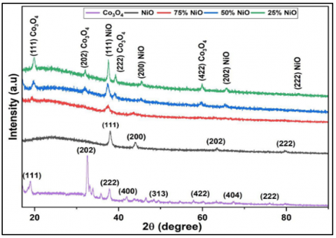
3.1 X-Ray diffraction (XRD)
XRD of the (NiO)1-x(Co3O4)x thin films prepared by spray pyrolysis at substrate temperature of (400℃) have been shown in Figure 1. These films showed a crystal structure of the films was identified to be polycry- stalline [10], with cubic structure for nickel oxide in the planes (111), (200), (202) and (222) this is agreement with the study [10, 11], and the optimum peak was observed at the (111) plane, corresponding to the anatase phase of the Co3O4.
According to the data, there is a noticeable impact on the crystal structure during mixing, which causes the peak location to slightly change and the Table 1 shows peaks position for all pure and mixing films and including inter planar spacing (d), Crystallite size (D), Micro strain (ε) and dislocation density (δ), We notice from the Table 1 the crystal size increased with decreasing the dislocation density and micro strain. The average crystal size upon mixing increased from (10.02) for the mixing ratio of 75% to (27.18) for the ratio of 25%. The presence of the two phases of NiO and Co3O4 in the films confirms the presence of the composite nature of the two oxides [12]. The results of XRD examinations of the prepared films are consistent with the results of other researches [10, 13].
Figure 1 represents the XRD results of the prepared films, where the purple shows CO3O4 pure, the black shows NiO pure, the red shows (NiO)75%(Co3O4)25%, the blue shows (NiO)50%(Co3O4)50%, and the green shows (NiO)25%(Co3O4)75%.
Figure 1. XRD for (NiO)1-x(Co3O4)x composite thin films at different vol.% of (x)
Table 1. The result of the structural parameters from XRD for (NiO)1-x(Co3O4)x composite thin films at different vol.% of (x)
|
Sample |
(hkl) |
2q (deg.) |
d-Spacing [Å] |
FWHM (β) (deg.) |
Crys. Size (nm) |
Average Crys. Size (nm) |
Strain |
Dislocation Density |
|
NiO Pure |
(111) (200) (202) (222) |
37.74 43.75 63.14 79.58 |
2.367 2.057 1.465 1.202 |
0.542 0.514 0.559 1.799 |
15.47 16.65 16.67 5.75 |
13.64 |
2.24 2.08 2.08 6.02 |
4.18 3.61 3.59 3.02 |
|
(NiO) 75 (Co3O4)25 |
(111) |
37.4 |
2.402 |
0.836 |
10.02 |
10.026 |
3.46 |
9.95 |
|
(NiO)50 (Co3O4)50 |
(111) |
37.41 |
2.401 |
0.656 |
12.77 |
12.774 |
2.71 |
6.13 |
|
(NiO)25 (Co3O4)75 |
(111) |
37.56 |
2.392 |
0.308 |
27.18 |
27.180 |
1.27 |
1.35 |
|
Co3O4 Pure |
(111) (202) (222) (400) (313) (422) (404) (222) |
18.88 32.53 37.77 46.48 48.37 57.84 67.48 79.93 |
4.696 2.750 2.379 2.066 1.952 1.880 1.840 1.537 |
0.734 0.342 0.498 0.898 0.089 0.268 2.883 1.921 |
10.96 24.18 16.85 9.62 98.92 33.85 3.31 5.39 |
25.38 |
3.16 1.43 2.06 3.60 3.50 1.02 1.05 6.43 |
8.32 1.71 3.52 1.08 1.02 8.73 9.12 3.44 |
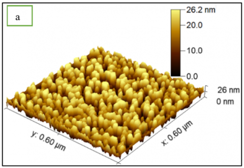
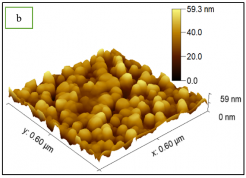
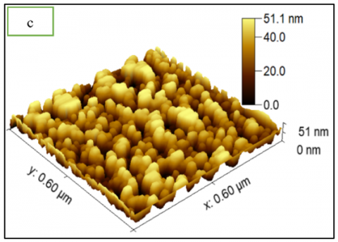
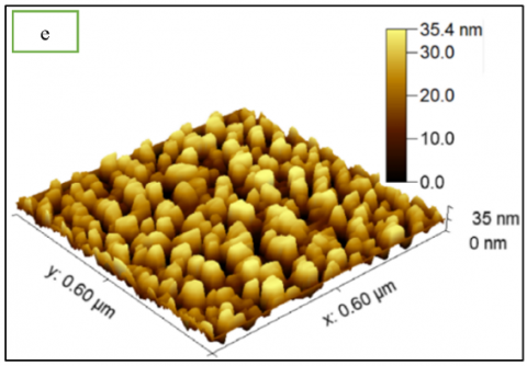
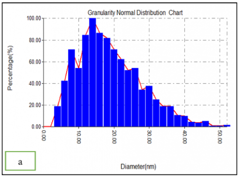
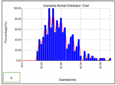
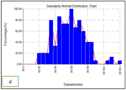
3.2 Atomic force microscope (AFM)
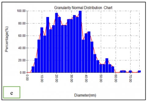
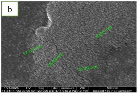
AFM images have been used to observe the surface morphology and roughness of the films and the scanning method of film surfaces for all samples. Figures 2 (a, b, c, d, e) displays the 3D surface structure and granularity distribution of (NiO)1-x(Co3O4)x thin films with different Co3O4 concentrations (0,25,50,75 and 100)%, The results of afm of the prepared films are consistent with the results of other researches [14].
Figure 2 represents the afm results of the prepared films, where, (a) shows NiO pure, the (b) shows (NiO)75%(Co3O4)25%, the (c) shows (NiO)50%(Co3O4)50%, the (d) shows (NiO)25%(Co3O4)75%, and (e) shows Co3O4 pure.
x(Co3O4)x films and (RMS) root mean square of same ratios ranged of (7.81-10.3)nm. At the same time, the average diameter of grain size ranged of (20.04-29.62)nm. Figure 2 shows the AFM images of (NiO)1-x(Co3O4)x thin films synthesized at different molarities. The average roughness and root mean square (RMS) and average diameter for (NiO)1-x(Co3O4)x, estimated from AFM, are given in Table 2. The sample prepared at (NiO)50(Co3O4)50 has highest average roughness of the film. By observing the roughness values, we notice that the surface is homogeneous and nanoscopic.
Figure 2. AFM 3D images and granularity distribution for(NiO)1-x(Co3O4)x composite thin films at different vol.% of (x)
Table 2. Morphology parameters for (NiO)1-x(Co3O4)x composite thin films at different vol.% of (x)
|
Samples |
RMS (nm) |
Roughness Average (nm) |
Average Diameter (nm) |
Ten Point Height (nm) |
|
NiO Pure |
7.81 |
6.78 |
20.04 |
26.2 |
|
NiO75 Co3O425 |
11.3 |
9.83 |
61.21 |
19.3 |
|
NiO50 Co3O450 |
14.8 |
12.8 |
39.83 |
25.2 |
|
NiO25 Co3O475 |
11.2 |
9.78 |
58.53 |
19.4 |
|
Co3O4 Pure |
10.3 |
8.89 |
29.62 |
35.2 |
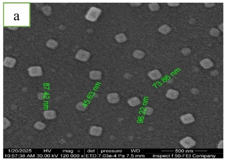
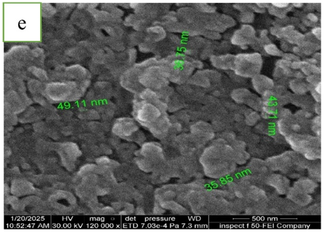
3.3 Field emission scanning electron microscopy
Figure 3 (a-e) shows FESEM images of (NiO)1-x(Co3O4)x thin films at different Co3O4 volume ratios (0,25, 50,75 and 100)%.
Figure 3. FESEM images for (NiO)1-x(Co3O4)x composite thin films at different vol.% of (x)
It can be noted thin films have a common surface shape comprising many randomly placed chunks or aggregates of Co3O4 on the top surface of the films and the images shows a cubic nanocrystal. The findings suggest that there was a tendency for Co3O4 to cluster and diffuse well in the (NiO)1-x(Co3O4)x thin films. As the concentration of cobalt oxide increased, it formed a continuous network with (NiO)1-x(Co3O4)x. The findings presented herein are consistent with the conclusions reached [15].
From the Figure 3, we notice that the size of the particles in the membranes is nano-sized, which means that the membranes can be used as a sensing application because the nano-sized size provides a high adsorption area, and this is one of the conditions for high sensitivity, this is consistent with the work of researchers [16].
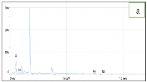
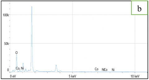
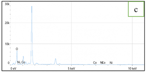
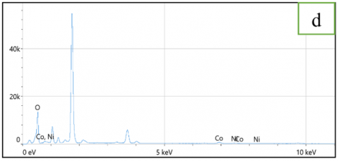
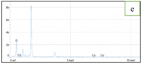
3.4 Energy dispersive X-Ray spectroscopy (EDX)
The energy-dispersive X-ray analysis (EDX) spectra of the (NiO)1-x(Co3O4)x thin films deposited on glass substrate are given in Figures 4 (a-e), The energy-dispersive X-ray analysis (EDX) spectrum of the (NiO)1-x(Co3O4)x thin films with different Co3O4 concentrations (0,25, 50, 75 and 100)% deposited on glass substrate using the spray pyrolysis technique are given in Figure 4. The analysis revealed the prominent presence of the components Ni, O, and Co, Furthermore, as indicated in Table 3.
Figure 4. EDX images for (NiO)1-x(Co3O4)x composite thin films at different vol.% of (x)
Table 3. EDX analysis for prepared films elements compositions
|
Samples |
Element |
Weight % |
Atomic % |
|
NiO Pure |
O |
90.3 |
97.2 |
|
Ni |
9.7 |
2.8 |
|
|
NiO75Co3O425 |
O |
87.4 |
96.2 |
|
Co |
11.1 |
3.3 |
|
|
Ni |
1.6 |
0.5 |
|
|
NiO50Co3O450 |
O |
94.5 |
98.4 |
|
Co |
2.1 |
0.6 |
|
|
Ni |
3.4 |
1.0 |
|
|
NiO25Co3O475 |
O |
86.5 |
95.9 |
|
Co |
9.4 |
2.8 |
|
|
Ni |
4.2 |
1.3 |
|
|
Co3O4 Pure |
O |
99.0 |
99.7 |
|
Co |
1.0 |
0.3 |
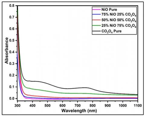
3.5 Optical properties
In the UV-visible range (300-1100)nm, the optical properties of thin films (NiO)1-x(Co3O4)x with variation ratios of Co3O4 and deposited on glass substrates were recorded. In addition to the direct band gap, the absorption coefficient (α), extinction coefficient (k), refractive index (n), dielectric constants (ɛ), and optical conductivity, the variation of the absorbance spectrum, transmittance, and reflectance was also examined. The chemical composition, crystal structure, incident photon energy, thin film thickness, and thin film surface morphology all affect optical transmission and absorption spectra [16, 17].
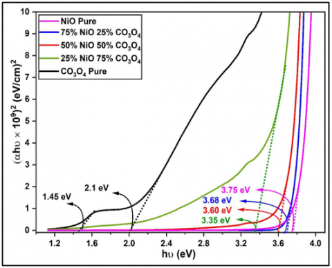
The optical properties of (NiO)1-x(Co3O4)x thin films as shown in Figure 5 and Figure 6. The results from Figure 5 the absorption was high and then decreased with increasing wavelength. In contrast, the behavior is opposite for transmittance and reflectance. It can be seen that with increasing the concentration of Co3O4, the films showed higher ability to absorb electromagnetic radiation at longer wavelengths than NiO, which means that Co3O4 can reduce the band gap of NiO. The results showed that the unmodified NiO had a band gap value of (3.6-4)eV [18, 10].
Figure 5. Absorbance images for (NiO)1-x(Co3O4)x composite thin films at different vol.% of (x)
Figure 6. Energy gap images for(NiO)1-x(Co3O4)x composite thin films at different vol.% of (x)
Table 4. Optical parameters for (NiO)1-x(Co3O4)x composite thin films at different vol.% of (x)
|
Sample |
α×104 (cm-1) |
k |
n |
ɛr |
ɛi |
Eg (eV) |
σ×1014(s-1) |
|
NiO Pure |
0.79 |
0.020 |
1.38 |
1.92 |
0.05 |
3.75 |
2.62 |
|
NiO75Co3O425 |
0.45 |
0.012 |
1.22 |
1.50 |
0.01 |
3.68 |
1.34 |
|
NiO50Co3O450 |
0.73 |
0.020 |
1.35 |
1.84 |
0.05 |
3.60 |
2.38 |
|
NiO25Co3O475 |
1.38 |
0.040 |
1.64 |
2.71 |
0.13 |
3.35 |
5.44 |
|
Co3O4 Pure |
1.38 0.94 |
0.065 0.064 |
1.649 1.45 |
2.71 2.11 |
0.21 0.18 |
2.1 1.45 |
5.46 3.28 |
One can observe that the energy band gap of (NiO)1-x(Co3O4)x thin films decreased from (3.75 to 2.1)eV with the increase of Co3O4 concentration from 25% to 100%, as shown in Figure 6 and Table 4, where some of the unpaired electrons combine with the unpaired electrons on the surface of NiO, forming a structure. This decrease in the band gap is caused by the shift of the valence band edge upward [19-21]. We notice from the figure that with increasing the concentration of Co3O4, the energy gap decreases. The rise in electronic transitions can be attributed to the doping process, which produced new levels near the valence band inside the energy gap. This resulted in the formation of bridges that allowed electrons to move between the valence band and the conduction band. Thus, the process of adding the concentration of Co3O4 led to improving the structural properties of the prepared film.
Since the energy of the falling photon is less than the energy value of the semiconductor, the electron could not be evaporated and moved from the valence pack to the conduction beam. As a result, the absorbance is decreased by increasing the wavelength. We note that the absorption increased with the concentration of the Co3O4, which led to a decrease in the energy gap values of the prepared films [22, 23].
Thin (NiO)1-x(Co3O4)x were successfully produced using different concentrations of Co3O4 and deposited on a glass substrate using spray pyrolysis technique. The effect of Co3O4 concentration on the morphology, structural and optical properties of (NiO)1-x(Co3O4)x atom contributed by XRD examinations to the presence of the formation of several smoky (NiO)1-x(Co3O4)x atom crystals. The crystal size of the prepared illuminant atom increased with increasing concentration of Co3O4. The (NiO)1-x(Co3O4)x atom contributes to the surface morphology of indirect clusters of NiO, Co3O4 on the product surface. With increasing concentration of Co3O4, there was a tendency for Co3O4 to aggregate and spread well in the (NiO)1-x(Co3O4)x atom. The energy band gap of (NiO)1-x(Co3O4)x thin films decreased from (3.75 to 2.1) eV with the increase of Co3O4 concentration from 25% to 100%, It was recognized that increasing concentration of Co3O4 brings about lower energy values that will start with. we notice that the size of the particles in the membranes is nano-sized, which means that the membranes can be used as a sensing application because the nano-sized size provides a high adsorption area, and this is one of the conditions for high sensitivity and also can in optoelectronic devices, such as photosensitive devices, such as photosensitive devices.
The authors would like to express their sincere thanks to the Department of Physics, College of Science, University of Babylon, Iraq.
[1] Shinde, V.R., Mahadik, S.B., Gujar, T.P., Lokhande, C.D. (2006). Supercapacitive cobalt oxide (Co3O4) thin films by spray pyrolysis. Applied Surface Science, 252(20): 7487-7492. https://doi.org/10.1016/j.apsusc.2005.09.004
[2] Balakarthikeyan, R., Santhanam, A., Vibha, K., Shkir, M., Algarni, H., Ashraf, I.M., Kumar, M., Reddy, M.R.V. (2023). Noticeable effect of coating temperature on Co3O4 thin films developed through low-cost nebulizer spray pyrolysis for photo-sensing applications. Surfaces and Interfaces, 38: 102849. https://doi.org/10.1016/j.surfin.2023.102849
[3] Louardi, A., Rmili, A., Chtouki, T., Elidrissi, B., Erguig, H., Bachiri, A.E., Ammous, K., Mejbri, H. (2017). Effect of annealing treatment on Co3O4 thin films properties prepared by spray pyrolysis. Journal of Materials and Environmental Sciences, 8(2): 485-493. https://www.jmaterenvironsci.com/Document/vol8/vol8_N2/52-JMES-2101-Louardi.pdf.
[4] Walsh, A., Wei, S.H., Yan, Y., Al-Jassim, M.M., Turner, J.A., Woodhouse, M., Parkinson, B.A. (2007). Structural, magnetic, and electronic properties of the Co-Fe-Al oxide spinel system: Density-Functional theory calculations. Physical Review B-Condensed Matter and Materials Physics, 76(16): 165119. https://doi.org/10.1103/PhysRevB.76.165119
[5] Shi, Y., Pan, X., Li, B., Zhao, M., Pang, H. (2018). Co3O4 and its composites for high-Performance Li-ion batteries. Chemical Engineering Journal, 343: 427-446. https://doi.org/10.1016/j.cej.2018.03.024
[6] Meher, S.K., Rao, G.R. (2011). Ultralayered Co3O4 for high-Performance supercapacitor applications. The Journal of Physical Chemistry C, 115(31): 15646-15654. https://doi.org/10.1021/jp201200e
[7] Godse, P., Sakhare, R., Pawar, S., Chougule, M., Sen, S., Joshi, P., Patil, V.P. (2011). Effect of annealing on structural, morphological, electrical and optical studies of nickel oxide thin films. Journal of Surface Engineered Materials and Advanced Technology, 1(2): 35. https://doi.org/10.4236/jsemat.2011.1
[8] Hassan, A.J. (2014). Study of optical and electrical properties of nickel oxide (NiO) thin films deposited by using a spray pyrolysis technique. Journal of Modern Physics, 5(18): 2184-2191. http://dx.doi.org/10.4236/jmp.2014.518212
[9] Falahatgar, S.S., Ghodsi, F.E., Tepehan, F.Z., Tepehan, G.G., Turhan, I. (2014). Electrochromic performance, wettability and optical study of copper manganese oxide thin films: Effect of annealing temperature. Applied Surface Science, 289: 289-299. https://doi.org/10.1016/j.apsusc.2013.10.153
[10] Hasan, N.B., Jasim Mohammed, M. (2015). Structural and morphological studies of (NiO)1-x(CuO)x thin films prepared by chemical spray pyrolysis technique. International Letters of Chemistry, Physics and Astronomy, 58: 102-112. https://doi.org/10.56431/p-7ul205
[11] Madhavi, A., Reddy, C.S., Ravindra, N.V., Lokeshand, P., Reddy, P.S. (2014). Effect of substrate temperature on the structural, optical and electrical properties of electron beam evaporated NiO thin films. International Journal of Advanced Research in Physical Science (IJARPS), 1: 16-20. https://www.arcjournals.org/pdfs/ijarps/v1-i4/3.pdf.
[12] Mallick, P., Sahoo, C.S. (2013). Effect of CuO addition on the structural and optical properties of NiO nanoparticles. Nanoscience and Nanotechnology, 3(3): 52-55. http://article.sapub.org/10.5923.j.nn.20130303.04.html#:~:text=doi%3A10.5923/j.nn.20130303.04
[13] Hassan, N.B., Ghazi, R.A. Optoelectronic properties of detector(p-Cd1-xZnxS/n-Si) preparing by Spray Pyrolysis Technique. https://www. phinxsai.com/2016/ch_vol9_no11/2/(331-336)V9N11CT.pdf.
[14] Shkir, M., Khan, A., Imran, M., Khan, M.A., Zargar, R. A., Alshahrani, T., Kumar, K.D.A., Mohanraj, P., Chandekar, K.V., AlFaify, S. (2022). Spray pyrolysis developed Nd doped Co3O4 nanostructured thin films and their structural, and opto-nonlinear properties for optoelectronics applications. Optics & LaserTechnology, 150: 107959. https://doi.org/10.1016/j.optlastec.2022.107959
[15] Soltani, S., Rozati, S.M., Askari, M.B. (2022). Optical and electrochemical properties of spinel cubic nanostructured thin film Co3O4 prepared by spray pyrolysis. Physica B: Condensed Matter, 625: 413464. https://doi.org/10.1016/j.physb.2021.413464
[16] Alfayhan, A.S., Hasn, N.B. (2024). Influence of pulse laser deposition on the structural and optical properties of CZTS for sensor applications. Journal of Composite & Advanced Materials/Revue des Composites et des Matériaux Avancés, 34(1). https://doi.org/10.18280/rcma.340112
[17] Mohamed, W.M., Hasan, N.B. (2020). Effect of lasing energy on optical and structural properties of (ZnS) nanostructured thin films for solar cell applications. NeuroQuantology, 18(5): 29-35. https://link.gale.com/apps/doc/A676650492/AONE?u=anon~cbf8c558&sid=googleScholar&xid=ec674bf9.
[18] Alfayhan, A.S., Hasan, N.B. (2024). Study some electrical properties of Cu2ZnSnS4 thin films prepared by pulse laser deposition. Journal of University of Babylon for Pure and Applied Sciences, 45-52. https://doi.org/10.29196/s4v42b82
[19] AlSultani, M.J., Alias, M.F. (2024). Enhanced optoelectronics performance of hybrid self power photodetectors GO: TiO2-AD/n-Si heterojunctions. Karbala International Journal of Modern Science, 10(3): 1. https://kijoms.uokerbala.edu.iq/cgi/viewcontent.cgi?article=3362&context=home.
[20] Louardi, A., Rmili, A., Ouachtari, F., Bouaoud, A., Elidrissi, B., Erguig, H. (2011). Characterization of cobalt oxide thin films prepared by a facile spray pyrolysis technique using perfume atomizer. Journal of Alloys and Compounds, 509(37): 9183-9189. http://dx.doi.org/10.1016/j.jallcom.2011.06.106
[21] Patil, V., Joshi, P., Chougule, M., Sen, S. (2011). Synthesis and characterization of Co3O4 thin film. Soft Nanoscience Letters, 2(1): 1-7. https://doi.org/10.4236/snl.2012.21001
[22] Manogowri, R., Mary Mathelane, R., Valanarasu, S., Kulandaisamy, I., Benazir Fathima, A., Kathalingam, A. (2016). Effect of annealing temperature on the structural, morphological, optical and electrical properties of Co 3 O 4 thin film by nebulizer spray pyrolysis technique. Journal of Materials Science: Materials in Electronics, 27: 3860-3866. https://doi.org/10.1007/s10854-015-4234-2
[23] Abbas, S.Z., Aboud, A.A., Irfan, M., Alam, S. (2014). Effect of substrate temperature on structure and optical properties of Co3O4 films prepared by spray pyrolysis technique. In IOP Conference Series: Materials Science and Engineering. IOP Publishing, 60(1): 012058. https://doi.org/10.1088/1757-899X/60/1/012058