Hanaa A. Al-Kaisy![]() | Rasha Abdul-Hassan Issa
| Rasha Abdul-Hassan Issa![]() | Noor K. Faheed*
| Noor K. Faheed*![]() | Qahtan A. Hamad
| Qahtan A. Hamad![]()
© 2024 The authors. This article is published by IIETA and is licensed under the CC BY 4.0 license (http://creativecommons.org/licenses/by/4.0/).
OPEN ACCESS
The current work aims to enhance the biocompatibility and antibacterial properties of titanium implants using chitosan/Na alginate matrix composite as a coating layer reinforced with various ratios of hydroxyapatite (HAP) and ZnO by the Sol-Gel Dip method resulting in a product of exceptional purity, a limited dispersion of particle sizes, and the creation of a homogeneous nanostructure. The coating layer is characterized by FE-SEM for microstructure observation. From the results, it was concluded that the precipitation of a bio-composite coating layer by Sol-Gel Dip was suitable for creating a strong, adherent biocompatible layer of chitosan/alginate with a thickness of about (126.9µm). While the average diameter is approximately (21.5µm). The results showed that the dip-coating deposition method is very suitable for making CS-based composite coatings reinforced with ZnO and HAP. From the anti-bacterial test results, it was found that the addition of ceramic particles (HAP or ZnO) to the microstructure for the coating samples revealed a uniform distribution of all types of the natural polymer coating layer on the implants, indicating a suitable preparation and type of coating process (Sol-Gel Dip Composite Coating), which also enhanced the coating's roughness property and effective at inhibiting bacterial growth. This work revealed the assets of chitosan/Na alginate matrix composites in various percentages, which have not been tried up to now and could be very important for the development of the biomedical field.
hydroxyapatite (HAP), ZnO, chitosan, Na alginate, biocomposite coating, Sol-Gel Dip method
As compared to the attributes of metals, ceramics, and even composite materials, polymers' properties, particularly their mechanical aspects, must be improved in any manner possible [1]. As a result, polymer-based composites, which are heterogeneous systems, improve toughness and strength with high performance through the distribution of reinforcing particles [2-4]. The surface and mechanical properties are very important in choosing the material for any application, and the most important of these properties for medical applications, for example, is the nature of the material's surface, which is the first feature that interacts with the surroundings and thus determines the fate of the material's performance [5]. Without having an adverse effect, surface modification approaches can precisely enhance a surface's bio-performance characteristics and compatibility [6, 7]. Changing the surface of implanted biological substances can improve biocompatibility and reduce the incidence of associated illnesses [8]. Multiple technologies are now being used to improve the performance of medical implants, including the use of bioactive and osseointegrated active coatings for metallic titanium substrates [9]. One way to increase osseointegration is the use of coatings for medical implants, surgical materials, and tissue adhesives [10, 11]. Many biologically effective alternatives are used to replace part of the tissue (bone tissue, for example), using polymeric natural materials such as chitosan, which are characterized by their availability and good biological properties (excellent biodegradation and similarity to natural bone) [12-15].
The science of tissue engineering has shown a great deal of potential in the natural ionic alginate polymer. It is very adsorptive and can be utilized to produce polymer scaffolds since it is renewable, antimicrobial, and affordable. Because it is compatible with human tissues, produces biological matter, and creates and maintains moisture around wounds, sodium alginate finds utility in tissue engineering technologies [16, 17].
It is anticipated that chitosan-alginate films with varying ratios of mass combined with zinc oxide and HAP ceramic nanoparticles, as reinforcements will offer several significant medical benefits, such as film development, biological compatibility, broad antimicrobial properties, biodegradable high hemostasis, and superior attachment to injured tissues and blood vessels [18].
Hyaluronic acid, collagen, and chitosan are examples of natural polymers that have demonstrated some inherent bioactive effects in tissues like cartilage tissue [19]. Natural polymers provide the means to recognize the biological system as a result of their macromolecular composition resembling tissues. This further results in the aversion of problems with toxicity and activation of a chronic inflammatory response, as well as the absence of recognition by cells, which are commonly induced via numerous artificial polymers [20].
It has high biocompatibility and does not cause risks of disease transmission or immune rejection. It is mainly utilized in many biomedical uses, especially in the arrangement of bone fillers, coating materials for metal implantations, etc. [21]. Evaluation of the influence of a sodium alginate coating encumbered with ascorbic acid on the lifecycle of samples was first coated in a sodium lactate solution. The samples were analyzed in terms of sensory and microbiological characteristics. The sample covered with ascorbic acid showed the longest projection life, which was about 60% longer than the control group [22]. The growth of ZnO nanoparticles in chitosan coatings is caused by the use of chitosan-based overlay coatings for antimicrobial medical uses, such as the reinforcement of chitosan coatings with ZnO nanoparticles in various weight ratios [23].
Ahmed et al. [24] improved the bone impedance and corrosion resistance of 316 L stainless steel by using electrophoresis methods. Various HAP-Zein coatings are being deposited. With improvements in biological environment performance and high-capacity simulated body fluid (SBF) adherence. Hamad et al. [25] produced a thin poly (methyl methacrylate) resin coating film from several bioceramics as strengthening constituents by utilizing the electrostatic deposition method. The mechanical characteristics were enhanced, and the coating layers' surfaces were homogenous without cracking defects. The composite coating was compact with consistent dispersants and persistent with a homogenous mix inside the coating, according to the SEM&EDX findings. To enhance coating layer characteristics, Hamad et al. [26] employed various bioactive reinforcements (biotin and hydroxyapatite) in varied amounts (5% and 10%). The dip-coating procedure was utilized to apply coatings to pure Ti, SS 316L, and SS 304 substrates. Sample evaluation comprises contact angle measurement (wettability), MTT, and a microstructure statement using field emission scanning electron microscopy (FE-SEM). The results show that adding metallic elements and those with varying particle sizes enhanced the mixtures in the alginate matrix, improving the composite materials' overall qualities. This had an additive impact on the composite materials' attributes. To improve and develop the surface properties of this metal, Hamad et al. [27] created a CS-based composite coating augmented with nanosilver and biotin that was deposited on a pure Ti substrate using a dip-coating process. Field emission scanning electron microscopy, atomic force microscopy (AFM), Fourier transforms infrared (FTIR), and wettability experiments were used to examine the surface morphology of the unique CS composite coating. Findings indicate that using various particle sizes helps improve the mixtures in alginate, having a twofold impact on the film's characteristics. Hadi et al. [28] evaluated two nanocomposite structures made from bio-epoxy and readily accessible HAP and bio-epoxy and seashell (SS) nanoparticles, both of which could be utilized in bone substitution. Both fillers had nanoparticles that were 50nm in size, and the percentages of the strengthening phase were 1, 3, 7, and 15 wt.%. Utilizing the Fourier transform infrared spectroscopy and differential scanning calorimetry techniques, correspondingly, the effects of SS and HAP on the chemical structure and thermal characteristics of the bio epoxy were also assessed. These composites are an intriguing prospect that could be employed in the orthopedic area, according to the results. Hamad et al. [29] created a novel coating made of chitosan and alginate that incorporates nano-titanium dioxide (TiO2) and nano-niobium (V) oxide as reinforcement materials to create a bio-composite layer using the dip coating technique. The findings demonstrated that coatings incorporating nanoparticles had an identical antibacterial impact as coatings made of chitosan and alginate. Additionally, lowering bacterial activity for all kinds of nanoparticles that target the bacteria, stopping cell proliferation, and strengthening the composite material's antimicrobial properties.
One of the main things to be worried about with metallic devices in human beings is corrosion, which not only erodes the implant but also runs the danger of contaminating bodily fluids and tissues with ions of metal. Two avenues exist that allow this corrosion to happen. Wear (as in synthetic joints) and electrochemical deterioration cause physical erosion. Applying a bio composite layer that encourages bone cell attachment and proliferation to the implant's metallic surface is one way to improve osseointegration and assist shield it from ions that could cause corrosion.
This paper provides a comprehensive, analytical evaluation of the use of dip-coating deposits to establish a composite coating consisting of sodium alginate and chitosan that is strengthened with zinc oxide and hydroxyapatite, to improve the exterior of a pure titanium alloy, and to develop dependable, portable, and useable systems, along with a review of their challenges, shortcomings, and potential. It is widely utilized for implantable items such as dental crowns, artificial hip and knee joints, and bone plates, that restore injured hard tissue. In closing, the author discusses possible future paths for the creation and improvement of innovative biomaterials with a wider range of improved biological uses.
2.1 Manufacture of testing samples
As a substrate, pure Ti discs with square specimen dimensions (2cm*2cm*1mm) were used. This alloy's chemical composition was 0.08 C wt.%, 0.015 H wt.%, 0.05 N wt.%, 0.51 Fe wt.%, and the rest Ti wt.%). The test was conducted at the State Company for Examination and Engineering Rehabilitation. Carbide silicon sheets of 200 and 400 grit are used to polish surfaces to get a uniform surface condition before the coating process.
The chitosan-sodium lignite bio-coating ratio that was selected was 70:30. In the chitosan coating specimen, 0.75g of chitosan nano-powder was immersed in 4ml of dilute acetic acid, creating a milky mixture. After that, the solution is put on a magnetic moving plate to guarantee homogeneity and get rid of any bubbling that may have formed. The NaOH solution was removed, raising the pH level to 6.0. Next, 10 milliliters of water were used to dissolve 0.25 grams of sodium alginate.
After mixing the two solutions of polymers for ten minutes at 350rpm, the mixture was homogenized. Employing a magnetic stirrer, the previously prepared material was once more stirred for 60 minutes at the ambient temperature. A variety of ceramic powders were utilized to create coatings for metal substrate samples. A sieve analyzer was utilized to sift every kind of powder within the 50-150μm range. A composite coating layer consisting of natural polymer-based materials strengthened with reinforcement has been deposited using the dip coating process.
The precursor mixture is submerged in the substrate. Continue moving forward at a steady pace for thirty minutes, or until the specimen's whole surface is thoroughly moistened with the coating reagent. The substrate was pulled forward at a consistent speed to carry out the drainage and deposition processes. The film is deposited, or a tiny layer of the solution containing the precursor is retained. Liquid availability. It will emerge from the top. After that, the specimen is dried outside for the solvent to evaporate and leave behind a thin film of precipitate.
The dip coating technique has been utilized to deposit a coating film of HA/ZnO-based natural polymer with reinforced materials, as presented in Table 1.
A metal screening equipment with revolving plate technology produced a smooth surface. To provide the sample surface with a consistent form, silicon carbide films were used to polish the bare surfaces of the specimens. 400, 500, 600, and 800 grit SiC films were employed. to be polished. 100rpm rotational speed. The specimen must be rotated 90 degrees between phases to achieve adequate grinding, and the grinding angle must always be the same throughout the grinding process. The specimens were disinfected with liquid methanol to eliminate any surface contaminants to guarantee scratch-free top polishing with rough diamonds. After that, the specimens were air-dried and cleaned in deionized water.
Then, we repeat the same previous steps to prepare the coating solution by adding zinc oxide and apatite hydroxide at a ratio of 15%. Ti samples are immersed in the coating solution for 30min. Samples are left treated with a natural polymer-based coating. The samples are then taken out of the solution and dried at room temperature in the open air. Figures 1 and 2 show the steps of the biocomposite coating samples in this study. FESEM, EDS, and the biological activities of the prepared natural polymer matrix of the polymer biocomposite the coating was made with different ceramic particles.
Table 1. Samples on pure Ti substrate
|
- |
Composition of Coating wt.% |
|
1 |
(70% chitosan+30% Na alginate) matrix |
|
2 |
15% HA+85% matrix |
|
3 |
15% ZnO+85% matrix |
|
4 |
7.5% HA+7.5% ZnO+85% matrix |
Figure 1. The steps of preparation of the biocomposite coating
Coating and composite fabrication procedures may be presented in the arrangement of a diagram as displayed in Figure 2.
Figure 2. Composite and coating fabrication practice
3.1 Field emission scanning electron microscopy (FESEM)
Field emission scanning electron microscopy (FESEM) was utilized to assess the morphologic characteristics of all composite films according to (ASTM F1372) standards. In addition, the microstructural analysis of samples employing FESEM. To fit inside the apparatus, the testing material was divided into tiny pieces. Each sample is first blasted with gold from the surface to the edge to accomplish optimum electric conductivity. After that, secondary electron pictures are captured while maintaining a working voltage of 10Kv.
3.2 Inhabitation of bacterial test
To measure the inhibition of bacterial growth and resistance to antibiotics, the area of residence test in clinical settings is used according to (ASTM E2149-10) standards. The usual diffusion technique with agar wells was used to test the synthetic powders' antibacterial effectiveness against two types of bacteria: S. aureus and E. coli. Müller-Hinton agar was used for the diffusion test. The diffusion approach involves adding a dense inoculum of the microorganisms under investigation to Petri plates to produce 4 mm thick layers and semi-confluent growth. Coating layer specimens were soluble in pure water at a concentration of 250µg/ml to create the test solutions. Solution specimens were placed on agar and maintained at 37℃ for 24 hours. The larger the Inhibition Zone Size the larger the antibacterial activity of the tester, and the larger the diameter of the inhibition area usually indicates that the sample is more active [30].
3.3 Energy dispersive spectroscopy (EDS)
The analysis portion of the EDS system functions as a function that integrates into SEM devices. EDS analysis was performed on specimens utilizing bulk analysis, which is supplied by "TESCAN Vega II XMU" and yields semi-quantitative values of the parts. These powders underwent an EDX test following (ASTM E1508-12a) standards to determine the chemical composition of their constituent parts. Between 5nm and 2 microns is the beam width, and it performs at elevated voltages of 2 to 50kV.
4.1 Morphological analysis
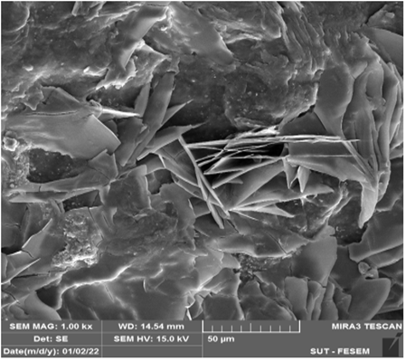
Figure 3 indicates the FESEM/EDS images for the chitosan with Na-alginate composite coating, which is reinforced with ZnO and HA by the Sol-Gel Dip method. A typical structure of a polymer matrix (a homogeneous porous film) was observed. The chitosan/alginate matrix of the polymer coating is shown in Figure 3 (a) without any additions. The exterior of the substrate was seen to be consistently smooth. After a uniform coating of Chi-Alg was put on, the textured surface of the Ti substrate showed no visible pores [31]. With no major defects and a small amount of porosity that helps to promote osseointegration, Figure 3 (b) showed a uniform dispersion of the ZnO ceramic nano-particles across the coating's Chito/ALg matrix. The incorporation of 15 vol. % ZnO into the matrix resulted in a heterogeneous surface appearance, which dramatically altered the surface's shape and increased its roughness [32]. As illustrated in Figure 3 (c), the uniform and smooth surface layer of the coated surface, the consistent quality, and the rather uneven dispersion of the HAP ceramic particles were noted. Surface adhesion increased because of the natural polymeric coating's surface texture forming with uniform dispersion and high bonding after 15% HAP was added to its structure [26]. The uneven form of the inorganic component and the rather irregular distribution of particles in the matrix completely changed the surface's morphology, as seen in Figure 3 (d) particularly after being strengthened with HA and ZnO particulates, a chitosan/alginate layer developed and coated the whole surface. The accumulated layer's morphology was substantially different from the pure coating. Particle size variations resulted in a rougher composite surface with preferred homogeneity because of the reinforcing particles' consistent distribution in the substrate and an excellent relationship of granular sizes among them. Therefore, combining various types of reinforcing materials results in a high-quality coating substance with favorable surface biological characteristics. It is simpler to improve the interactions between the elements in the matrix when varied particle scaling sizes are used. A preferred combination of particulate strengthening substances with an improved dual impact on enhancing the general characteristics of the composite film is produced as a consequence of the large surface area of ZnO particles increasing the attraction forces and facilitating biotin precipitate at the surface of the HA particle [33, 34].
(a) Chitosan+alginate matrix
(b) ZnO/Chitosan+alginate matrix
(c) HA/Chitosan+alginate matrix
(d) ZnO+HA/Chitosan+alginate matrix
Figure 3. FESEM images of the (a) Chitosan+alginate matrix, (b) ZnO/Chitosan+alginate matrix, (c) HA/Chitosan+alginate matrix, and (d) ZnO+HA/Chitosan +alginate matrix biocomposite coating layer at different magnifications
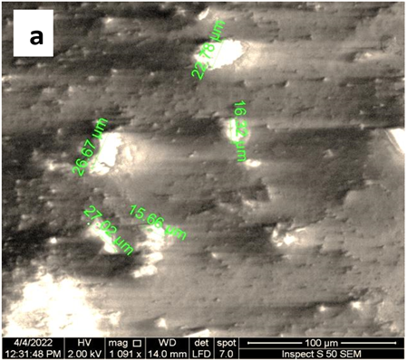
Figure 4. FESEM images of (a) average diameter and (b) thickness coated of the ZnO+HA/chitosan-alginate composite coated layer cross-section
Figure 5. FESEM/EDS of (A) HA/chitosan+algenate, (B) ZnO/chitosan+algenate and (C) ZnO+HA/chitosan+algenate Composite coated Ti substrate
Furthermore, a high degree of adhesion with the surface is perceived from coated sample images, as presented in the Figure 3. The accumulation of ceramic particles in the matrix leads to the formation of cluster-like particles that form fine particles on the surface. It can be depicted that the reinforcement with ceramic particles (ZnO/HA) increased the surface roughness of the coating due to the formation of the microspheres on the material's surface during deposition. The composite coating thickness of ZnO+HA within the polymer matrix sample was measured from the SEM micrograph (Figure 4) and was about (126.9µm). While the average diameter is approximately 21.5µm.
Figure 5 indicates the FESEM/EDS images for the composite coating of ZnO/HA within the polymer matrix. FESEM of HA showed only a few small particles of calcium and phosphor within the polymer coating due to the few additional percent, and the result analysis of the HA composite coating showed (11.27 wt.% Ca), (0.0.39 wt.% Phosphor) and the result analysis of the HA composite coating showed (11.27 wt.% Ca), (0.0.39 wt.% Phosphor), with 4.51 wt.% C and 4.08 wt.% O, as shown in Figure 5 (a). While for ZnO within the polymer matrix, the clusters of zinc oxide within composite coating film are formed with weight percents of Zn, C, and O in EDS analysis of 2.04 wt.%, 2.57 wt.%, and 5.32 wt.%, respectively, the C percent was (8.78 wt.%) in ZnO+HA composite coating as shown in Figure 5 (b). While revealing more particles of hydroxyapatite within the natural polymer matrix, reaching Ca weight percentage of 22.75, P 0.43%, and decreasing in Zn content to 1.48%, respectively, as shown in Figure 5 (c) [29]. The EDS leads to also clearly shows that the C % significantly decreased. This is likely due to the raised depletion of the ceramic powdered substances, which will allow for greater diffusion and coupling with the matrix. This will give the coating the required protection, but it may also occasionally lead to an upsurge in cracking because of compressive stresses being generated at the coating layer [35].
4.2 Inhibition of bacterial test
Figures 6 and 7 show the size of the housing area of bacteria for the composite coating layer (ZnO+HA+chitosan-alginate).
The findings in Figure 6 demonstrated that by reducing the level of activity of bacterial growth and proliferation cycle at the composite's surface, the addition of metallic nanoparticles as strengthening elements to the polymer matrix improved the antibacterial performance of the entire composite. The development of bacteria was limited to the composite coating, demonstrating its capacity to prevent bacterial growth with a rise in the hydroxyapatite or zinc percentage, but it was significantly reduced for pure matrix films [33]. This outcome is brought about by the polymer composite layers' constant hydrophilic character, which outperforms pure coatings and promotes reduced bacterial adhesion. We further infer that for the composite layer (HA/ZnO inside chitosan+alginate), its antibacterial effect versus Escherichia coli was greater than that against S. aureus. Nonetheless, the bio-coating clusters on titanium substrates generally demonstrated strong suppression of the bacterial development zone, demonstrating acceptable biocompatibility to be utilized as an implant coating in medical devices [36].
Figure 7 illustrates the diameter of the inhibition zone (mm) for composite material samples. The increase in the diameter of the inhibition zone is due to the double effect of the ceramic particles on the cell walls of both bacteria [37]. In addition, due to the nature of the microstructure, the additive ceramic particles precipitated in the (chitosan+alginate) natural polymer matrix due to the large differences in particle size and surface charge, making the composite surface rougher with preferred homogeneity, which helped to promote and prevent cell growing and increase the antimicrobial performance of the composite materials [29].
Figure 6. Diameter of the inhabitation zone of different composite coating samples
Figure 7. Size of the inhabitation zone in agar plate different samples
A composite coating made of chitosan/Na alginate matrix and strengthened with hydroxyapatite and ZnO was created using the dip-coating deposition process. The matrix reinforcement with varying particle sizes enhances the characteristics of the composite such as (morphological properties, and antibacterial activity):
(1) The polymer coatings were enriched with active ceramic powders (HA and ZnO with a natural polymer matrix). It was characterized by continuity, and homogeneity, and consisted of a lamellar structure with flattened particles of different shapes and sizes.
(2) The surface morphology determined by FESEM analysis of the bio-composite polymer base coating showed different sizes and shapes of particles embedded in the matrix with a uniform layer of chitosan/alginate with a thickness of about (126.9µm). While the average diameter is approximately (21.5µm).
(3) The scanning electron microscope (SEM) images of the Chi-Alg formed composite coating boosted with ZnO and HAP nanoparticles demonstrated uniform adhesion among the coating and the substrate, demonstrating the suitability of the dip coating technique.
(4) EDS mapping showed that there is a high concentration of Ca, phosphorus, and Zn near the surface within the polymer matrix, especially at (ZnO+HA), which gives sufficient surface area for diffusion and the creation of more coupling between the coating layers.
(5) The size and diameter of the inhibition area of the different composite coating samples (HA and ZnO/chitosan+alginate) are larger than those of the pure matrix, indicating their possible ability to inhibit bacterial growth and effective biocompatibility.
(6) Although access to information and time are restricted. The results of this study suggest that these various coatings can be used as a starting point for more investigation and development in the area of biomedical surface engineering.
The authors would like to thank all the operations at the University of Technology-Baghdad.
[1] Park, J., Lakes, R.S. (2007). Biomaterials: An introduction. Springer Science & Business Media.
[2] Yoruç, A.B.H., Sener, B.C. (2012). Biomaterials, a roadmap of biomedical engineers and milestones. Retrieved from InTech: http://www. intechopen. com/books/a-roadmap-of-biomedical-engineers-and-milestones/biomaterials
[3] Isa, Z.M., Hobkirk, J.A. (2000). Dental implants: Biomaterial, biomechanical and biological considerations. Annals of Dentistry University of Malaya, 7(1): 27-35. https://doi.org/10.22452/adum.vol7no1.6
[4] Anselme, K. (2000). Osteoblast adhesion on biomaterials. Biomaterials, 21(7): 667-681. https://doi.org/10.1016/S0142-9612(99)00242-2
[5] Faheed, N. K., Hamad, Q. A., Issa, R. A. H. (2024). Investigation of the effect of thermal, mechanical, and morphological properties of bio-composites prosthetic socket. Composite Interfaces, 31(3), 331-355.
[6] Bashar, S., Al-Kaisy, H.A., Al-Shroofy, M.N. (2022). Preparation of bio-composite coatings on titanium substrate by electrostatic spray deposition. Key Engineering Materials, 937: 129-138. https://doi.org/10.4028/p-224uc8
[7] Jayaswal, G.P., Dange, S.P., Khalikar, A.N. (2010). Bioceramic in dental implants: A review. The Journal of Indian Prosthodontic Society, 10: 8-12. https://doi.org/10.1007/s13191-010-0002-4
[8] Hornat, C.C., Urban, M.W. (2020). Shape memory effects in self-healing polymers. Progress in Polymer Science, 102: 101208. https://doi.org/10.1016/j.progpolymsci.2020.101208
[9] Swain, S.K., Bhattacharyya, S., Sarkar, D. (2011). Preparation of porous scaffold from hydroxyapatite powders. Materials Science and Engineering: C, 31(6): 1240-1244. https://doi.org/10.1016/j.msec.2010.11.014
[10] Islam, M.S., Todo, M. (2016). Effects of sintering temperature on the compressive mechanical properties of collagen/hydroxyapatite composite scaffolds for bone tissue engineering. Materials Letters, 173: 231-234. https://doi.org/10.1016/j.matlet.2016.03.028
[11] Gammariello, D., Incoronato, A.L., Conte, A., Del Nobile, M.A. (2016). Effect of sodium alginate coating with ascorbic acid on shelf life of raw pork meat. Journal of Food Technology Research, 3: 1-11. http://doi.org/10.18488/journal.58/2016.3.1/58.1.1.11
[12] Dhafer, G., Al-Shroofy, M.N., Al-Kaisy, H.A. (2022). Electrostatic deposition of poly (Methyl Methacrylate)/Titanium carbide coatings on austenitic 316L stainless steel implant. Engineering and Technology Journal, 40(6): 918-925. http://doi.org/10.30684/etj.2022.131478.1038
[13] Hamad, Q.A., Abed, M.S. (2019). Investigation of thyme and pumpkin nanopowders reinforced epoxy matrix composites. Journal of Mechanical Engineering Research and Developments (JMERD, 42: 153-157. http://doi.org/10.26480/jmerd.05.2019.153.157
[14] Scaffaro, R., Lopresti, F., Maio, A., Sutera, F., Botta, L. (2017). Development of polymeric functionally graded scaffolds: A brief review. Journal of Applied Biomaterials & Functional Materials, 15(2): 107-121. https://doi.org/10.5301/jabfm.5000332
[15] Yoruc, A.B.H., Sener, B.C. (2012). A roadmap of biomedical engineers and milestones biomaterials. InTech Janeza Trdine, Croatia.
[16] Devi, N., Dutta, J. (2017). Preparation and characterization of chitosan-bentonite nanocomposite films for wound healing application. International Journal of Biological Macromolecules, 104: 1897-1904. https://doi.org/10.1016/j.ijbiomac.2017.02.080
[17] Cervini-Silva, J., Ramírez-Apan, M.T., Kaufhold, S., Ufer, K., Palacios, E., Montoya, A. (2016). Role of bentonite clays on cell growth. Chemosphere, 149: 57-61. https://doi.org/10.1016/j.chemosphere.2016.01.077
[18] Archana, D., Singh, B.K., Dutta, J., Dutta, P.K. (2013). In vivo evaluation of chitosan-PVP-titanium dioxide nanocomposite as wound dressing material. Carbohydrate Polymers, 95(1): 530-539. https://doi.org/10.1016/j.carbpol.2013.03.034
[19] Ambrosio, L. (2017). Biomedical composites. Woodhead Publishing.
[20] Boccaccini, A.R., Ma, P.X., Liverani, L. (Eds.). (2021). Tissue engineering using ceramics and polymers. Woodhead Publishing.
[21] Faheed, N.K., Hamad, Q.A., Oleiwi, J.K. (2022). Tensile and stress analysis of hybrid composite prosthetic socket reinforced with natural fibers. Journal of Renewable Materials, 10(7): 1989-2013, https://doi.org/10.32604/jrm.2022.017573
[22] Al-Hasani, F. J., Hamad, Q. A., Faheed, N. K. (2024). Enhancing the cell viability and antibacterial properties of alginate-based composite layer by adding active particulates. Discover Applied Sciences, 6(2), 70.
[23] Boura-Theodoridou, O., Giannakas, A., Katapodis, P., Stamatis, H., Ladavos, A., Barkoula, N.M. (2020). Performance of ZnO/chitosan nanocomposite films for antimicrobial packaging applications as a function of NaOH treatment and glycerol/PVOH blending. Food Packaging and Shelf Life, 23: 100456. https://doi.org/10.1016/j.fpsl.2019.100456
[24] Ahmed, Y., Yasir, M., Ur Rehman, M.A. (2020). Fabrication and characterization of zein/hydroxyapatite composite coatings for biomedical applications. Surfaces, 3(2): 237-250, https://doi.org/10.3390/surfaces3020018
[25] Hamad, Q. A., Rahman, H. J. A., Faheed, N. K. (2023). Improving some mechanical properties of green biocomposite by natural pumpkin powders for prosthetic socket. In AIP Conference Proceedings (Vol. 2787, No. 1). AIP Publishing.
[26] Hamad, Q.A., Al-Hasani, F.J., Faheed, N.K. (2022). Comparative study of biotin and hydroxyapatite on biological properties of composite coating. International Journal of Biomaterials, 2022. https://doi.org/10.1155/2022/8802111
[27] Hamad, Q.A., Al-Kaisy, H.A., Al-Shroofy, M.N., Faheed, N.K. (2023). Evaluation of novel chitosan based composites coating on wettability for pure titanium implants. Journal of Renewable Materials, 11(4): 1601-1612. http://dx.doi.org/10.32604/jrm.2023.023213
[28] Hadi, A.N., Mohammed, M.R. (2022). Performance of biocomposite materials reinforced by hydroxyapatite and seashell nanoparticles for bone replacement. Journal of Nanotechnology, 2022. https://doi.org/10.1155/2022/9156522
[29] Hamad, Q.A., Abdulrahman, S.A., Issa, R.A.H. (2022). Investigation some characteristics of biocomposites coating for biomedical implants. Key Engineering Materials, 936: 3-11. https://doi.org/10.4028/p-nt9b2f
[30] Ullah, I., Siddiqui, M.A., Liu, H., Kolawole, S.K., Zhang, J., Zhang, S., Ren, L., Yang, K. (2020). Mechanical, biological, and antibacterial characteristics of plasma-sprayed (Sr, Zn) substituted hydroxyapatite coating. ACS Biomaterials Science & Engineering, 6(3): 1355-1366. https://doi.org/10.1021/acsbiomaterials.9b01396
[31] Kadhim, N.N., Hamad, Q.A., and Oleiwi, J.K.(2020) “Tensile and morphological properties of PMMA composite reinforced by pistachio shell powder used in denture applications,” in Proceedings of the 2nd International Conference on Materials Engineering & Science (IConMEAS) AIP Conf, Baghdad, Iraq, March 2020.
[32] Oleiwi, J.K., Hamad, Q.A., Rahman, H.J.A. (2018). Tensile properties and morphological test of heat cured acrylic resin reinforced by natural powders. International Journal of Mechanical and Production Engineering Research and Development, 8(6): 325-334.
[33] Hussein, M.A.K. (2022). Preparation of PMMA/SiO2 composite sheets for tribological and mechanical tests. Misan Journal of Engineering Sciences, 1(1): 58-68. https://doi.org/10.61263/mjes.v1i1.22
[34] Mohammed, S., Karim, A.A., Thamer, A.Z. (2023). Experimental study on the effect of size, type, and replacement ratio of recycled aggregate on the mechanical properties of reactive powder concrete. Misan Journal of Engineering Sciences, 2(1): 38-52. https://doi.org/10.61263/mjes.v2i1.43
[35] Issa, R.A., Al-Shroofy, M.N., Al-Kaisy, H.A. (2021). Al2O3-TiO2-PMMA Bio-Composite Coating via Electrostatic Spray Technique”, Journal of Engineering Technology, 39: 504-511.
[36] Mohammed, M.R. (2022). Mechanical and biological behaviour of 3D printed PCL-based scaffolds fabricated by fused deposition modelling for bone tissue engineering: A review of recent advances. Misan Journal of Engineering Sciences, 1(1): 33-46. https://doi.org/10.61263/mjes.v1i1.18
[37] Faheed, N.K. (2024). Advantages of natural fiber composites for biomedical applications: A review of recent advances. emergent materials, 7 :63–75. https://doi.org/10.1007/s42247-023-00620-x