Molecular Detection, Sequencing, and Expression Analysis of ALS Genes in Clinical Candida spp. Isolates with Antifungal Resistance

Fatimah F. SH. Alhashimi*![]() | Thamer A. A. Muhsen
| Thamer A. A. Muhsen![]()
© 2025 The authors. This article is published by IIETA and is licensed under the CC BY 4.0 license (http://creativecommons.org/licenses/by/4.0/).
OPEN ACCESS
The agglutinin-like sequence (ALS) family is crucial for Candida spp., particularly C. albicans, as it aids in adhesion to biotic and abiotic membranes and formation of virulence factors. This study was conducted to identify and measure the expression levels of ALS genes in Candida species isolates from clinical specimens. A total of 276 clinical samples (vaginal, oral, and blood) were obtained. The isolates were characterized, genetic material extracted, ALS genes detected, nucleotide sequence determined, and gene expression measured after antifungal treatment of oral and vaginal isolates. The study found a significant difference (p ≤ 0.05) among the number of isolates; 135 (48.91%) Candida positive isolates, with C. albicans being the most common species. Also, 15% of isolates were resistant to nystatin. Most Candida species had ALS genes, except for C. kefyr and C. lusitaniae, which were not detected. The gene expression analysis before and after treatment with fluconazole and nystatin revealed that C. albicans isolates from the mouth had higher gene expression than vaginal isolates, with gene expression reaching 92.82 and 17.52, respectively, for the two antifungal agents, making them resistant to antifungals. The ALS1 gene expression was highly elevated in antifungal-resistant isolates, while it was reduced in susceptible isolates. The study’s findings highlight the prevalence of ALS genes in Candida species and could be used as a marker of drug resistance.
Candida spp., molecular detection, PCR, qRT-PCR, ALS genes, gene expression
Fungal infections frequently impact mucous membranes, including those of the respiratory, urinary, and genital tracts [1, 2]. Candida is a primary etiological agent of fungal infections, responsible for candidiasis, and ranks as the third leading cause of sepsis in Europe, with an estimated mortality rate of 37% within a 30-day period [3-5]. Several Candida species, including C. tropicalis, C. albicans, C. krusei, C. parapsilosis, and C. glabrata, are responsible for more than 90% of infections. In recent years, other Candida species have been identified and diagnosed as causative agents of infections, including C. kefyr, C. lusitaniae, and C. guilliermondii [6, 7]. Currently, invasive candidiasis is a common and widespread disease and is associated with high mortality and morbidity. Individuals most susceptible to candidiasis possess a compromised immune system, which may arise from various factors such as chemotherapy, use of broad-spectrum antibiotics, surgical interventions, kidney failure, and organ transplant surgery [8, 9].
The bacteria that colonize the mucous membranes, including those of the mouth and vagina, constitute a varied population that influences human health, particularly in women, notably during pregnancy. Many studies have demonstrated that an increased prevalence of infectious disorders is caused by any alteration of the microbial diversity in the mouth or vagina. It is estimated that 75% of women will suffer from vaginal infections at least once in their lifetime [10, 11]. Approximately 90% of these infections are caused by Candida vaginitis (bacterial vaginosis). The initial diagnosis of vaginosis is mainly based on clinical signs and symptoms and examination of vaginal discharge under a microscope [12]. Given the widespread prevalence and clinical impact of Candida infections, particularly among immunocompromised individuals, there is a growing interest in understanding the molecular mechanisms that contribute to the pathogenicity of these fungi. One such focus is the agglutinin-like sequence (ALS) gene family, which plays a critical role in Candida adhesion, biofilm formation, and virulence.
The ALS gene family of Candida spp. represents an important gene group for the genus Candida, especially C. albicans. This is due to the significant role of these genes in the process of adhesion of fungi (yeasts) to moist or wet membranes such as the surface of epithelial cells in the vagina, mouth, respiratory, urinary, or genital tract. Furthermore, the ALS gene family also plays an important and significant role in biofilm formation and other virulence factors, such as colonization, invasion, and pathogenicity [13]. Candida dubliniensis, Candida parapsilosis, Candida orthopsilosis, Candida metapsilosis, and Candida tropicalis have been shown to include genetic material and nucleotide sequences from ALS genes and the proteins that arise from their gene expression. Although the presence of nitrogenous base sequences has made it possible for researchers to identify ALS genes, these sequences have limited research on the processes underlying the gene expression of these genes, as well as the reasons behind both elevated and decreased gene expression. According to recent research, ALS genes, in particular, the ALS1 gene, play a significant role in modulating biofilm formation and, consequently, the connection between these genes and antifungal resistance [14, 15]. Research on the ALS1 gene and its relationship to antibiotic resistance is scarce.
The present study aimed to detect the ALS genes and evaluate the effect of antifungal agents on the gene expression of isolates.
2.1 Collection of samples
Between September 2024 and January 2025, a total of 276 samples (120 vaginal samples, 120 mouth samples, and 36 blood samples) from patients ranging in age from 3 to 70 years old, depending on the type of sample, were obtained from Rusafa hospitals (Imam Ali Hospital, Fatima Zahraa Maternity Hospital, Ibn Al Baladi Hospital for Women and Children, and Alawiya Women's Hospital). The samples were collected from patients following a clinical diagnosis by the pathologist in each of the aforementioned hospitals. Before culturing, the samples were stored at 4℃.
2.2 Isolation and identification of Candida species from clinical samples
The culture medium used in this study, Sabouraud Dextrose Agar (SDA), was prepared according to the manufacturer’s instructions. Swabs were collected from the mouth and vaginal areas of the women and then cultured on the prepared culture media. The cultures were incubated at 37℃ for seven days. Then the colonies were identified microscopically at 400x magnification according to the taxonomic keys [1, 16].
2.3 Antifungal susceptibility test
Two antifungal agents (fluconazole and nystatin) were used to test for antifungal susceptibility against 60 isolates of Candida spp. The isolates included 29 isolates of Candida albicans and 31 isolates of non-Candida albicans, including 15 isolates of Candida tropicalis, 10 isolates of Candida glabrata, and 3 isolates of each of Candida lusitaniae and Candida kefyr. The isolates were identified phenotypically, microscopically, and using the Vitek-2 device in accordance with the technique by Liu et al. [17].
2.4 DNA and RNA extraction
To detect ALS genes in 5 isolates of C. albicans, 2 isolates each of C. tropicalis, C. lusitaniae, and C. kefyr, DNA was extracted using the Promega DNA extraction kit (Bioneer, South Korea). The ALS1 gene family was selected for nitrogenous base sequencing in some of the isolates. Regarding gene expression, two isolates (one resistant and one sensitive to C. albicans), one from the mouth and the other from the vagina, were selected to evaluate ALS1 gene expression following RNA extraction using the TRIzol™ kit.
2.5 Molecular detection of ALS genes in Candida spp. by the polymerase chain reaction
In this experiment, the primers whose sequences are listed in Table 1 were used to amplify the ALS genes using polymerase chain reaction (PCR) following the method described by Sadeq and Ismail [18]. Additionally, the ALS1, ALS2, ALS3, and 18S RNA genes were amplified in the real-time reaction according to Ahmed et al. [19]. The forward and reverse primers were used in the PCR procedure to amplify the ALS gene and produce numerous copies of it. The reaction was performed with a total volume of 25 µl [18, 20]. After completing the replication process, the products were electrophoresed according to the method described by Green [21]. The PCR products were purified and sequenced (Macrogen, Korea) for molecular detection of Candida spp. and analysis of genetic relationships.
Table 1. Nucleotide sequences of the primers used for the study
|
Primer Name |
Seq. |
Annealing Temp. (℃) |
Product Size (bp) |
|
RTALS1F |
GACTAGTGAACCAACAAATACCAGA |
58 |
318 |
|
RTALS1R |
CCAGAAGAAACAGCAGGTGA |
||
|
RTALS2F |
CCAAGTATTAACAAAGTTTCAATCACTTAT |
366 |
|
|
RTALS2R |
TCTCAATCTTAAATTGAACGGCTTAC |
||
|
RTALS3F |
CCACTTCACAATCCCCAT C |
342 |
|
|
RTALS3R |
CAGCAGTAGTAGTAACAGTAGTAGTTTCATC |
2.6 ALS1 gene expression in Candida albicans by relative quantification
Using the quantitative real-time polymerase chain reaction (qPCR-PCR) technique and the standard 18S rRNA gene, the expression levels of the ALS1 gene were assessed in two isolates of Candida albicans from the mouth and vagina before and after treatment with fluconazole and nystatin. The procedures followed the reaction conditions as described [19, 22]. Relative quantification was made according to the following equations:
Folding =2-ΔΔCT
ΔCT = CTgene - CTHouse Keeping gene
ΔΔCT =ΔCTTreated or Control - ΔCTControl
2.7 Statistical analysis
The data were analyzed using the Statistical Package for Social Sciences (SPSS; version 21). Statistical significance was set at p≤ 0.05.
3.1 Clinical isolates of Candida species
Out of 276 samples obtained from patients at Rusafa hospitals (Imam Ali Hospital (AS), Fatima Zahra Hospital, Ibn Al Baladi Hospital for Women and Children, Shahid Al Sadr General Hospital, and Alawiya Women's Hospital), the results of culturing on SDA medium revealed the presence of 135 positive isolates. As indicated in Table 2, the positive clinical samples (48.91% of the total number of samples) comprised samples from the mouth, vagina, and blood in varying proportions (50.37%, 39.26%, and 10.37%, respectively).
Table 2. Percentages of Candida spp. isolated from clinical samples
|
Clinical Samples |
Total Number |
No. of Positive |
% of Infection |
No. of Negative |
% of Infection |
|
Mouth |
120 |
68 |
50.37% |
52 |
36.88% |
|
Vagina |
120 |
53 |
39.26% |
67 |
47.52% |
|
Blood |
36 |
14 |
10.37% |
22 |
15.60% |
|
Total |
276 |
135 |
100% |
141 |
100% |
|
% total number of samples |
|
48.91% |
51.09% |
||
|
p-Value |
0.04* |
|
0.01* |
|
0.01* |
|
Person Chi-Square= 0.033* Pearson's R= 0.148** |
|||||
* Significant differences at a probability level of p≤0.05.
Table 3 shows the results of the identification of Candida spp. at the species level, where C. albicans was the most prevalent species with 63 isolates (46.67%), followed by C. tropicalis and C. glabrata with 43 (31.85%) and 20 isolates (14.82%), respectively. Meanwhile, C. lusitaniae and C. kefyr were found with 6 (4.44%) and 3 isolates (2.22%), respectively.
Table 3. Types and percentages of Candida spp.
|
Species |
No. of Isolates |
% |
P-value |
|
C. albicans |
63 |
46.67% |
0.001* |
|
C. tropicalis |
43 |
31.85% |
|
|
C. glabrata |
20 |
14.82% |
|
|
C. kefyr |
6 |
4.44% |
|
|
C. lusitaniae |
3 |
2.22% |
|
|
Total |
135 |
100% |
* Significant differences at a probability level of p≤0.05.
3.2 Resistance of Candida species to antifungal agents
Figure 1 shows the Candida spp. sensitivity testing against fluconazole and nystatin showed that 56.7% of Candida spp. were resistant to the antifungal agent, fluconazole, and 15% of Candida spp. isolates were resistant to the antifungal agent, nystatin. These isolates included 4, 2, 4, and 1 isolates of Candida albicans, Candida tropicalis, Candida glabrata, Candida kefyr, and Candida lusitaniae, respectively, as shown in Table 4.
A
B
Figure 1. Susceptibility test of Candida species to antifungals, A: Isolations Candida Control, B: Candida isolates were exposed to fluconazole at a final concentration of 0.6 mg/mL and incubated at 37℃ for 24-48 hours
3.3 Molecular detection and sequencing of ALS genes in Candida species
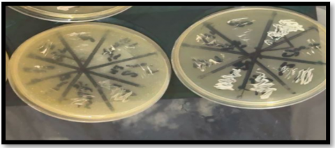
The molecular detection used to identify the ALS1, ALS2, and ALS3 genes in the Candida spp. under study revealed that the molecular sizes of the amplified genes are 318, 366, and 342, respectively (Figure 2). Every isolate of Candida albicans, with the exception of one isolate that lacked a gene, and every isolate of Candida tropicalis, with the exception of one isolate that lacked the ALS2 gene, had all three amplified genes. Additionally, the results demonstrated that all isolates of C. kefyr and C. lusitaniae lacked genes. Following the electrophoresis separation, two isolates from each species of the genus Candida, including C. albicans (isolates 14 and 19) and C. tropicalis (isolates 6 and 18), were chosen for nucleotide sequence analysis of the ALS1 gene since ALS genes were diagnosed with high frequency. Table 4 highlights a comparison of the ALS1 gene sequences in the Candida spp. with reference sequences. Isolate 19 of Candida albicans and isolates 6 and 18 of Candida tropicalis had the highest nucleotide sequence identity (100%), whereas isolate 14 of Candida albicans had the lowest identity (99.62%) because of two mutations (Table 5).
Table 4. Susceptibility of Candida species to antifungal agents
|
Antifungal Agents |
C. albicans n=29 |
Non-albicans Candida (n=31) |
Total (n=60) |
|||
|
C. tropicalis n=15 |
C. glabrata n=10 |
C. kefyr n=3 |
C. lusitaniae n=3 |
|||
|
Fluconazole |
18 (62.07%) |
6 (40%) |
4 (40%) |
3 (100%) |
3 (100%) |
34 (56.67%) |
|
Nystatin |
4 (13.79 %) |
2 (13.33%) |
2 (20%) |
0 (0%) |
0 0% |
8 (13.33%) |
|
P-value |
0.001* |
0.001* |
0.001* |
0.001* |
0.001* |
0.001* |
|
Person Chi-Square= 0.663** Pearson's R= -0.087*** |
||||||
Non–Candida albicans (n = 31).
Table 5. Nucleotide sequence of the ALS1 gene in Candida spp. showing mutations
|
ALS1 Gene |
||||||
|
No. |
Type of Substitution |
Nucleotide |
Sequence ID with Comparison |
Country |
Strain |
Identities |
|
14 |
- |
A\ |
ID: OR664373.1 |
USA |
Candida albicans |
99.62% |
|
Transition |
C\T |
|||||
|
19 |
- |
- |
ID: AF201686.1 |
USA |
Candida albicans |
100% |
|
6 |
- |
- |
ID: AF201686.1 |
USA |
Candida tropicalis |
100% |
|
18 |
- |
- |
ID: LC791630.1 |
USA |
Candida tropicalis |
100% |
![Word - bbرسالة الماجستير الى دكتور ثامر[1]](data:image/png;base64,R0lGODdhGwG7AHcAACH+GlNvZnR3YXJlOiBNaWNyb3NvZnQgT2ZmaWNlACwAAAAAGwG7AIf///+UOgAAAACUMQB7MQh7MQC9QgDFSgjFSgCMMQClQgCtOgCUOgiUMQh7KQC9SgCMMQiEOgClQgilOgClMQCMKQCtSgC1Sgh7Ogi1QgCUQgBzMQC9OgCUQgi1UgiMKQjFWgilOgiUKQCMOghzKQCMGQBzMQilSghKAADOSgilIQC9QgjOSgBaWlrm3ua9KQBrKQC9pUrm5hm15hm13rW1UgDOOgC93uZ7IQBSMQjm3kp7KQhrGQCEQgi1YwhrMQDmrea15kq1rebeSgAhGRnOawgxAACMvd6MvVoQlFo6vd46vVqMvZyMvRkQlBk6vZw6vRljvd5jvVoQa1oQvd4QvVpjvZxjvRkQaxkQvZwQvRnvWgjvexBjCACMEHOMEOY6EOY6EHOMEK06EK1jEHNjEOYQEOYQEHNjEK0QEK3mWnO1WnPmWubmEObmWq3mEK3mEEK1Wua1EOa1Wq21EK21EEJze3vvnEKM796M71qMlN4ZQhBClFo6794671o6lN6M75yM7xlClBk675w67xk6lJxjlJxjlN4QlN4QlJyMa5xj795j71qMa95Ca1oQ794Q71o6a95j75xj7xlCaxkQ75wQ7xk6a5xja5xja94Qa94Qa5y1czrFWgDOEADmezqtrRmt5oTmOgBKEAB7Y2Nze2sxEDrvlCHmWjqMEDoQEDpjEDoQQjrmEHO1EHN7EAA6Ojrv74RaQmuMQuaMQnM6QuY6QnPvtUqMQq06Qq3OOghjQuYQQuYQQnNjQq0QQq1aQjrmnHvvrbXme2utnK2EOhC1e2vme+bmMebme63mMa3mMUK1e+a1Mea1e621Ma21MUK1tYS1vbWMjITm1rVzOgC1UjqtCACMQjoQAACMjGPvEAjv1ntrlGPOrRnO5oTmMXO1MXPv97WMlKXvtRnmvYSUQhCUOhC1nIS9EADOrbVrlDprazprlBBraxCMMRClMQiMlDqMazqMlBCMaxDFShC9OggIGRClQhCEQgBjY2vO/+a1ShD//+Z7MRAAABAI/wAFCBxIsKDBgwgTKlzIsKHDhxAjSpxIsaLFiwgBaNzIsaPHjyBDihxJsqTJkyhTqlzJsqXLlx0FwpxJs6bNmzhz6rwpc6fPn0CDCh1qsyfRo0iTKl0K0yjTp1CjShXqdKrVq1izkqyqtavXr0y5urxBqh8AO9JI9rvhQu2Nk9OmmZTWtuQ0fXVHupBGzGQ/YmlHsv37ViQ1AC7yhtyrke1iF2tbjBOZGACxyXrbSgvc0cU0ajek/SWmWKXYltQEjHNBhLNIYHIF5zBJTIC+kqXw0mZXWDARWKVBjtPXInjHFnaItQBmXKM0WADGtSD5HAA1U801wrqBjtT0kNO2E/9hPtLONLL6bpjtOI0IMWr6mNxQ5/J0SyamqBUvaX4kNTvYlXQDMaVkt5EjsMG1nl7sbFeSZ8D0BlJcxNzlWmfQSXObXsBEt8eFHelDH2CUtUBNKQ6KNE6Bwx320XMuAAfAgivZt9J8m7WgHkmSkXRDhw9Ks+NI06xmkgt9lUTNOHb0QyNI/Yzjokh22OEZiBzdkOF3IlXnwn4htZAWlhxJo82ApTzp0TRNUtOceuqpI+VLNpLEZpVVtoAccaTg6eefxCH356B2lELooIYCc+ig3i2KqKOD6tNnn5Dqo8+di3rXwl2QNtpopYJSumgL+tgh6qF6kkJKqZCSammVp5r/2gIpbN4laJWxoVTnSLBg5OuvwAYr7LDETnSPaQK4pI1ARqDwSgldvCJtFyhUi0Kz1RqhbbVdUGvttthWy0O3XWxr7bnWRnttuOuGO4q3KGRj7rnNamtvtvOi0G2103J77bf0XpvNwPYWDK69Atgr78IFr8vwwwTjS7ARDBtM8UAUUyxvxu1mnE3CBA+8scbaDgyyABOLTBARyLpEigAopPOCDai84MkLNuN8DQcq4KyzzzaIgrMNPb+wsw0y26ALzjfLPLPPL6jgiQ2eCG2DzxxEnQ7PL2R99cwqlHANBSpk7TXOKqRNNNRpPy30C0nbcLXcW8tttNNkl6B32ioo/wAPBxyUkDYFJVSgt96GqyBCBSKI0DfhiuuNQwWUl/CK4ZSLYIACA4jwbOOFNy4CBQooIEIJOODwyuSMvyJ6BZ0zjjnllxc+gOUJNP4s7a/wcDntA2C+egI8fMxySruKVIoAr4hSDxdDXM2BLoBTYAAH17NQDws2sMCBDYDbUA/gHHgvt9K6WC3394Ab0P3cQ7DAgi7ncw/+9+6TzwE/CmB/vQEUKN/2/ue973FPfvZ7X/eExr7uGYAFD3wgAkEAQNJR4IIUsAcH4KHB6pGNAgPAIOkAyAHS8Y8C54CHCAbAwgFo4IIhHMDmzKHCAagAgyoIIQjhoQB8tNCHLIQHBf/4NwB8UICG50hiEjHIQgsOQAEX5OE5YiiC0pWOdD4EIQtFgAIBHE9XyWrJ8q5hgyF8wns4s0fZOgg4e+iCevdbmj3iqIt61OONeLzaG1+AR13wQ2l2xOMd/2iPeryABXPkQD1swMb9ZRAe/GgjP66ngOvtr418HBr9utfHmU1ve5E0gAEmyQ9dBDCS8BilPeBhxAzywx784OEF8QFJDOLDivxT4RQHYI4n8jCECtCAAnrJQnzc8hycAyHp4BHCW8Zwh0WE4Qtb2MRzWLCHy/Sh3/zmQhDSUgP40MALNcBKEKpgFAIwRctaYgcBlGAI8eNjKSPJAQ3ygx8vsMfQ5mj/DzdqEJZ3fOMdoxfQPlLvjUqTm/bqqEGBzsyOi5yjLtxHz/3Vg42kvF4H+3hH8YlPF/CsB0EPSsH9sYAfBewgPy64v1TCI21qtAcAjXnBG/LQmLe0ojWLGENzKOAcHjBmCMU5zCfa0BwUEKpQn6jUInIOmeHEh0+R2kJw4iOo4GwmPF54S6XetHQaCGIPB8ADARwLeWFkyRiHENFLblCfhRxfPwupCze2sZ517WdBdcG9O9rxlXkF5PQA11HAHVSkXbNBJCsaya6NT67aq4cBNJjPuAp0kXKrI1sRqlfJGmB89dwgSxWAyiHC8pUcrCU0RTDFYJLOA1jkXC+Nuc2r/wKRl+FUJjCRydRbstKZSZ3lWBUQVM4ZkZtJjGYlRxlULbawh1D0wAlpqoBXCGAP61SrACogCvFZsp6r7Fob3VhKyv7ze42dHj8VaQ/tgaAeKugnI+FoR30elL513OQiyfdK8EVSrpLFHmgD/D1UfCKz8RPfEPL7RvWKEoKA40c96CnhWK7UHvi4aAAhyY9WCjWsLOyhBzxQunA+l5ZdBec5eslVowqRhSw2wC1zSYGb3tObTjVHcY3I0xBX8J7Q3WpSoYsPfpCYdD3Exyv+8cWTJC8ky3vF/fY3StSu8p8Shus/86q/CSPSr9rTHixhqcgBf5l6Zd5vXT87PgmLcv+yhSTfRespyv+eFHx2ZCREt7e9OgYScO+14yU1OMR7cgAfMsbioUfJsw7n1ofgHOsRhxlMFxYzrD4ssgfMcUtO+22ZwwTnC3vIP5yK2JgrdOqIfdpVc5DTb/go4UxrAF0X5pQfnFhpkqFoXXWi1SXtfAUqJjxJe7z3nkK0p0ov6kdi2yOA+7OnAYYtUoi+8rT9zPK15zrhZ0d4wt/2QLdJ+EpiS7ceHgBBoCVrR/fZoBOoGGU93jtsDe752/eMJSxZycMOQzHfIm6uVUkb1KA+0Rw+wMc5NMBbSMNYqhowhwWMKUxW9lDH/PDBPaVbXG8eE6f46IEPpKvNc2S8udb/SLiIjUzkJBdh5SJOgFmzu5LllUCxHWYmLVVY47+h8sqrTC08hg5QnolWBVZ+6RDt6AF4fECIsVSBNc8hAiGOeJL81jcks67RVSaRpkMXItlIyY/BXRC1NS3kEBxZ46A6Gh8f2GlS+aGBBFTAHCk0h94T4OpxGvMd5kiABgBvjh6cIwEJ4GWl35GPd/TgHQnIxwl6wPAiBjXxPdB7MczheMhz2ohSHcA7Rj/6zSuR0/zAhsJxCvnM9+ADOmb1U5W4YrznwIs0V8nyUGA5aVXgAx/4veguWQ98sDb4lHuH3j5QgkK+UnF5E8EHpJ+5/YEgloVLAOCpDny9neP6/Aje/+97z/wKPCuVyP4AtSaHAxiorgSO6x8sCbe7051Ob+m4Y0tf8YEdrC51KMADFcBKBvcOMAADO4ADOwADPMCAODBFrjZ6B8gDOXCAo8ADo4AD78BV5/B7Fuh+P+APMDAKBaBjHjAA/dcFOTAKI7iCRiCAOqYAiMcDO5ADDbiAOMADPLBi/PAO5/AB79A7GOh+qoMCQEU6vYR4FdAFPFA4OBAvuPdrYiQAo/AC8AID3WI5zAcPoEUBr6AuvZOB/AdT8PVS8PCDlsOE/Dd9/QRJH4ACGYgD/pCDPMB8HwBuUoeBDcgD3MJ/8CBdG6QC5YKBo3CBKPABtwNkSWU5y6c3z/+iAtdQXkg3CtVSiKNgg6OQREiHDxWQAJaoLaNgBKEogEZVeD8Qip+oLTDwDpbXgSNYiKKYAwUQijhwVefwDv6QA3BIBEaQA9uSA69QRL3UAyPYgp+YiSsGfPxnLUN4gRUwRVglejvwDuNSLQnQRQLACrmXEu00CtfwCs7if1/IhHpTSFEDDzhwgaPwCjAQgPwnOKrED444ju7IfEhXT/wgAtqygpTIL88CAiV0hkzYgBcIjq4jAvekAk6ngzuwAzTYO4fDASCgWPCgN9InLRgpNi8wYfBgDuoHA+yIgdXyAWHzYuaAiisIAyuYDdVSArckhyZgiRVIidjikj70DpT/6IIokAO+SIXRlAAO+YoqSYHbAn/8sGJ6WC3+EIob0wVx15C9swNz2I7iUgI8kFTJp4PmxwMlYzzbiBLL0gUwBXclAHwf8ArmMAxF0IZnCHlA2IFrOAzGcD0ZJALXcA4q8AEddg5lyQnD0AlqBHcOuQMJkI7P8g5rEAwXpQL4oAI62H9ACHddYA3B4AGBaDk++HVUVwLwYA2f8G3XwJhqhJdikwrWMAwSdoaspURAOIIfQJmcYABniAMFsH1B+A7FgA3mkGuBl44mgAG1SXr+EIQS5kNAeYGr+A6Ggw3YkAPWwAn4UJg0OHodGDu++Aqc8AJPKZXYwA94B4TFMAr+/2ANFfkK23cOhseXatgFuVYBk8MDCZBC59CLOfAPUQhGwAYzzWYNGKaQKcQJwcCf9lAExrdLQ5dCSaWW2CNT21ZI1hBLIBAMXDBvCZlCA0B1KfQBPlAOqNlSFLCalHMOtHQOnMAJpxV2tDRL8GADw0AN1IAKcWUPH7CR0NBPfoQK3EANwxBtQhR2SRR3nckN5TAESQVjSDR6ReCiLkpit5gAPXACenehHhAMLloOnPBUrBR3oQcPOUoN4sAJRxSiUwRJjckJ4iAM1BAMHDA6SWQOR1lOZoqmwfB2WZRvfumiVAoCYTVbkMQN6uAC1uAPwNBkJvFkILE8XZBXuhB2Qv8ERdDGQStVY6C3QYY2RFRGT40UWhnUT/jQhteWby/1RJfEYSL0bJHKoMTmSNcnYIb0AhJmRz5TV7L6RizwAgG0qZG6Ujonm7OUVB4gTFlkTGWnRkd0SxHnasYUX/dEnkUUVvx2VZx2Oi8wbB1pRDTUTLQES6gQDJ8AAhYHRWGlpUbED9xqAUFGWtYjSpyACi0qDEWwpl0FRUVQDsFwC69gDeJgDF95Eu3EA34EUABXqVfGYTUwbBFGZdWjSKQUbf2kqfo2ZjE6UZO0XH/IYbF2T9d3bIlWT/MmZ9jzNzjzShK2RwFlbVO2qXN1oiV2QbsGeokWdqylTODkaFhUYx//ukLi1Fuck2QvBYll2Uyk1UOplFRhA38iAHJFRls0dTqM42GnlUHXEzbXUAK2mm8eMEo9+wIlcC3QIYXa1QVdc2GkmqyoVUuUOlMJy0He5lYcq0Zih0rXBl4aVGSVBEUydly+KrJUVjqx1KkSRj6rNFmuVE9qK1L5tT9+FEkKMLDEhnSVlFNJhWjWlGQdFrlJJWTINEWYNnTGBYFhtXDQRQFcRZ49l0REhmStZJd4OWr4UAMdt1RbZEysZkKyGUmKMzYU0FzS9WwVyXxGcJ9OllYrsSwocEMc4De1BEUioEg+N0TwULWINkQZVGNj9kqnBFfT67yEZqqOVEJQhK6J/5ZTtwRAJyS0/1ZCq6SsHBRAo5RF1VsPywVAMjZWMsaR+8M/Jca6pRNDT+SokspMFICINIVU17pFvNQDLCSiIlpMWoRUwnVLBrdFU2ShnhZNFnREk3ZVwsRqo1S3ZwdFQ2dLRFd19oBOhFoShvoRL8MDJdRKIHxLPXeGCnlBVTdkRtR0nyZ2KHtlPedKxru+uuphGORT2ARF2mRCIjSuITyukdSpjbpSsXa9dIu6VnRFWzdkrIZTRYp3F9rFXZx4njfBKHShZzgA2td4+ZCEekdMa8xLIhqlsfeMXdyRlTdFUWVEexpxERc8T3TBoDdkOpdLSUZTAwAPZXXCWyG8Kv/RTl1QQrYUXABcyLpkx1tlxAenTNbUozB1QRi2SjW2Qal1aDWGTSjkTTXbSs3EshisQ8YkdkinQqy1TTxrQY4cS4UMXVyVopzDx0aFx9TUSxMsek4an8S0S6xVxpAXecPMdwkcVojXS0noQoPnxkkEO1TnxSwmusx0cC6EeLglut7EObLEsscErkV0S4e8ryaBqDdUZFHETFVnQ2LMY+JsVMpETDtENiE8dKk8OkNnajjVOP/cQhYnVCnkQ+VUTDnUaT81AEG1QpD8wKRcY7HETL9EaQksusW8QhjKxWasfUmIeD1gd4DHxyR9eE7aAyqtzJk3RYhXABqofYjHd4H/F5+8dKHVnEQ2XccMx2kngHgrFnHJlFUEPcGp5kPWVLPo5Gv42RLEq2vJGkIpxM/8S0wIPXVjvFPW1MphB0OF3MoipEwKkEOFXEzbXExkDMs790xFlGrDxEq3fFvYykOHNnQ5V0QvJrQwVqSdU3XyGaKNN9PDHNgzDXmQlzqIZ9g9UACPJ9ijVwApbdhK6HgnAHnPqJysycwLlw8z7UII/MwwxkKJ58U5XFVAtMsUgE5n1dQs8TIoYKuI1jk5NHRINMZnXcZSJ3UdmdE9V8hYvUvNCkSuds5NBGIN3UJFeoYfykwTPHVAlEU+1ZHO1Up9e1y+lUJQVEzO5EvAvDgf/4BiP3h4ogfZ74ABKr3YtVnYMj2ckMfYrdd5rdcD/qDS5uAP7g3TPqh374ADdveWMo14+XB4hmfGLu3SvMTFFZCZcoyhPWbRU5RU6KSNXrsS7VS807VzxyzBZPyDhYxCohPMOYTJu0RLXsy/1GTHwK1waZ3ALKTbF3rMOwXXCew3IAZj3ARqPNtMW41FCneGKZqh7wB3GIrS59B4I114tQnZ7K19tTnfiw151TDfnMd4j1ebjyeCSa59twh4j9cD1jDSo6d9CFzkMr1iZqycfGd3YRXYHSjTPigCn5fAW91LJqwmI5HCHhFlFZRUt2ANjVOkK3R8VMdacCfDU7dCB/96hksUQwfKWr+cRI0DO9ptDiuEd9v8g0P3dMGnnBea1RYZy6GHW76NwUTM0EUauT3ObxjKSrcIfLu0cDWdzCM9nMHJ5P7wA6RXDQWAAVE+0gXw6wVQDYjnD8Su6wXgD9o3eu6pgY8X5pB308I82uH9AdrHcP/9eAnupJ4n55Qu6UVkwurMHzADQ6x0T4Gu0+HdOHEHpI4Oz00UQ4cnx12sifLs6D8ofNBexo5+ocDndLTk6MlsxtJH7e8QfMJc48Ik1Q0dXEPlcLdYyN+twFIHd65OTW48zI6n0tUwel1O7DCAAb/56xgw8j+w6/MNnMPZA8Zu3/P9Dr/u8riYOu3/Z9/HLtOQndjI16Q+KOYqncwun946zTquvkLXCLyFqsgpsTxGIDiKfoawXJa3+INxl+5x1+6XC0Rt7s2ZLNVO9+IDH3fx+VQo+HshylqsJXUFP0WMN3oeCYQcn8waWHnkBMy7nEVh5U1rbnc25PT9TntKdKF614GAB3jF0NjNTuw/YAKKjwEhWA09MPIYMJyF7w/C/ngh6A+RXwAmYN+4aN49UAxzSPMJYN9yOPoiDXzUqdPundjKiezJHHcVMI04AHwioJy/y9TBqywwo9sfTjlkP+0kmaGuXsbN3UId+IzynqFHy8+N86PeXqDBR/Ba7nTAl5cuPdNTftiG7YOG/32hO4tEX8fGzUpMck57Qj71XkzMw8kPHQ+cjEfrxH7rNI/5kH/suHjsSX7rv+kPJhCC+E/sAIHBn78ePar5K1AAx7seCAu8g/iuwsRz5/xVeJfAX4IE+TK+SxjxY8QEGUcJuAdA5UqWLVcKEOBS5kwApASMokBhwIBzInhWKHHuwzuhQj8c/SBCqAimIj70xDigJI4EPXs6hfcBXkWuO3kOqGDO3AARFYZ+7FERXtajPTNynIi2JEZzCaKOPafz69atO82d23lOw1gKgHt++Fu0olSQOB6+M1eMYUGDPRI8TgjDYTUMJn4MBOkPQw9z7wT+yPHDMwbWPz4LRAijWP+PAgUzFkRYjeNjs3Z34Nj49qPGkAMfXuaYwIgAVjRpwnQeHYCdm4V1ikigVKnRo/g+qEha8UMF7k/bQq4a3unR7XZ9BuYJWClZt3DHmxXv81xJjRIhhn3nvgAlOgefAeD5y6uxzNlKvrB20gAeDRT46imhyJPInIZgY0isHhjCoLZqJgMJBtP8OcgEGEwoYKAdTHzHn9BggEG1gTBQrUWC/MFhB4IgokrGhB4qoKPkLiKpyJCKdKxFkEo6yCB/fliOiH6kawk6LGcqRQAU9NqKPPJ4ckqoAJFiqiLyxBvqQqLMasu8cyb6wK53vKIPrO2c+gjOo+hTir+RMnrKO6T/dmgrLwa/AgwefBjsKoEBxtKAUfIsnKikcyBq8bG0GHqHM9Yoy0chhH5oMcQpZbwxtHwG8scEExCCrSGNcIPBxxYdQwghj47rs6SMZOQIB8dAegghUDstAIblTNmSJS2jbYk6FM6Bp7DFDGPrzKPYMqsCp9bzM1HzkJoTI0TVfVBSxcBK18KhMIpruCEfC89bdOf7S1K8ANsJnq8mFRifngYYKiueRHqooJAsm4w1gVrsqLNqWkT1oAJY7HSgD3H4rEZYKTMu2QJ42OHDd2DA+AeNQwIyU/5KIw7Ukn6FqAeBWFwuJWoBmPZnlbo04rqdSsCvLKSW1srMj+b8wNij/+C8FL+nMEpgIf44uhNqpSESlisBk2NIowpwOBsiec9CF6OKMpqzq77gG8vAbbkSayGwNeSU0w8x0Nhh1mQ1YfDPftg5hyZPRWjWD3/wcaDLPoQBOCKJo9WhD5WEaL8KdtgaoZIKyEejy4CLscUelttD6KCF1sZLsraL6KgSSkiKaUzp3fQdvdEOMKgPEFWqzgoeqmBh/qpGWtiy14w6OK15rEDSt7JL96OphaV3Tf0WGxhgnjiqKIHBIMatbBlzLqgzzwjSWWJ/Uhu88B5YfGeH34A7laGLRPOHZnQUJIgoizYFmJjqQPKkkoxnB9Vj3Ds8YpAE/AAHLAPJDyBjhP9/EMF1MRHaSmxiBBUMoDBMmUtQvAMeFbzDUOM5ypvsVL55IeVN5jGW5+Zkl5KUaV6Xwcx9xgM2ZPEnL5DJz32cl5G4TMQnexrX+BiTgNJIhGuSKkla6tKx2kAENjub1aqmhCrCES4HAOQfywZCIyHNKiHA0YhDLlfAgwAxRqxJyBW3lhAUbWQ0fOTjZTLyrA+GcCXUMYJP1gIvo1QEPI4aytKql65NJWdTSHnFuXwSIDG5hXdOGd87XlG5FoULa2/BDPakMoAdIG2IetOUKC9llfkUBmGZKttHCuA2jgygBwuzjOp0FKqBVKMYncHRquTnmhzQqJmpER2PZORM1Xj/pmOo2kjJwEYbHORDgw5hjcYkGEDg4EYhmmkIBXvVg4UUoEpXotbrftYlFDTlJ/gpj+7OQpfxVLKS5bKQCODWFu3M6R2gzF1EJMe9jBzrVJ9xW0NBgpEmCUtQRmFKfBg1lOR8aH2aqopB65WQ+40mH7gZyKwwUAxZVbMzZ9yYitg4uGaC0Zkg61UBPgNIZXHqN5vjYzoToipeEYmnsGSWQ5bTnJ/Jk1qIFAE8VKAfgubuHOD5QCavNpf9EMWgxTsXPOZzyjX1zi5LUcqZ7hUc/mSTjwL0xwcUIkgiNUZr3rJK13gynqqgz3dErArXcHmbhsD1RDZyH2pyMIocsDQH/ylS0axQI9PDDSQH72PZZ1SEOIxpJgG5yqZHKfYRjbmmVwtsCIia5BjLfaYHJ/FgU0FoSHpSwG4+eUpWyGXDzlVSak0si1DYgtE5UaUqE6le9eqkO98NzzEYQUtDHFNYUv4GbHPdmiChZp4BSAQ/Y9rPBPXmHzuRBG0yMidrUNSQXpmgGs9cURlZ+j4coSa+oxGNayTb0mbKCkc6LYD+fNTOXpWuSBy52JRgRrHNgSp/OAAc4lAF20IaEgCxK5rACmqe9URtXsirE/CmdincFcV256CZsYA0HhzAEKB6ewzLTMepXk0MNCUbknAeEx/zMDEB5upqAiAGGQJB5C+Dyf9Ikfw3onTGqCEb2xkM+lsNM9pojWVsrJQJF7IANjNXlR2IsWD1RjUK6iGAcxKxeBXAXJZENA7DAREEENt4zjaEI4wqPsQ6u305DS5wAhVVmEaRDj+lu3k8DqICtINFx+l4l9mp77SmGZDplFidCpGQyVYaihgFugts4Lk6kmQcbAprQ0Fb9bj2oWrgpiCzgRWsVLOiWBUjB4u97E41E8CUenkgzFzRivQXkWrQWnWfWRVJR8LFuZ7lREQqnUZG4z/WVdiQ1BmFCrbCILAmJVBBMh2PquLd7YDLqlOLiMoeIxK14W6Iw6rA+vL3adaqUUfvCxWnDpyRB9KLU8A51vD/zAK2UtkFuuGSNFHQMkGdOenJNWrRZWUVq2L+QMqmhVUZVeSPYhzkgsFWo2litDgNQpyzDkFIaisXmr+m3CG6HF1VzEFh2VoYAEQzT1+08p0LQfdXguSP3pTW56kx2sf/AZK+1cbRui7a6cPqz6kwxmwh5fRJ7OYjSJZ2HNLMSd4FOEt2iUOyi2UG5RaXVTOn5N5R6Ne9OrV4rNR+GhkVY42gfbKOqgEc2BinVyOazA46BepjbYzRaflQ6WCIAgFAq+YWpk42vlMCpWSFuOTdjaC45h+z8MVzWWvnmY6HkToV0LMffbmxftMiuQKRI53RaWoQjV6go1whwvlQ1h6i/+gkKznebq5cr4z1mIcOCWepglV87104xLX0faJZad9d0zF/+IhF0C8ANHWKuYeMCIg/6tu68w62/QhKbyfxWZ1tTjThIQhhL0xKBXJ/4ANrXiL9zF7v/dNidi+QSKgKtwXyKQwCFtzIAT6yMYIojR/pD0CamWCaqADZldz7AGUxFZ7qvQ2ZkhCxDSIZIxU5I9VAO1mhsoMwrc6ghxjBJoq7IAAqAFHpDNdAHIQoJw1xM4oZttRpkVfBHNKoC6qIiPNrPGsLodhBgbVwlEJDiuQBjIYSFDjhvzMxqCXxPQwhH9+5lRtLlpFLGWRRlo3II53xjMIJES7LH67xQATKvf/yepI5og1WWRn0Co3sG6aywSMUWTcdRIhcKyMpozLXUK+EeL43ihUaoZFUcR+WqhHAkaOPcjCNUCMTzBxUEQ7iUAjSgwjYgqdocapoGaGdiKqlKbGCSo7SM6Izub+rMQvWyqIfu5rfGLjHCDfowkFkUbfPALy1Q5xjYhEL8iz9aZLa+JFdkjRB8Yj+qBxugoE4mpj3UaBOQQ1UGQ1z2hi3G8MyojLP4AwUwRE3io2U2rUAyhUz2kCdORxe0YjFCTAL6pRqQBWKgYvR6SmqWSoiFBpriaoCwZZ9LJ65oA8s/DGusCK1GbgAsSjTiYunkIgWw4gDqx7OGS3/AzAluS//5wujL2wvDIgIjyg9TvGI4egVGVEx1egBEcQmhsiRMXIZifGvELk10TCj90o7KgunauKsWbm+F3GSF8m4E4QNZMuV3RM8ZLOc1rBAISsgBBqzXbILmlM/C6OnqBIBFSiQRCkTgaKI8gpIqPkr7zobjmKitNEUt6kXizK600qAVlNDWtFIHNC404C9BIqRg9Ac4QiV/rgiGmS5hIgct1wj/rGcKcmBicFFwJGV4GucWJmVbPQvwqkGUZGsFXkp4ADKWQnBxGo+OEzDHQm+jeki27gMHsEjwHuSKrHHn8FHp2gh3WKLfipLKhKWo3ggghSWV5ikHapE5MqP8vsP2+sU/ygBiQRLlrcsI1QRoKkTnYGkDI4ojSqyNHbyHxnZAQ2iLodwMwzINbbsjJmqtIGwNfeaH/AsQRRxry2buGADICmzuzLamdMAsBpzQZwqHIJ4DMvREeM4nY0wzcejLQFIpLUAj3egyqYBj+3AJZDKKkSZF0YrtTcBi/vbmx+7E4QxOCXJGmy6ld1AuY14IDbSjGxMjWr6QJdqNbVZCFaRUMzpG9vwGB+ZQ2RrHBkkww+cuNj40MgiHBqBSxM4o2vURh4doykBrcOMrz/Ur8MBMNOiGD76jGDUtzzavISoR/4MIaK5Bj3DB6wKD7ZIq79CmwZitEHzj6coie46pZDyyv+gezThc7heacCBkM2USjuZkjjYaKk+ukTfCSRpGh3Xqk85FD6f3JhsdC0FM0khvaxt1LLNyq+1q5HC+cOJs5GGqFHNeAeNSw2Jw8kyShbu26mG2RyO6IHSiZh3olKhoSetWAt8gL+nWA+lMTUgVJtWshCDIgos4rfqKTWuJCIBzCI4epLgyBnripEYSbtqAq2UQjvRcEfFSTJU2Sm3IieFKhJGI4hHPVTNoqnMWBHEmh+3lBVFVLtVEU8S7Lhbg4EnuxGV9CjFzDVDvMgcDSMgUpWTorjUkaB8qIunwYFsGMJTRU0vUQGduKqlyK04yR8bUjW7QBeDGY4gG9MHuqT/T1OyJEMUW6Gx2wMOwWMV92rWapo4H21JDAi+YaE6Ihk+kPCoqGkRGqGHGGwvwgmgmGopzTIBHNDU89SMnTHSMrqycPIMKaMRGLVMjVPMw1QRPAKkEFHKdJ0YZEElsPGHnjlNaiGanDAhbrGKCykBQhEPgYwL4VoMuDGo5JBQM+2cLBLAFUsdBeMR6pOsx1Q71VA7HA3XKNG0ybEM5MDD4xgKaeJQ55IpGnm+tTsjubu1muJRAdI4zVgRznivW9vOA0SRybqsxDXEGnGZ93rUyHLXo+XFiTTUbErLfAPDh5KRk2gdgKUWI7QOCOm5qRG4OHE0t0EYvhKbelGbrsJE/5VxGIewIMFL0mZJqkPEzssSGf3SDHeQWVmhQqhbH4fIFQlEFqE0FhvhVJ3a2MPhXtPaqcPJ0fgiQYtsplTJPv9KjUOElcd6LLejW0lVzPkqrUZ8QSEbVYYoOxPUoHwYhQ6qWk/0zxISmFCyKIJkGtyBBysa27hAmEZyRa8MQrlCi5zKL0jtRRX8EdAyzKH12RjpL8JRMtfbQS6Kkd+rPv+JEUgsThMoVtBoFpW8Pp2yJjnFyRCdU9d4ETIsQyqzYdeQW5dB3jGcNX+gh7Xz2RYpu/UaPI9E4gj7kf18ymvzkhIQmM/Lrp2QCEU7sd7JlNLop3OxoaiZCyBcn2QpJv/oM1ZVMWM+6hGOxV4yZMzIuizRSOE8yg2NOIg4NAc2swxYyRpWAZxkKtRrWrv54UzFpENNjRXOmE/FrKbHjGOQZUxGPszLcp8wkrg5fkf8FJLJQI5x0hEncac5+98toSdty4smDCwkAowlJIqmkYq7Cpt8EqkT/Z1OxTRRSR9RzinWq8DDkZhz3dlZEZlpPI7uS0uBUGZT8R3jKKoFgr4ZfeGEqCxoFEYFC1rjWA1ZcYcxqkYjfkws0zialCzoM5yWAhnKZa+k6qJ2lkWQUGPfmdIoDiFrWSS9AJhKGQungK6qmA+15aayaiT/YIx3K+HfMLoWAU95kxz/E8cwY1b/j52vG9mZKUkw4QgOZsGmhsCmIYEVmzlapn3bX7O0QkzXANJISXYvawzp1nCvx2LMo+VhEjyIx2RJKVOvNVLmN1QvYfmYIkG5xMuiFnFKTrQzVBVYChABncBa7bCe7+oa7HjYYnFFMF6I8UmAHTCrH2sxRcMjVAnMkeOVGBEZ02jJYjimY1Kv09hGoP5kL/xeJa6fH5CuMvYM7zUqnMqpyImRXzNkSB7f13BMbCRsSn4sIHUfzjgm0VDrGHzBlg1JuvY/veURHAjV0WC0ot6STtwSDCuBqaKAbFmL4vEhVv2AnbAes5kLerGdv50rsRjTcUO3rImYFNFoS7tmZMvG/5Wa6JdlkdFIZtarnojAjF5hmQ0hWYgTPrSZTl1MiFx8h9l4w6Ri7uo7I8Ce0/C8bkjmbka2mHAypt1Ga9goYllpuDukXI/eFSUL5WFMswLQbCzhbCyxCRSYKhXQs5zoia1gj7MIioM5G7qAmu5KD/F4rlstr4lYN937K46eWXhtbxaZz2Vt3t+mjNQCO6PTPdQRSYVAoJ2xHJUhsNcwDhJpYSQukQL6W9WQSdVIJgmX2ccChoYYbzh+TGCoDRxHtu0cM8eJH2U5PNNAQDkCpMQjpoOAYqO2OWuxB237DgoY2KaQqhh6JIDhKrXYKHR7iFSrxIVAm0t0Ho00DpCTOP8ZvmsAijUw8q9UUWt2E5QkUx//qSw3Gxa5WjuNUZZyUu/oyyYaM0wSlJiVlJjHQjlslJjHTMsXjBUSL2cWYbLZmLY27EGDiJIQySWDYAhnIWXWjRZ6sgd4aPJssQ7DEAqq5IuB+bHO24qyKPCh+BDdRR4yHolgUqCQ/I0dwRjRGLwiUa/0PU4xAr9OSZkeYLSg86idshW3PrvXACq1pGbAWeyMRmKbtujWeEx3uB+JtiZvVMqdLqa+jmYZPDuBSGFhuabHHNXH8QcDk6BMn2clhzwveYHQBnV8sK0BMBCx0o4BoEpQdJ49QTEz7a7yabEC/ivSeZIGK47/Oi3aYFr/nemphzgmal6V0mIN4g49FxE8IuoY9vo3k0FiBJou1dlp7OSM0SomSFZa8PbOSy6GR80x2hDyaUulghhDkekVEgcVy9CQ1wCcxxSRuto8E0jyzT7qeRJYDsgWbWPqgMHa1+XHjCgN8Umy7OK0twCdtiog/viY6PFdlUXuURFWtZU6VCkVd8TtC6IYhPiNqCEJZHHee3Frs10g6KqjVosfHM+akujG99Ll0yjPcAWGSN55sLe6uYheS8sY3VjJd9AgYMHji4HcUUk8y6AMGPBXOoN3lugHYugHF7gS0HcBF3AJI3wBFeCAa1CB1Y/ya6AADjgQBXAUjWpIriHwR/uI/4HPvbGPrldBoBhhNP0B6mFZMgXzn4Ypm2cPp8nY5F2B5re3bBAppoV+Mt8fDizciANqH45ZH17xw0BmMhafH4nJBxwBhvfpwC5SsLry3SHZXBMQMv0NsKYTRAQSFc7IIrEQFCrZ9CgmhlIACBewYEmzQ2waqXEtiAEA0K8hAFICUKQbousFxiEvtmWsx4ECvgEDzokQeS5BhXfvepxLqXKAuQoJ3iWYWdNmjx7vCuxcmSAfTZUqZ/KsVu2HP3oqCxBFSdNfPp0FemAw0YPeDp0qte4sgMNlTpX+ihbw96MAhrEY3vnzl3bqynfnns4cSzOnCRP+EkxN4LZq3rEJev8UKDvVXFSd1QpEPaszAYwdhYFWm4ljx12d+cZWw0DVnzmp/nLq7KHWRDW1nffepOk3tREBrCDSrm1bgICG/W7AAmCnhT47dmCNIwWrlJ1xwl34FtDlBZc7we58unOKy6lgt+7wo+D9nEhzrWmSnDlyZE1zcmu6fhc6aA++ezGUTSC5Z/yx/j7Y3cm+PU04jEZfWz/F9c4Okvmj0lcfnEPYYIP1kFpZm73lVmFB2UTUO5tl6I8JPOkE00og5gVXWRgsNhNQM1H1DgZIhcXTXisRJhSLrg3omVHwsdXWaG2V5U9qqeVTjYY2dfhDbEQ8ZBuUEOEG0TiwuFAQKfoQU0r/KeMAU0oL08gCDHN2TPQCKreUw0055dwSTCzc3IHKCyrAM8CdIqH3zgAJfDBABSaZA95JNYHXZ01MdRgXX1HViMMHQ42Flj8wfJASDk8t1heSBThGWA/5uEhTgntF+s5XNtGHllSD0ZRWDmgVVk18Og62V5BM4drnOeLtlBZSk6aoYgIaJApjWvQ9ltNgotLEE198vVMNDj94hoFn8fWgnl8F7OCWWWUhKaqo7LHHE03VxGZKlO0CMKVD0hDjwjjMScMcMf24MO80BDVUypmiCIzKFgOjIgpGJcADEj4UnEOBObzOVUEFfz5M0qEJACrCST5Bux9QQPWAw0xD4ZfW/2WXBsWUiI/VJJVP7eWU6YHvpHSTtgmYo2xXpuFAlYqMDiVgtxrqJJN4KTEbV2EuImlOaCwaltRb9Pmkmcdh3VRjaTgv9c4PVm3Wk185pWYuUFDHl4MA97gbJbwQPambbS4w9+9EHHCAkSfXpHON39e8QEEJduKjAAUKwBPST31WwHFJA4hQ0jm94lN5SeWGyvRNRSupUj4FwCBUSoWKaBO08UUoIXmo1qheBb12PEA+55BMWk04FDPaTlcxpVWfhRnml1PczlXuXJaZXBrUtPO1mGffpkaVouKJyHuEe7XqbItxhY5taSd5SpWz0k61Q6QFjCLAHm9DGbf7UJr5nP89Kuytwgv18GMABSpQsPD/FBCSkchEA4c6x+LgQajKjYQkDvqAknrwA6ItSCoUM5eE/EGyBFSuPS1S3U+iZTaenMNYBvIPg2JXuXw4z1yZ0oqLelAMwlSQPecIV4egprOjVQCE5hodhCyTI5FFpTD6KQ2zULKgHmJQVIzp0F4elB5RmaYsLOHKWELEEyNWcTAw+AcR4lcb+ImRNgCjHz/swQF7GECN/HgjBxb3v5BQYCQcG4ABBwASQkHOgZNzkAhkQhTttcdchfTYgk4ikwLQqiZQURbuViUiX7mKPQ0M5Eg0wBLiMUVnNslVWBZEM7/8ID46dApPxLPJx+TwJ3v/wQEO3pMP7k1mLDrRYH0WdZedICkBtJql8NzTJ1V2ZUiZKcwPEiSgqZXlHU0qo5RyA83aaGMi9rPHC3ShRr2t8X8U4EfDBIgPFSAQHxGr453goUB8fKCd6LnjXEw2lVphMCiukYlQUNUtrh1JRUjSmmlAVZNZLmowDVRZegbgGkNJrmPxaSZi0tOVoIUQRyaTkLQ0hkduyQVqVLxJUVzULRxsRmOIOZJRGBOaWiHFoMwC4V4Ksxb3TGhYngIXUgrwzGmScZoAQ4EN1vgCeKSRAmo06je9E0AFJvBieApJO6OqwJK8g3IwcaRIT7chXVWAZK7ZlmZcRhqsZWZpUdml/03A8wEF9qlDowqKQgWZLqAUQJUt8UvI0mMgVr3Meg/dliddox4nPrRRYcmMW7+6knx4ZiiJMQ2LVAfXElXjBMWIGmOxpZf85DQ2s+GpNKcJEYC9IqhB5YBHOGCA/VHgI3HEkzrV+bCGeQcfeWonAhuoMT7BRKEcclFU8hofZ+nEeFdNST7M8U++xOdBmO2QqMzBlJNI8TwikEshO0exjuGHJnh0nE9muaHFLgg+0s2oxorosp+EammkiRoHYQKewfxuuXFBDGIqE5XIRmxQ2lqsqoyiReLtZKfQ7Ck0f2rabKKWBRxQAP/UCA8OGI6pCkSnHM9hToeJhK2FCs9V+f+ktJuIp1eVVFLnNtQDDTyWlck7TE7wW7LKYQw8p9QZLO1JsXdowGiugWVyxxoVRBWmsMnN61kNiJOBGmuVX5Vx7QbQA9qNtZImlhZi3FO+hSZmVE40gWah8tX1hRG0EKHGOPpBjHmN417TuMGbbWMmFFzjIi9Ihw1sgM29YaROcQSJ/9RJARHUsdAa0JM57sQx8Gy3joOiCdJ29hPqKlSyN4kdB1c3k6hFraOQhRpNuKVCBtp4oVNeKMXUkyQO1ipDaiMfj581z4XS9CcniK6NjWdAUUn5J00zx6FXPBMN9HeHS6Op9kKT3B36WmdErExciHQiaIXawGWMGzEccQP/YPRrGt3WhimmcS9p3OAhCraIJzwhinTz7QWi0MU17MGPj9QWcSBBpwJEotHzfIByhzqPngrVIg7mUTw4U2Q716OZHg/mBB26ZchMOFzX0Jhy7G3u5wwVsWHXer8rXewTjeXW9o78JNuapcHdgzMO6tBG/uUxe1gIbE3W5AQ5SS6jAOtJCZFGuJBZCVqqQqDQMSYB6xJt3KQhC3U4ol926JdyYGEHYNhBH2RqDgpEgQrrcGELXCDYwO5QjxfYwx6tDWfDzOGdAeDDnIfWwADVejG2Nm9QiCqW3cFzaPiCJ0CxIw8LQ7VfECaXkoXXmc0uuGRonSe+1K2JCZvcovcg/4aGhiL20mASH2L3Nx+cN3iEJv6yHsgE0z7RGec1X41Os2dbLVSb15jixLXcklW0/+AA2OYkMzdkGmQaBzHgLA1qTEMdTEiI05lzxm3cghvObz43ttP8WyDMHkR98OFCIsABIE4DHMZjnhC4sMUJyu4MP8EJ/n0eYNNYY5WLXVvdI3GdZXkwEaXJkYGdcORqq1x8aolIbBc+pcfmhFAFwAVODMpgsNihyY7OXFXg3cQsTZmwsVizzE7z3AVmIUafBJ54iAwlmRJ//YQshYZ6wMALkeBXDcD6fNaBhVZtzE1tTIMdQEQ1ocA2bAEqRMcpbMEp3MIpnMIdbMEQpAM8GP8AB9iWAigA3MGdAOXJd4EHPPzJwswWBNVEeFig6wWcnmAhr8BDTJwDXVUAi8kYsVFZxFTOigFbiWkItyBR6sGfzVxZr5wA6JlLRG3LO/CDrxReGuIRSqShkkVMsEkZ+tmhePXYSIhHxKzYV2kAzVHgrZFg1IhhaNihSGAiJG5arewAyTDcSUTNAFibGCFYGc2ZJ2yBKgpMwbCiDyIMPPgP4ogEE0KiSOSRSTlMw0TO2g3RdwFbsfSWAZ1SoQQKjWnIXdjh27HYXbmewN2dexQKpEBN5aShSQ1TCXnXybXVy+AVMLqHa6DfoV3eLRLcLxKb5/Vc4NEOAzGiKkVXcmn/gM3BRI+VC/1B3oqxkDwemRhGSFS4I9REDI+tmO4hHQyK1hl5widsxx1YB9dhxx2Igv3MUb4NEDo5DAUcmp44TJ7UEaAQnKFoFLFpJD0CW5+YBAONHlzMHDCuX3oEpHghj7PYzAe8woed5MYlFDBqFBsynHrgl2Dx2Cx5Xq9sVAnZGOdhIebtEIvp4ykdGY8dD7D54S3iEU18Xrk45RpqDAiJR6ocpcagHgsKQJm9oGiZkXOkIhfwoCoKoXVswTbgQ3e0nRxpwJ3oHcRkJKHgycOwnQLk5AOeh/rp0XfFo+yA2IEE5EjmGj0mFztqnsH1najITq9AzeUAG8ydRBkC/9uK4RE6DiO3QN5G2WIeGdDbbdQAeMA4jiQ+sBjt9InrBWSHZGM+xM5InmHqAaMONSJLsNAEUhFW1pNTxONYsgvvnSUAVNMrbIPAiMI2PCdzNucWXARIGED24VHbjUTbkaQD6RHctWY2oqb7Sc4d/eUtQiL7jUSeqBqoFYuxFGWfjCRKEJv8GdQpOcg5OIg5gWF4/OR7GotJhocpUVxosqZ4GMvbQd41YmGJQSJ6jiQbwkTUMMXGSZlnbuKh8FhE4d09ImjLjcSUsVCvTJ539ZcGkJkMxo8pipFELGd03EHXfcJaioLX3cE2fNNcLiH3sV0gaRhMeKQCgUdIhMR8/f9o5bgfIT7MoRXeLRLK5VRAxLBQqG3iAzYPsb0kZKZYTXwApFQVqUnonihosSAeisnEUc5cvpXm5SEGacLmv/WYCR0ZmIYGrcwmg47kIj5gCRlQSsDkBY5plLEc3nXMpikSn/CAbBgkcjbE/HjCKThf9EnfHbjJHWiE2RVa25kTiIlEpradjYUE3DkMfOqJLd5ixPTWofwJF/KKRu6dzLGdOXhAb03StoQGyVDS/8UToXTUhoxmeCwUg4LHX4bEzLXmVRVLCYHpMsrjSHreUP7E4akNm+LXaRpLb5mDsRqcY/aAHWoojZXaH4KH6ugWn5gDKaroQfrURKhbK0pnEKr/4jbYz8KogB4pwMPMHEwsDh2R6o7SJacmq9rt3XfJF6A0UN/11pRdo9sxYLFoqlVOJpymnEASG8YY43y9xDC2Zn/BZoxNWdth69vhg8a0JqieqpSZJLHZ4a3ZXLNyq8tmGcq5p+elLNQ05aEJkAcQ4i6ZYD7YXE6e0h9aKXWZ6ViW5bWla4IJwCuom3NCp9MWjC6sURKCxPZlZG/ZFp6IREYOkNoJJvmBRHjAQ7XqiWvcHd7h0Ypl5BmSCwvdotvhl4TaRNLETsS4E+aQ2E1oZL6hnoOu5nl6ps54gNieqi3ig2rSjjmcgAWiLCQOZZQqLm4+LiRagDnYq28WC4+V/2SoeJLa5Cz9mRDb0eMU6YwPmFNrkpmiLippeQJ2rKVbtqLXbcELUJgcCVBFGivbge3i5Fvoai22SuF3cl+rbpTbnQf+mSjevZ0C3Fo8nuFfesAsQWg9guVgMGaGdg7MDdMZyq21xqfb5RrhGm6siu9fQqIySi+biq7asCy1siEhTqUmfRfc3Vr7NqsyFiYe+Sj3Shkfwh0MCIBxmqXqOococMEtHDACM+QdUF/UvtGdNMw5PG8daQDiKGHbgYT3EWbDjES+eeTl2GvokiQhIqsttiQFJy5nTiLcbZTnRUy+FZvmZVKtrpgGcMIJlKRpuu9GBSSI0qM4ep5J2Otfnv+AB3jAdQoQS+LpMs5c40YF5LKY4s5cAqhmGXZmosHdzP4oG/Ysm5ImoWCphDroAPzv7gkwcv4Uc3pd1xWMAb/rNsjbRyxMB++o2iWOOOkJ7+qJ1iIOITahd2ak2FZlqQqsRvbxSSkjvs5cCTEhsaqSSbXtlFmpGFsr4Xpm5SKaR2qSPBJivoGwOcUqHzoaIXoAJmYSHgGxJiEG897ag45j4oZK4kIiwG5i5a4wqT4o4kIMogwKaxLbEq5PAB/toiZnwDynJ2zDMR8zc2KE3kSYOL1w4hzOwnCqHh2K2glrRero1kZMw4SE2A7uLTpMBYNq6LbmbRIiqIYsBeudwbH/6V2g43nqprWe4caWxAX3LbXCHbbuKCTCUgQfWkaSYd2ZxJJC6ASu7JSxWM4262g2YTqzIROCsIky8YV2IKweGhVHzPq4zXGeZYvmoCoWzCqmccF4BDxcKlHVlh0fTidvJJ7Y69Z2M/epHe5u8NeiM0BzKhOqLSQa7glbLXqa8wnrW88KWwLYIeM2oXx6KHoKr7B6cM56QPLikUey8BbChLGKozonrjiyH7BFceQutE+XqmfuKSSa50gs9GiyoS4H4pAa6wrHRvt0tGg16g/eAjkg8HW86HQmYTe7EThVMMNkZB0NKTwgjuJ45/ZV5M0S2wbT0QkfjndmZ2MzcuW2/6Z5cnXlAtsAFYvgNWVPGlC+/fMm9q1Z0+KhZWvfmrMwZlJT/xsTv6/i9mwZvl1oB+hgqGasOqHGhodYrzDHaKT2AVuaZiQhUq1SA5sRgFHqnnFaRkdeR/cp5PUtcEH13Rs/qJPZCZB3KM4db6RdZh+hdSRrG1AddfIFcx8S7/ThiLaJUrBDyyqzKq4CZDSENqlwz3SaCmMeVSVLkqyDJjGEEvdfeuHuel8eLeHCStnNYitmCxAFa2wJ3yaA4xEVR/Th/qWOuvdfbu2wcjcja4Bqoi5dq2sXMK3T5uBaFkzfwJF3nLRtrR13i4A4WS3YKg6mJg6esh0FvzT3ZaoE6/+dsdbi4aBzkR8i+hUxeqYpXAfjMio444okJS/msKY2kfet8BquppoU1ECMhp9wqLKmOZnndu40cX/yardmEe+2OVAu5eZwEXu5hXNy4jJyNEe42pG4GbuAHVCDC5SbOvy5OrTLT6kbMqdbMqebMdeDnmV3Gj2M4oiAbd1JdWaq8FKtXHYzhMd0Yhtx20V0avIhHjHhZrc3a+MD5Zav+fpALc90hyUyTHh3e/O2PgvxZcO1cF/jANjhdkZMEUs2LwcshNtre1N1h1sAJE5we4utOBFbzg5QffcX9J6ABfiAaoK6B/BhLU9lzsKEBWj1dvo09+EAADc3LBDBlmRJcnz/id3cwN1AhJm8gihchyuy+DFfx2nJm3Yn1TkYsQIEdr1m6mHfGz7Uw3CHuIJDzKy7bcCaEz/I6o+POnZGOIST7KwfrsbsXZte8gqvcHvvd8dnJ4/7N/rV4hIS28P3QM7u6RJWbkTfrMnDd5Eb8Y+PbwnXomPD3Zs7+3aeMBE79IQ7Oz2woQfYq6eLxP9ytBkXBHBMAxGMQylI3dNpgyxIA1ouZyz0QyxsfT+IQyw0JDf0wy14Agu0kfUpTtsJLtXCg+AC0L0J8Y8fDtXWshBLNgYPbPdtbR4dtvhSsO3acYQb8RPTc83KMkl6N762PO8yDMnaLhM+MNFDza9bdsp6/56py7ySG27OLiHnz3pEu51Qr/Dfc35qfnzKol8PFLk+U7wFFHGSaz62djsZp64LTMOatQDwlcJBBAcxwEILWP1oKa0ohH0/FL84HH/xA4A4cAFGzFt3fJMCcAA/JDxIgJM3Lcxbg584tWZhe3j5YnbDf7eIf7rtZvqnex/ny6NqnkAtpqdnXmh2fvenmzNmH44FsLTc44N10qP6K3gt9ixAnMCnAZ8CfB4KajhxwsMJhR40GIRoQcE5gwMUZBxgjqE5fBj5FUyoIOJBcxA1aMjn8IRGfAYkDjxhTqHIgjFHCSACgGdPnz97ChDQ8wY1AC5cADAKQFq/G8CS+iwloP/LtS2nuJzCuuUON3HltlzzxMGAPXsU+HE4S4GCAgr42FLgAM8DP3hwDWrAaM6gW3xwEWIsuPdv378fBwwc0PYtwb4K+PItCBNyw4UpzW2ETIEgwYJv8ybOCzmjyIgK+KWG2XYwzdOBB6pUYIEvSQ0QFzKceRufBYgeaoskSdJcAoeHDQvOmNFy7oXAS6NmrdexhYIhFXjwgEOAKaDffwoFD7Tfd21Ur13blp69WE+e0r+wwc8AWgPw5s6Fd9aeWgr2UlvrI9DwgWexz0JqqzTOFnRruts2Yw2ylB6LTrTKLLjMHAo8ckuv4AhEyDEKMJLIrb4ogElFkzJqDB/XgNP/i7SUDvqIo4UsiOghik6zqDSPUlrphBxLKwnFvG5z7jmD+PGLudRMzIgyBWAQ4J7xxhMPyy1JEeCV9+BrL71XxBKFgzPpU+C+uf6jYD83UYNHTrhqjOuuAe5iKzW5bqrwRJE408Ai0CRajC2YPlMTphoy6uxDzv4aYDGCJDwJOEhJ0s6iEktkjbHs+kwIH4cyIxFU24gbQCEKqUNIO9F6I2mg5jY8cbnoKINotpTyJAsveBSgCyEDroOrSX54EGCPLb/Tklnw7KDKky2oFWULLrYQZRtPXthiCLXw40cucfuzJ1w1DVJQQrje4rNJtDiQy8lbUZxSOIOApalAFTjg//eFElT4T4Qp26LpHL2U23TAvjTgy7qITrrpNAoC5peCF1TA2FcOauhtysdOalTNuLK7lS3bajuNL4Lo4cicDQMmix8RRCALLYrUZSveMzlgwa0BVDgHQEQRUu0lHv7Z6VmfnF36p6le2eYUbri545aqbyHH6lu2qEeX+gyo78xgATQrWI0oEAEetgJm6xq54BE3LnyaRK1udkeueLm0SyiBZgX5GWIIUWx4QZQXXmBBvoDRjVTSdAvjK7GTS8twuOX+ImsIGwp/4T3DtyiChXjrMaAGD0xH3fSXXtOuExY6GaIeFmwwgAUVtbuNr9+o++s0/DpP5xoVVDDA4r0NeP/hmhKUZ554Edrm4CB+fKP7OgpQ0MnpoIba/mmqpObmlvGz7kqcWO4QhYUUX4JnzRTtVhOfF+wB+BoR2EtH/3Q0Rkvcl/DmPnx0TC42qEfhOCCKehwOcTYYGz5EYINT3IELFbxWBblwhy30y3Y0kZSBbgIPHd0rIQy5CUUOYoAh3IGCWbnWVa52hyEYgAP1AMENb9gJD4CgE5woggeWU4RPaAVb2KLWEItwOsfM5iQMgZh13MIPak0xW1X0lgrcx4LBwScdYEqPJ2pHlnrIz3oFqmGylLa9pnkPAFADnQWriJX06SItKZIL/JpkgLTAhAMvSIcuRDE4wVnLWloZwln/6vgY60UxPzb4xC3gyIVPXKUctxjCACkwhFiI42rio1osNmlJe6iABSJRzl0M4xk69aVEqMkOfoqwSazdQgfc2GQ/bmEmtBQBFT64YRHqgYoi8NIHxDJAEaxGQRZq5RTjQwVliDQkh+jKN74RAQuoNr47aI184hMFv2zQFXIwE4MvLAJZwiY/IFKgLvUQQXd4gpSeRAU8a/RetF5hg21tY1ue2Ia2tpUOG9hDbPw4S7z0eEcOiEsXurjaLVrIwvGJQxyoUEF/FFShk0nvYqK4BSchurWr9SMWM0TNFm4Ri/F58mri6IcMOXANG1hPAwQ6iKwUoCoEBctAxgyQLrZg/8uVxkKoROXCfBaqnbClphM1OGYRVGQAUZwCFdQqYgVZeDvI5Igl1rEOEEMmVVtyQwdEteUtuTEEFqjAE3dw6fnOB0pQiuMULzCdUz1QA/qkJi0l0F4/xgELANiBGNKYBjFKQYykyBMA9twe1DwxOM9xC4zcGmh/eIYmDihgs8akWx9F4RWiPpSWsSApF64xgABJD1HiIhZC7eFI8k2QfBD9hC76woJyVvAU1NJKIO2BOOu95SbIIQhwEmO9d32GnWkZAlaMyAVCWmsI9DmUQcIGNgPURX7bVasDX6CLBQoOBJtdToYo5JuM3M0AnUhmBpupFapx4Vt+3IJL5UpRiv/2ox+nSIcHWMAPvdKnLkv167KOQoQbaGMc0riHPlpQilJMQx+lSIpjnTaVEgTyn6LwhCg87EdR6GI+dzToQsVVmrCxaRtdEV8zxwfjO9hABWkB0MmIBcSXoAUe0LPBFlDh4Q+DGMRg7I2briG8JO/Pj54wwHzscQ0HFfcmtYnY9dYLRO2gDh9mecEQuBWfdJRgeQErLnNaxCEVjUxNxLOLCmjGrowq5GEPAyJMPGCBAXDABof78OA8TGQW1COmU73DECU5wbFu4QU9q0eB+QHE1ICgAkk7CjHsQA07HBYWLWiBPvRBYVhcuHts5AlkX0jFbBFyc/3iGUZVQyw78sP/cCD+54ffsy0/nuk/e1IksezGFuaRmR/XiCn/iscBxwCnxzT7G+UOBewTSQiIilRxirTTJO1Ujx9YdJOcRIAunJ73JRaom4IGOEDfJCY1HiBUQRBSuSEBsXoGgaIC8EdmfQOMeHakoefYQ6YveoJ/7turHtOSJg+8Ap79UCxTbtAPaSCFGIBtQVQwvLSpXGOqteXmHZqpQUKbZVwy06Nb9Igm4sVHBSXoF/Ea3ST8dNk+k8ELY2pWH7RAby0GbQtBLKBXpc4mI4yiyABW9xZYgaow1zmRUg+S5y37oJody/ZLmHPvxyhVj3q0AEws4FW8CKe4W57NkE6idSLdBnXL/znNbCjioCf3EeZ9W14JKPABGooLxZHGswGSdaXxlOco9Mz4s6AmNdJCVHwPRUXMP1FH1qaIPjumYT1KJzaK3VHnAPyftA9FwkudzD457VN2pA73sFPkdE4du8hyivqbSKhJA9yyb4bkA+2cecvVjkjbbc/lFcWd6IXZG71KQpuG6Gq7oUK9dopOdIrc+SWmy24nXi4zzlJ+ofUwW5NygmA1ltrUGt6GVVVNRBBzjh+oAMF/OBsnd98HBGnJfPV1hh9i1e1uJ1oosC0FUE6jQLbKQ4bj9rSD6vIq0hjFXsaOMwykNqRDRehjuzxA99hu925l+fJsgGLC7PTI7PoC+v+qjTJuykTgjSI4ogPZjkcyBQFXj1Fab2T8ruv2SjX4KkUQLgRNBwcszXsOj1mmogsMp8NsLdB0ob4ISk0AJE26LU/Kgj7OZAkpj27qZv+i4zEQZTrszTfeDt50RTt2x95IwgJ0jwfJaDbupV1cabP0pLu2rEYmo/IMoCQgYvh6I+y2ywJfIgwj7cyY4yZ2z6uYY9tQzwfyMAW3Lex0T6m+TlHqjzKsCwTqwh7KK24UxQKLgB8qAJ6AkPzYCGoU6BoSR60UB3FgTQcnY2T6rk3MgqCwLqHqo0VaRGIQQiRWo/bYSQ2Xo/9E5AQQ5flW7yAoMS0kotrQDS9caakocKn/+IHqVOQt6kIDrYNRSkbLpm/oUK80qq0P86pPtIMhfuMvnCMOn2P6MOQ2go4fKBF1xoXAwIZceq3duAsE8KH7QIDhBG/8TK0noIbPBooFRkc+OIfXmBEmxOVd6iJeLsosUmOz+I6vXAlQDAPrSG/aECLu0gIw0ExXFOB0tKwhwu4hHaQvgKUt0oRQjiXSKlCv1kePdK/6Uqw3tg3qUC8iTgf1fEMDTxBU4u10DuIE+GEmiitDnMMLFeA4tjEcRzIHR2ap1CRNenEjC2ypHq0e0IiNgnBLogUFEieQQgyMAkkXeqYC9yoqu+4/ysagZrE+7KG7VCxRlJHspIRe6uZW/6DozA7OUlpkswyuZO4j/gqD3mhIZgLMAEDgIL1RTaBP1iINNRZQIjpmqcxOOzoGMhFFr9RRA2hjOTIyJXKDA+9tqayu2nYmRYAlj6KSA3bPmK7LpzqhHuTCr9LIabYSS4YQLIlsijpMcPoDH0CAs6wr5eKFH7yvCRFObrAuTT6jRlwJStAl0jIyCwnkvEoGNcLmMuliu4zRHuLtWIwJXYblzpjxdGzo4BQFHrcsRZwqbLCuLihAr/jo+SKNT+ItCwuCIuptJ5vDIXSkevYQUXrjJfJIWOjQCTvBALjtWLiOHwIMBJLFOz6xH3niPF5BFLggpIgIK6BLcNCpAsGmCf9r6D5cq6BSzC3MS9qgZKHy5PRypDM6UEIk4qsmMD3XKyR07jGlBIBaazZs8EzCZsu2y6kOLsDQov7qDzv9LiWj0rXqYUnHpTQ/8iC8CuhODkN8YN5EYp2W8zJRz7rg4UmzCx5zTDvMIlxQR8DC5kk7cR9tExS9p0sutJkaT5u6AqK2QBcIKh5ZC0CelNf2KkVKxwlfi07gM6H+T37szSEKkRshswuzbQ+rkh3ds8CwTtwQEs9AlBnthjsDtOtMzoYGteuWs90OzroqkLtkD/psD+6grxBbghHDrjWzq+34akWYca9AoB7wqkYChg71aFc9oBM+wBP5kULxaRsySEP/s2K2Akk2jckAMM+gymsKDZIdPUCz9s66soMxFop9gG1WFEIcdcwtbjEmkE5abyhNgrTAPACqpMTv4DJAG5TrdtAGB3VXR0dd1xQETufyPKB00KJTUfUMt1Q1q0nqEjDPMkL3wk4kHjRN1YQFKNECI/FeefUhH3RnDM442fGGNEAnCO9NKbSNBMArQYyKrMWIvIV+imepKHbk4sbGgFSPRlW7GCMhfSVHMYczEqMhligLZW8njXN2CCxNa6Dv3BMhs6MCq09NgGmH3FV1VATz9o7Q9OgwnWpX15UxCww7QTUq5ZM75ed0dnLeem9WE3AyUOeGMi+PlrT+9IrQUuMw/4szEuMFLTBvVzGvCDqRFbQSTrenKzlnCKoF10CsofT0WxWFLNQCQOYDymhoTHfw/8Q0UP+vPuiGNbQNQhS1LYilRixQTcfUMkG1HjoBNfAqSNNpTJ+UHW2ISBPz4NR1V+nDqR7thtoWW7OtAQk0W89SRSaTBJOSIbjqq7YNOFHPAhfycoU0LWpIdT40wICUPkonTKG3+2wIBN7JTZfmNscDagatZ0bnTMDLgewB8wY1wOaD0MRITANsjxx0cqXQQdUisyhPUUbQVZ5PdfwHd1IIJNMEJAvMaA8TDUfVPuyXLCI1a7NWfm0oNm+IYt1WWEtHSG3VOOvCOCNRO4JTOv8zEvd2cjYakRIT8zpC8GIVoB33KsBuxwPsAR/OU6tq9SVxMH5vqFhr83sF12nOowtmxwYGrSCRis/U13ZEVV+fjD4SkwN0AQQGzXZAtYb4YT4Sbj/Q5OTq5lwz4qsIVMBuEFvvdUn5oRMqcNBS9SCHkx0zd007wYxvp3R2FTFpaNCKYAgSEwRkB4qXFHWe14+hV0xxVwqjbiezUaki7WKDNa8GrHT1qD0HbYd0dVo/Nmwe1Hi2lz7QOCsnlELlNHEG7VucGInd12tqyBRlJza3a6HKd3Zux4Xpdl8H7WsI7R3RSXUHcYCya7vm+D4CVj2ZWG63y2il+JHzqhhB4Cz/kNRBP7aMZyfzuLb+eFWOcSiJxdQCJVWvBHhMb+d2dwgfdEidGMWDMW+Pb5fA6jadPFjA5piZKZaVg5XQuLYuKLhWCSwxGU5Cj7Ufo4UIv2yQOIcFCEd2hJgfdGHQBg2K41ihNZnQHGjQbGiX1YJ6z0Q2s/hoy27o1JXAnjQxEZkFAnZ21xWJ9epDg/WArbe8DAAVZOd2OqFindlBqzk2bQh2ntmMbyiFlSrb0vQ87RmqhgUku67rAkyOXbJu5TM1iiAxhxl1SueS+QGqjBOJobgGAmyHakCHbCer06JNA7dkh3BaVrZarEitXFkgBdJqYfbJhvhxs3ZaDxPzHMiJ/zEPnaBSQYHodB4y5Si3BgY1QRMUW62ag/VIlVnXKoN0dg8zZl3abW/HkvW4qmFngqE4sQ/OGy3To9m367659+iZm6+vksu467J6o7Otl3eodEf7mQktwF66gmvAr5eK4cSPZHkC0wCAFJjABexAGohhHOJpZE/NSwopgzCIt7DFFBF6dhxNoZ85rcfXduwvu2JrPliAoNQaPRcFNSIRSsBGdXh1D+VTVH9oTcN7xfZuYsObHSEZrtsLdVEBp21HX1GXiffYhGG3Hf1YSKWZah/TBhkzbChvXct7ex94SB94nh/UX9+WErt5sR9burc3pP/VAF7hB/d5GoDhKQJr0/+AYcLGoQWmgZ5M1ioyiBxuYYK04r26Bq0F8nWU2EFHp3Zoh5kNk6Botn2fe34RMxLFk9b4JVihl50lm4ceuRgRiqkDFmb36IFB+qwdqOv8lQWy2rFfub1yWlqzM3cluQaK8cARU5IT8yNTdYBUgwKW2akaOY/rj8oTdLtYoLzVgsd36ILXVSBTY1ABbJiHOgRp86t54gYcgRr0wQ6AwQ7soAXsINT0ARiA2x8FQD0wiJywioK2IHL1OK2hmHwxzx7SWpXPZNBwvKLJks9EvWdCsLy/jkhl8Ukpek31aKkd/GMVO02rmZmd0YXbPGxiU5UTs75dWIzafG+Z2plldxr/K1ZaU5uD2zEqzZgCnXqDWy+vlsqM91yHZn2HKFH/wNjB/dpqnxTGyzvZ65eZ/7yTlUIdAOAG1MEFqIHdpUEdiAEWIk4qiNtargL9ishbaKdn9Ji1pbgeZMeVMw/hRuepY2tvD6iKC9NjpRMe3TdF6q99a/WpohpJl/yew4ZvBRKd3DZrb0dfq3njZ6dfCd65cd3L5faHPLj3RJWS93A1PZU7X1rA1hbBPXWYuPZjn/TRQDrkxRSHZHxdO/iCuXOqOSCfAZ1ZpIEUgKIrw2twpgjfQUytzBuP0dqGPKCgvS+Kp9jUbdyAEj6KNzXlaPdyxXR7cTpbfUiDqbbNcZDE/zydmXfV0WA5oRPTjEHdmavr7tV64zEekYO1iWUyti+zbiMNpFsYLfLqAhtfrwi0HetB1tPUkh8N4d61/jhY6yOadJVdWpnZhrp36cuPKgzIBgaHc863IB30mQc+5KurgfaIqbPL1R20P1rbnbE1VRGK+xaKqauVjrpZr0Zadyftgt98dKZbWi+Ygn+emcvYhY2TpmdH5A2THYcJQECyHVX+8rEZm9sTO+mZdlNnyzYRnfy6LoqAEw6zvBOZl5fKbT/UhFFHPmPbzoM06bUH3UufB2TmhgCCg0ADLATWM2CDhY16LOoxbAgCREEW/BryA+HBgIGDDDk4FFiQYUWHIP/4aTSp0YBHfhwqmqRo7+BCA5061TDAz0ORiBLrYdz4sx7KhgRrdmL5st7RTh6a4hwCoh7UhgchUtToIaeCnFE7+cRYJCuFrGEvNj3roUbTmzgxKsDnwQfamx5AaGSasW5dpfx8bqyX8SKITgpwUuwkF2dGFiBqfMWJs4QAIgAqW76M2bIAAZk7ewZQSgCKlg1R2VVZj4MN1fY4KJTK8G89Gxspzu4bkWFsiB6j0j5Y8aTwrBwM1L04dPZADwc11oga1Xh0vUTr1eh0krHSgTefN47OgnnuitodMtRp0oMCH2GjNkWJtSQ/k/NzF+FXI33a48aNn5V/n3Qp6fQVP53/lHSYXV5hZJJadul1HHLG0VfSKwKY8tlnm2XIIQB2CNCFaz555ZBJHNijiy71qGgPYyywQNBqKzUk0EJDkGRXVSOlptFV+KRk2EX1UNAXaS5lZ89PJ5UEmEYeHRXRTYPR5FMRRWF3Fz87YRQRYzw5hJ1PJFHkHlro6WUAg1spqREFHnmAD24/KRAYRS15QCZLLfmUUUpCReTnnxkdlZ9J0Ak1XxGAKRpRXY3lVAQOAtzTYWYbVupZaK8U1xePKqnGgS42qLiQiFTC+CJDILzQGm4vChZVR8VBVteEBAXXV18qmdRcQxaFh5GjZjVXV3hpRWegUkoZBpSX9qT5VW5Q/4GAynWo1MRRRRDCGRd9XNmlFlryYfQVn/ulhVSaKGVUHE85NXWfhO2muV9jAypp0H316CufQ2FJygqml10qMGah8XDVRhTg5FFqutgz6o2+6ubrSPYIKSZ0UPk0hAecOiXokvBxWpK6FFlFYbCKgQVoDeGygKVXNNUAY8byGVCDaRnvdN2BYZYkkZA/hXvWhLAW/dJ8BJIrrkY+4aeYYkIBhWZTZPKcplo74cxlW4m+KxQXTxvLj6REuFAwAASnXZmmtqlk2Gwpqki3VDASRFBPJ1N8mkS1GvCscW+fdBCTbTEZkaEIlgjudGkZB2VUz9VQhFxZpQl5RAdKpeiBmv/v5BAqRYge0RBWekUm4vwUlhd/TgdbsgKFKWDbzc/x13pWCgj1mAKJ50QXdqDbRDl7TfHUOFjs+QCWlnuBwAEPF7K9NtsfotAiP62iVg+KKTpE6kgTE9RRjxdBdNpBnBK6q5GY46n9nhLhJFNFD+aIpmAn+125Y+OBUATTeQkq2MoN8ghSBM+hAlZf4lPs0HWR/qREPlvJS2FwAh/A2GUw9AkLW7zUGPjBLDAYsY1jdJIYvSTuVtrjhw+elhPIiMkDFZgU9TjDNsto6gUG+YhHbrQah63IIy+KCozo45CT3YggEztQsmB0PyHlqUsm4kBdbjIQrKRphYmjgMtQhx///NhFaNHqyuiKUASYSWUwUPEIBWxTE+/sTVGMahB1HLWvxhzRWOo5S0Gw4ijmoMQC+wkX4iQnnfRgpBMval0NLEBILj0nLISrldYU6ZMuTOaGObSMNkQzEB/uSYhCUdFrXDIrikAlb8FBVR518xOWiIlPbFkcJkGwMJzQRZDaAZTgHEW+A3lsIcqSSEOGgB0QEDCAC9pXEXKWQI5prkAR1CC7WoJB6PTJJHD5j+5imJ+20MWD9WqO5KK1K5oBLWtRSswWlwI1LKErlvKxEGXSVr20kUI0L2DJSS6XGyKqyCEE0YVrWhJBXfkGKFXxDZGA0ybAaMdleMobRyDKHLY0/0VmBBKMdPLSGM8hyIhFlIgyS6fMBDJSJx7oBBqRZ0QSIYuRYYQM5lKCHUu6rFD52WkhYRqXsLQURo5hAc2Y87TuAOo8ngOmCKkEKAjRyQc/qpWFAoZPHHbyQ5uCmxaRUh/zmMcGncog+U7TI7bEBmc4s8lQIMVWYc3qUxvJi3+2yEEvGQhexcoRjtbogamkFCI7KSmgNLexwcAsTHg6Ks3YhxwtjZEgx9PjUjRnoGBJKTBraUtjo3U1mhxPLWocjAaZQi/GLOUgZgokC0SQDUpltZNtE8ArSnSSltSATgdJEkMS0r2HnCZZBzoMdjJr2vDghWYpSdrtOAIj3PkNI/90+RJP8ArAQJ6GkQtJFeqgpUxGxgamIByMaRmpudx4x2rwgdK+bvmg8FDEQB8VZk1+qhHoIDc3C6pLe9ozEJ601L/V8hJr+/ifg4hgk7OlLVenBSTxwINhQ7QHxPhkkVudTEpAyRtlz5KfoOwNKP5kqIKU8pNHdclLePoKU2wSrKMwBiGoYiRjpvLdNE1LJDc+EFSQWUBANdLFJVnMdR8DJab8pLyRq2t7VJg3u0DkZ2HZyfFAZhP5NmqpKI5SY5RVkxres2D5LJimBCKYWmUFHkicTfd0UZv5nQxXRuXaySBDXebuZq2GIS+0ZNZW/m7QvBJ5jucmaVKiQMuoX+r/TU3Mi2KlHCiBpjupSK87XfkwRnGGwhGCHDVFI6oXLNKZDk/KW+jBGNpRivVZXlHilc15h4DhLUE2MNTgTp7ZJbFqCac2wpohsGRiiyvirkrWGA5LcJtde5WUSZKd4ESplxKpSTAjDRHA+MQxj1UKzE59UgK6iJEuDSAIuABAZEJlSzBFaY+W4j8NHzBNMCPtdl1c6rrEmmY3imOUF5RtnZh0MFlWtZQMSG7V8qUgseUkbUPThSPyqCLF6ZNDbGDh1nRkX0sUU0po9pzUNig/nrtzbij7kHvND7uyxNGzptLdLeri5MVd0PwMOPOTwmzcMP1xSg2440pTpFo+A+Gr/1TL6EWyNqQ615yPFYtiILsU3AlK5pYI+GieFBZGK+2Euqfy2ls7fKu2lWDS5EOBN0alYRdVplBetGm8HsVpi7VfbpidRqDcZEzRWgzvumdAf46umNCxj83VqGQfw1JRizUKtibGAmSOSKSLHSwaExtuxdLsIjthipVKiNh0m9vVCQSgSiMSwBbv/EDYnTQyA4hGZrre5zpfMBH6kescQnzGwWlJ34R0Le3sjyMb83oRh9CiknpbcC4zgAKu40AxFXdJPG4pbVyyRuShkSRimhZJl496hmzOdOI3qemGIMDx18B0Xjc3Ssn/6QR+e+pGdTxMk6VOlPLE6yllJuwJDv8oj4ZeANQVU+d0aMQFpgN77Hd+ldYJKjA9ucc2bpMq2DQrbydpJHVWN5IxLsIx5iExTqRaUdJSZcRIgpVoL7F+j0EVqCds4YFM0OF+C5IUADglMFN8kUdwRoEK55dGAORSNyJ6Crhu7Md/r6dul+cVspd1yHN+PwczAZSAQVhcXRItQPZSNUdpSlgTqLAFsAd75ZZAAVRDskVmWqV7onENbsIPL6ACHPCGbvKGBvACdSgQFKACPAQPrVGHNmADL6AcdTgbf2iHcKgSKvCGLHENHOAmPGQPbshDBgWHL0AkHPACEuGGlggq3QMPjFgQneiG8QMxBXENKkAQAlEcqEj/RApREJr4An7IQ1BhD8L2igVRhynyAiqCfKyiC1DhhwfFQ6j4AixQBJbIAhDjEcI4jAnhEQagArPxAkOgEDRhA2/oGi+iinAWG6ooI6thAP32h3fIARLxhxOhEAlBASggAHswdjm0TwJgBPGIAkYwj/QYj/dIjyhQj/IYj10wj/ooj/ookAJpjwFpjwA5kASZj/M4CveYkPT4Cv9YjyjQBfQ4Cgk5kAvpkBOpjxGpkP2ojzyQkBdJkP6IkCGJkRuZjx7pjyZ5ki7ZBRX5jyQpkRIZkxiJkEYgkwspki55kgT5kEbwD2MmMGUmMMCwGUmplEvJlE3plE8JlVEplVNJ/5VVaZVWmQ1XqZVbyZVTSZSYYpSYMg0toA9laZZniZZpqZZryZZt6ZZvCZdxKZdzSZd1aZd3iZdp2QJ20I605Zd/CZiBKZiDSZiFaZh+GZaHqZiLyZiN6ZiPyZiJCZmTSZmWgTYAgHuX+RmaWZmd6ZkDg4afKZqNqQ/TIA3A4AI3AAzjIA0AQAzT4JqwgJk3MA03QAynyZmjqZuKKZm76ZudBAt76QgA4AIC0JoA0AKyyQTZUBnqQATa4AIuAAu5+ZvV+Ze9aZ3ZySHBSQQtQJzG6ZqlkA22+Q8AQA03IJ36AACwcAPa6Z6dhJ3vKZ+YqQ+sKQvnCQzS4ALT4AJ2MDcN6tAC4zANr3mbLtACxzmfCZoh8amgDeqgD0pbDAqhE0qhFQqaXYmhGaqhG8qhHeqhHwqiHRoQADs=)
![Word - bbرسالة الماجستير الى دكتور ثامر[1]](data:image/png;base64,R0lGODdhHAG8AHcAACH+GlNvZnR3YXJlOiBNaWNyb3NvZnQgT2ZmaWNlACwAAAAAHAG8AIf///+lKQCtKQCtMQAAAAC1MQC9MQDFOgCUIQCtOgCcKQCcMQC1QgC1KQC9OgClMQClQgClOgClIQC1OgC9SgDOOgCUMQC9OgjFMQC1SgCUKQCcOgCMIQCEMQC9UgAxAAB7EACtOghSSkKlMQhSCAC9KQD37++UOgiMOgB7IQDOSgjmWgi1jIQIAACMvRljvVpjvRnOIQClQgiUGRAACBC1tb3WOgCtjLXmjIRra3OUlIw6QnuMQrU6QrWMGXvm7xmMELU6ELWMGUpjnJyMc5QQQntjQrUQQrVjGXtjELUQELVjGUpjc5zmjLW1zhm9zkrvpULm73s6QkKlSgC1MQi9hEqUEADvhBCM796M71o671o67946nFo6nN6M7xmM75w67xk675w6nBk6nJxj794Q71oQ794QnFoQnN5j75wQ7xkQ75wQnBkQnJyMxd5j71o6xVo6xd46c1o6c95j7xmMxZw6xRk6xZw6cxk6c5xjxd4QxVoQxd4Qc1oQc95jxZwQxRkQxZwQcxkQc5wIQhm9pUrO3uYpKRnmKaW1KaXmCKW1CKW1rYTmrYS1Qgi1YynmGQDmznvmayHOWgi1SiGMQuY6QuaMlCHmteY6EHuMEOY6EOa1teY6EEq177VjlCEQQkpjQuYQQubmjOYQEHtjEOYQEOa1jOYQEEqEa2PvxUqEjGOESkq1WlJaSnOMSntjlGO1KSnv5krerbXvhELmxRm9hBDmWnPmKWvmKebmWrXmKTG17xm970q174S9pRC1WnO1KWu1Kea1WrW1CDHmWlLmCGvmCObmWpTmCDG1zoS1CGu1COa1WpTmSua1Sua9xb17Qhm9Wgjm77XmzrXmSilaKRBja1rvpRCtra17EBCtUgCt3uZSShkxShm1GQDv1uZaCBnma+a1a+YpCBmMnM5jnM6Mc85jc86McxBjcxCMvWuMvUrFGQClKQiMnO9jnO+lOgiMc+9jc++MczFjczGlCACMnKW9MQgAAAj//+/O/+a9EACcQgC1KQilIQgI/wAJCBxIsKDBgwgTKlzIsKHDhxAjSpxIsaLFiwkBaNzIsaPHjyBDihxJsqTJkyhTqlzJsqXLlx4FwpxJs6bNmzhz6sQpc6fPn0CDCh16syfRo0iTKl060yjTp1CjSh3qdKrVq1h/mgCwlWtXmFVZmpBSA4AIHSVzlB1pYi1JaCKIlDR0zWSNQ1LAlcwm4p5JHdcMjcyGFvBXjyaumTCU4/BHaDk06hAcMgc0HVLqhiScWO5IHfcMnQ1p4tC1GtDuSZGCFiwBmiKkADgkgqQ+Aq1FZpNdUgeB2mzJSXEMsoaUQ8Q/XqMBfCS4481Bxn5OQ/PHGjQAUKMB7TM5AIYIUP8jzT0bXpGraxBAPvJQqhwiRFD+eI9GDX3koEFz6zIsS0Pk8BUZSYfkVtwhNCSH2D3sicSgFNn8NSBJOWRmlxSehQRNYLFl6JEheYlGEogApEIAfx3pc0hZp4kUGzXxzQcShoak4oqCGoFoHor9vTYTNfowZmBlPHpUwyp/FancDScNKZIORHRXUlsjogWNjB9FlhiWH6YCXmAiuaKXkhvpEFk29+CoUV8AQKOPSFuZYAI0aqbk31456KAnmjX0mQ1hegYqqA5//jmooH/6eeihhBm6KKKoAfpooI1Oimg2NTg6aaOaPopppp0OWiimknr6KTQ3lHppoZam2ieolv7/CQ2rmxJKKqGH5sDlSHeSdA1GwAYr7LAEtUCsQMYeq6xDEaLU60g5IPvBtB+QQMIHLVCL7bbTZtutttVOe6233167LbnUWustuuK2S2225JprrrbowhsuuPTiW621IPBLAggcgJBCv/9yYO298lqrsMLiXruwwwZny/C82iaMMMILu7svxum66/C+3oIAQsXJkgnSsyJF+wEIJ4wwAgIIvIwABwicADMHMwwwggwxw+yzyz7HbAXMAyxAMwcc6Iz00i4vEPPLEsB8wgBESzAC1QNIELXPHJzAs8v4OGKABAbM4PQMSI+wwAI6j+AIAgs4IjcCHQTN9gheDzAAAwYg/9C3FWSHjc8ABuBjwAAVOKK3IzIMIHbhKgx+eAmROzI4AjorLoEjazdOON96L7445myH/TLVlGtdggGHQzAABI4wwHnjcjfOuiOJy26AIyrELvnujrge+t5YqzCCsVKehHJIJn7wDz7/OP38CFGP8M/TA5zwzwk5Z521BP+8PDQ+L4+AD/drI5Dzz3i7TD0C1UOtNvzuRy3BAPhQgs/TQIfvvtz7c1nOFrAzSugNbJQYm94wBzaXZY4Sissc1cZ2OHxY0G3GcyD+ZEC+fzgOehKAoCNcdj/ySVBuI3hE5UYoNghKAB8cdAQlIhc2422OEqszXwxnaDm3DUAGvRscPv96p4Le8a6GQ5wEPh6xArfRTm6L0x/y7OQjlVDjWrFQAczsB70t0s9nWdPeCcpGtZu57WX/GGHNalY0s7Vvf1bLn/7Mhzn+XdB8lFAb+YBmNfBZ7YwdlOHLKHE/7Hnwj/kzH/n+iEfLhS2PJdRZIvNHPgBaL4aDS2P9rHe16Vltbi4zQDUEZz69RU5nUtuj+cxHQwP84x9F/MckJiFDxcEQfxYsgSJPEEIjyk0FFUig6e5nAAjuD38qmMEHCJA8kywPJLMQiOXyiABHxIJqr5SaG0cwA7fpjxKTMFvOTkBO7g2AEtGrmSrbRj70pZGQLtMeAsinSvJRQn8zgB/5EPD/PKSdAGf+nIEMBjmNasBvnicYmvrKJ4GB7nMEIOhmzczGASuMwABuwwfOBgBQ9XEvBRwA2wz6ZQWRdSBgKSgpB2iIDwnMwFows0IKtIEAKyzAeBwdWL9EhjaF5m8FjviHyLTBr3AsoKQgoEQTC9dSe9TUZ1aI6ghWUEQVlBRwQyPb2Nwmtxm0gDtUXIk+oCGQFAgUbRLF2QzQt7YZpACka8XZP5NmswWgrV9mFdgMRqqNGQxuBDgTGNJAUNK9suxq6huYFaxB2A68VGT8bCcItDGwaJwAriKzqCQmcTO5oo0DOg0p22bGAco6VmADI0FGzxdas1LCrv1i4ACs8Aha/8TCGtw7QSxyO4PnqWABsaBFFWKBt7W6z4cISCo5i3FPzt3TZgy4qBVaQYtWVOER9xyobi0YuRLYAB8ViIU0qqECSmD3HxKI3AqE+rx7Rk4FtKhcEKforCqmhAXRIMcHUjowkL4VptnjpsJ0ujAQLMCDM5tBBwQWjWi4NQXTQsBAZ/ZWyrrVrQqz6MsC9rCDFSyEI5DpZa0RjUe8NRz/Ahg3kWatFDCWBC8GqfsWkNq3pgDF/JqnGl9qrXDcOLXacBxnp0uLWZjgBDAmsTUSaAXLWQECVZgFOFiRYmv8Q39tS8EJtBENcLQCBD4ORx0JZwAr3JMF4KhCNKzBZUoYFP8BFShBeqshgUY0wq0RHYEkJCGBxM0gvRyghT4ooY0RoLOPkjgBAcBaX5Zcgxo6aEGFGQsCa/ALBEBLbb8svTCaSYCXCKCEpdk86pUBbABou6y4yMEwkYHAahR+V7tIUNFqfvQD+iXAB2hArYAlza4n4Ba2QmZgna111dQiAAmI6je0Lbhi/WoBCGSLuWmJYNEEaEELSJBcK1gQASW4ljWMJTEQ4IOweouoFVpwiGrZK7kkUJwV7EECAoxb1wI5GAgq0FJ80LsT0VimQD4wCQO8um/VCHYLWBGObZFgf1ZAtUCaWZJnKucQh5AYviydVI1yeF8HW1lUW2qFLWPrWrr/Pvm/rjbYcnVLYdZTn7xe7LCIEvKfKfgYCXxcLTO3ggFkO0E4dprifbFCElfDsMLCYQ1Lc4AS1IWA0ThsVvFZgQQziEYrHLG1/oI2HAkVKCuo8YgSAM7gJOgA3ihb0iWQfWZGExkJcJ7ziEKjEVEzQMAYq2V5HYIFqvBHCVaX83+R4K1WSOajrTpQgrmaBPM+fCOiMfGwriQHNNi2tnBsLeq+ba2GP5ilV8YKWoSvpnL/ceFBQA8cDDSq3FsrJT4K5lQ0woBUcyw5tfxWDoSjCtSoQs+45zIZzMARjygmC0xAi8NpDW8yOIFj8/gPSeijFUV7qwzuuYB7wowS1Egz/0ZdxoEQSqKlJ9gsDgDQCPy5D4bVoAQUaFFRGYCjEU4NGwDnL75WMEIfjRBx58M2EjAJ01AIFaUN60cN+DB4F0UJ0lBNlEALjUANjaAPJtAKL6QCk1ANNkALtDB/+DA049EIrLM7HlANkhAJlAAJFYADjZAKszANCFB5jXZ5urZWlnUCj7BWaNMK6ERAaKNlD4ZaJCBcHnQ++IA2MMNNR4UD1IB9EuA0leRmMiQJ4NcILJBGZURA/+CDSAN+kmAFuoRqBIRlZOMIqtAIsfBqI9RAYRNS6IRdgEM17qMz95M1FDgA9nA4ckNAkTNGLzQJj/AI6OU5qKQ3s/dnI2BVEf8XRNPURdOFA5RAAvagAnozCZTAACrQCCwIAhdYBSN3OI5jPPjAABSoESywAH3IicCkAisABZFAC0BVBdoRVXzjCCswCSC4ArQAJAAAgLFAAl9FcSRhcR6hMjijPjOgDaCFNENzSQm2YWbFPRxQDejlMuGzffyEWAjQCglVOs/jD+ZjS5HzCFG1OYh0Op9mhyFmNaFzNaYjUiLTNlRzYC3jCPm0P/YwNEVjPlpjOYRzOTVVAoQzQm0zPa4kAf3YUOMnN1CkNWEjAQKoNxY0OAOpOhRJhrHDVHwTTCPAAIRGWFqlN8WkAh+5PUgGMHLDOpHzPDFQASswRJQQVSUgNkP/ZACTUAGTsALYhWfqwkyWpxIqQ354QzNBU0hmc1DrAz9vCDP7RD9TSEKz5Y9FEzVZQ0KFUwL2QJHw2E2cNIVWIwNaI5E7A0cv1FIDgF7zNE/UozNZIziIqDf3o41Z00MYNXgU9DaRRJcWVDguIw1ug1H6hw8QcJMq4A+keFFFw0CT4EFikzUYVTjDEzoRlDWrozOEiVFkEzXa4FSvwzq4xDpyVjhj448MUAEneIJ8049RZVFWsGvGyCv2hRKroGyLRD1Xk4dZU0d21ZsHljWv9Hw+cz3UcwJL2JtZGTNqBD8LAHFv6UGIFUda445X8zKNk4f3g5VUWT5Xc07UA4+5/wmPfYRKZ6RAZMYALfWGhGk5ZTmaqkOOLSVnDSg4f3U/peM5WGlLcLlIeYiQMuA61CM5I1Q4mFNBrWOR6qlAfzmQfFM4shM2h8M6gXOTb2MsJvMRyNgR0RJvJoSH9VOWDAQ0qLZAGLWW7/M06FU0vFSd1ikzf/SbbSOWaYlghTSjuVmiBEQ1jBM91UlMpGhC4Wg+J0pCdbmW9HNASspAnFmXfYQAgvcPlLmd5FMC0jmFcMk2/4iQSopYhvNXesM23UOXi8kA7HRAJwqXpRQ6hmmaLRk2mjmZROo2sbBMsykSG8oRRKBs/gBJb4k58Ag/7Xh6ScdAMfNHW+M+TUg91/+znNlTotQzWrIVPej1fFQzhdODh8BpjwK1PcrpQTOKOZpUlwDpNy8zNUd6nW0jPi3zMidKpVrjQa8qRGr5R1c5QjuaUd6jpoRzSJx5AndzN4R5NWlKOIyJD5NwNTIQpKGzO67jpm+DP9IaNYbjCIp2IkOZEr/yAVAKa8EJNz7TMtfjM1gZUuWzhOyDNeuznf6DqQtQlzZDOi2zAB1Ao883QuuDhzBDQLgKrHVDM7wEl/djkNVjj++alXAJP9vjNNepNojlNPCTlSVAOvXTNu75lqV0NXbFNhykM+pZNOGDNffzPAPZNpgDrNgDj1hDl4RzQCaUUYfznYo0mZLjOIr/YzlMpTMgsGh3GhJ5uhF7CnktJTNYIzOC+mnvSjRmIzhLCWvjipRNyE96g2AlOrL4uK9B0zIK21LvqptAEzPZIzXpA2pWw5lruZbaeZ1iSaL5hLBelJAopLAGyzYENKwHdbYaSzpSOzVX06JqkzUde6TXI6YJq5y7ObUCW51AM5nGWjTNajiAKzcuZULns5b0pTy1eRK/QgKEqptTCDdmkz1Rsz3uOpUyUz4INnUE5Llb9GdjxkDh+E/kVDNac7D59LnlOUB19D4Hlqg3up1/NGaI1aUt1YT6+D1ryktqWUZq866jxUF/C7Fl2ahOw6JWQ0BEw7DPiathqjMD9Y+N/7O60umwkhRZaxo6n6Q4WqqZ3nNRhfRCkuQyspmtKOEbD+e5+COqo8W6qOYzduWPfgUzOfNZ5vquEkAzK9qbX7k/NGNXTbg9mNO2mPpFzim85SOpqvqnkSpRBPS5UHmdzKmxmpp0Wfk2XXOH6EWO82OrGdyFz2c0WIuyV0m3xre66aM3OSOzu7NKHgRLAuk4h5uz8cg6F/Wd27lAq7pMGRoTmWsSm9tD+LCjdPtpstXA8ao+0pMz9uM02tM1+VRHPVNGd4ufUeM06XOw6UQ1jFhGWkO2Y3Y/X0xCsvVJp3OwJ3u9A0WeoaM+JUquccRLqno+79m3hXSd45tMjRlPXf/jm4DsRo5bvbwUsI6znU6DkHcTqWD7l2OjOXBJis7Hq2q7O14llDdIlARADv7TvoeKWE0DrvnEARCLNll5ukAzPwJ8TmzEiGAUR80ZrzJjxgxrtIlqPUgKrEWLlaPLSJBKQn0rpk5zvWtZvWwTPaH6NCMaqwfUSSB6o4IkPWf0aXUzNZiDvDLzT0HjuO/zPsV7uGmbkWOTh6QIP1PqSmOjS2PDhcVIvyehMiUwtDNaqAtgMxRlNC1jNh5MuxAbtXaT0PE6smUEQ60cM5CsYETTt9aTtFZTqf4QNds0nt4DPrubt024NnB5tqP1uXbIdSnamBfkz/O0ndRzOAg2vID/+jL7e7a8CWvwk8N2OL6zLESHc7DhaawVhIf+HDjnpzcOqEB5OAJKrM8m0aHkuFVjJqmf6zQ4Q34SxU1WwIQou0Utc8W/STTdCEb5xMUQu2EzI2GnmtH4eKlyW0eBemAGbIZUA7Wgm7TAqkeGqqVNo69FY8aoxqWf9nzhQ4oNdTokSpWHKrBF643j/LC66Z5yRkzwSzaEQzZSiksDMLHtW47s3NSrmgI2iLks0aEjYKVYaTMFzTbyBDcgFXdtNXV1g8VIOboJBrFWDK5PutYQy7AE3AF59NsBLdH/aNNN60okdD0B+5uRDDcExLARS0cyFI/0pKoDdMMjsNE9EzOV/11LCwTdzYuwZJyiTa02U9jBYLubaiSRJcQ6/YazBkCf7vk8R0o+pLiyoWM8+pjPpZwSt/kB0MOybkPNfDwzn4Y043rWCV0zMJwz4wrZbWUzsEzFPtMB9MoBGO5Ga1M+ay196rNGgIyVygmVIRurzruqn2vTh8q3zzMJ32uSrrOsjPPYAJmVf3RIyNpJ4sxG1QvYOWyr1MziDvSVB2up06rJDUhBFdTSmYPe7ndL0ZPBx0PKpo2DHnrJQFPc/xjJbUs08lRX2Ds//Ru80E0zYdtWZVRXXTM1xV0zfbs0+7o2aL67Nqo1RKOjWAM36c3Ha8Ow39ObJPudjfOdP7yjNf89lbzzpzxt0+XMQOtsAHQ7Nw385/eoqePMpn2EoN6zOg0orSwb6Edc0n7NNv3NaFduyqp1qglLM+P0M2sN0klpM9sDy8cnqG4JPyjrwFlDgFp7UgRUV3DTwBe+lLPbMrauyu/q5t2ExKpKNSjLUHptxoO7QTIrOjpTOrqZm2mE5Pc4vGpKgLqOh5EMvg17N+MM07u5VZzdspjZ2akdnlIqjyq9x2GUpftzuc7UxGmha0hjh3qzNFhNWhPVwfkkThBL4VbQAdn4Pm1MrknbmIG9sUgDrgxdM9Hz510shI0JtnCMTW205xq0RetLOgldP6sLNw4k1HuMn+VIneXp2HX/TL6e+50VvboQcEYQ25sfSp5k4/GcaThSKqWA6XwNi0tlWcjuOAOlve8sEeA4g71vnU8b9tUMXUYK/MpoA7HPY6h8PmY5/KfSTTObVOTSx4SznT4OZNOPPTPu57g57OD2Y837KrJfPXyJuErIvJYjdMTP19SAGtaaSpctTLXyeIdHL7Av6z1jg5FqaThak6b3w3XgZkBDI52S5DgcoO8Vx+8UomuMQ9Z3CzRRv5RR/zPUe+b/avHSzEBch6WUjM3takIxcz5rBMtiDuZTuD41j0pxJNgaO/n7I6QBXdg248FeegITe7KaWUhu+ci7+pZj3YQTD67ZfKlY8+OK2uta/4xLXhqXg/OeLdWZ82Sxl/pCOOSiagoC/p3qKRG0d+i6pwdscINNNgPXg6s+Fg4zpMvnwErtADECn4QTAxYs+HcCgYQREhwa8DegIYKFCgcgODji4IwFI0ZQ/HiwI0WCAyQuMDgDAQeJFRGMoDTin0OJJj1iHKByQcmPDHculPBvhE2TMlAulDhUIcoTIgcUbFlwhiNHCExKlDBAAkWKAwx8HHpRa82sEgxIwOd1K1pH+PA5CupIYM2vA/CdxTd0oVVKMwgQgAZA8GDChQX/NZw48TUCH/7ltfoRgUKuFDlY4YCA4wmOLytrFtvQ4QytQLUqHNr55EyHZk2TrjpZwv8CkJIhe6RN2aNWggxJo/x4setCFXrzPvXIcHZS4XovahSYdjcCuSd2m7Ta1DrtnbkNWs0Y2mZW5yPaDiVvYKHHof++ljD7eOBbFTMHXiU71uZXhyr8AlYsQMQCJDAHAsiR4Z+97JpopQVIUymrzyiaITMEjJLMM+EYwmemf2bajTYOZNitoYZyCk0GnZLKireZwOOAgwd3mk4rlDIq6Z+LHOpoOawcEkgFGRiqKawSTfIHvKuUi6wnyg5aqqOhzAMOK6u08khI8gb4Zye7xurwrRbTItMuFdwyAL678KrLvOMYHIs8R074AEACDRvwzsQMJAGfE4ISzirSLCQPH67/UuJOJQ44ok1HExGwAq2BJpKANM5oC6u1tBb6aSuDUDJJOYE8vUkssQzdaqiMkMoKIezE6tEhkEgbC6PrBnDEJNrQUnVJkrisiQOFsvrNIFxL3FQmm44L7qhQ63oprMi2MuAsswy4Ky38HHrLSIkMqOkqfGZogYbA9CQsT3QJW6UxmSR78sGlIEWgw9lekkE4EQ/KLFYOZXXIH5po+pWjOLEEi7bJnuMQPvR0PGmsI59q6VUvxcLopYHQgk+r22wNrckS/IFrpHBBfmmkEaCaTKOSytrxZGP1miGtf84TT6K24FwTULe0MmAm+NqyamOiCLJKrhbsXBcAdZsGwMAP/5TaazIOUuBAJor+ZDJajrbyegQSv+OVtdZ4Uw4tU4+cyyqMWhyBv9bwYpHUIh9yriVVdeTOo+PSMutE8sKqyVkOJx0v7huvgm7bTxmSizxKTFLIIs9mk1Q6k6gac8q2yLQWu7y4HcAfcN+6Uqw/F3CEEgnKPbfpp5uWumYFKVwpxhNkWMlDWcn6E7WPHstK4LPRXm+ntFsraEsiUYQ4so0l+HxnGz2WoASgosMyv7BUwi0pVs16/sYpp4yz2KS1xfwgU49Hz/qkF2BJMsLHilxiuxCAFiWGvmrLVzQ2CUfAByIBzEqbgDaUt3BgabFb1+zWJTWJzCtjtuJMjLgSlP/eKG8GDGmIFYDCraDsKjQglJRWhDKQl9iIIRhx35QoEjQDqKAtcOEKJQAnmYg8xFfeu8iNygK3vADOWaayynJatBZH+EcsrXqeaOaCFS7xb2Yo6lVZRkUkoQxABnDBFT6OuJC8DG1n+KhP9twSt2/hxzybswIBzAU1pxGAjoNpl2NEE61DVShGCuMKQraiIN6MkCaOSmJD8DE5u4BQJiScyauSOEkrAYohZxqA9qzyFQZu8TjZy1RNdEQRL00JOORDGOa4cj4rURFwSSyIKbOyJi415SqRuQpwxmYWfJTgLby51cYY+L+4DbGXReol3AwAAbvkS2eacwS5CDALOkr/EF1Sa1C0ugSejGFqQ8Pi0rsGdhGIKWdJZ9nQwKwFsN14qotGEtxCyvnIXHbrZiowi354Mx7m1MpYRCJSR9TTQoEUdJZx+2VPiiSXi6BJVyYhzVzykq+OMAhw1bqLPsO1H8J1i5eTINwsW8Mhk2DUAHBJoCP+Abtq2vGOUXOXOUmDE1pZpUuZ2Y2OgMQfE3HQRDV5SVmCVpqWcFBTyeGPXdzyQsaFUxr3ARd2RGii+hTJJG+yqsfyIiP3kWc5A1EBJUoALKKcDKyO4CSvGIQWcNUFPz/bnF5AhT1sSSAiY93oUBh6lxZFdQA6BJxJ3KPT/eRKW+AqwQjqs7QatPSl/zAlgUC+lhJYEUthCziBglAilLPpKJIwa5VB0BM4Q+kFW8CS0FhDeaWtuO9GM1BWT9vTmilehSraytmXRlCCghxkWvhRamD1wtRJYQuMatMrXyGCH5QIs6znO11aAjgficEtK7kqKXpe9dy6JvAtbSXT51Y6R6hZU095vAmFcoWALnEWJE1528ISay1CTikrEAOf4M6CHhqdiGG2bJFONSI+FMFsrH69r37mmimowAloaLIN4Ux7LbhhRy5iCyKcHEWTB5/FrdfBbVypNwIrLIABhIsqaAUCLoRyOKRX1RZD8bNfr3QSjVZ4oGNfKrVRFulEINmKFT70KX+NClxpA//cfY9CnvmCpEeO0+6f7lObFl2JaCxCWLjAgt2GwmkEzIwTJ7tnVV7lk23Hwurm/uYxrzSyxoEFFOFAFZ31hEoia0SrUtvIxmehB67issmUvuLXq2BrBnWCILrMeycer0Uonq1MFzEXnA6pRVS97CFaopeUYo4MhIvjjhfDpbZ6DZioHpNLh478xuCc6KTrNVaN3XhVsxw4xDZpSwmIxzi4gi1EjfQHQvUc1dNedS7soYp5qDIACEgnU9dFa1qwtVRO/rJbbR60f4uGbWnhMi/lamx5XXpHPt2kJw4Z5YlG106ADtGzXBrYz1gLb2EKpEc0EujY5ipKFk3HOrf0HpH/fimzpn7LTQnW1YmK6RW+bjRXXk2LwHab6zYvcNoopZu0vnSfKb5HNOI6MlsGrbPrOEIarqvxV4x3F0W6dUuGyrG4H4ve0XFHnr01mEGHOJECr6daepVLxvTrYqR8xybrGQ5OKPLni7DEfaFaWbQa0hSmaKysjEPntrFUHOoJFp15WUCJs3XntwblTKAcdRGz1SIwxQlmX1kqL9HXyBKMNS9u7ZJwBiCNavCmiF+vFnzOBC4ZfDeaiNYxuQkQWfv5WlYZO0mqRAMz5xSqNoIKTWLHmvdT8ps6/h1LKQPVEcyeaHViApaO4ItLtR0tgegzUocQoEO1TBE3IsVHvojE/0zltFUgYNZPiM1z0o2hNYHZZXuvvOIP7Sk8l/YOF6hgJu1vjZ3tA1gseWU3bjoSYfGrkx4lSQnIZuLjqa1GTyxHcJmPyMhYgGs4WNhOpGgdq2JK9Nv5Pskb4OwbIzJSkMDBum45LKUQnFkaHeQqtKXSD95rHsQZgBNLuYozCQnElrC4u6G4q4P6CqZguL/JG4/Yq1jDEmmLG/eYJfwAs3FhrMSjI8b4AJJxFMnaiYKIJfbqCem4sJYYicejGIUJonpBi8ExuC2RHo84Mr35KXtjlTtrC9pQnRfCiD/RGL86HevRGaJoCBrTrU3BCi90lKj6rDeTARYrNiAJq++4C//qwK7Tghy5kDGuARdMUrcPWictC5WfMS4Oy6o74wA5SjQ9WTQC8b4POI71aLzUS5ygyylcqpGpCy15uo+I2SV+MkIvqZKDe6EoEizzGDAyYo7f8kTcCh0J4zj0qRIqkiG3mjYIwxVHyBcRajg0Ca/ieLV6mcSCKwq4Uwt02iJRSz6Qg5ireri1Ioor8Q/Ek7mXYgwS+JCFGLQr6Ypd2bBjnAgFEQhEshv3QpPPApVpOcTQoxxSKTMpSkBgYg+B4B0igQqS8is++7MTuDAFFI83CrT9yIoSmDZsMS4WA52B60RpCywZICC+Ciwv+jBf8jA0eosyYSDuKiZ8YCaKaJ3/qIobhHITZdy+xyo3boEPXpmIP7GvuJmprtgNK7ACwXmR3VgkFbAChYSbUsmnd2S9uRMIoOCPEmiLLRGXwgMLWxINonGPFcNAllsAVpQwWSspZpKxbPMlf6gA0wkaacgVqjAACSyO0iC2uKkWXCGgn5MOuOuuiPgZ+PAHCMu2uvguZuJHBjkTNDI48MK+E2jBZVS8PtG16REusHkh3JoM9sKSExAhtCi19eCW+diYgfC9tZiI5KgosRGj/JGO48ALMVI4JaGkjtC9s/w5hWTI06kLXmJAtzijzbEtM+RHg1xIBmC4MDqpMcEuTNKWoTApgbzANOEYDzMgtTEuN2KA/8NallxpJzO8qgsEAUB0Qahplz7hq93EiA17CUcBlZmKM8DxHVwcwkEjH2kTQhByO+C6DbVaup3hOdxCjw9rrTtrDfc4MBjrypL6ChVgk2UxiLqAgNtqI2ZSTAb0LtYsAZDClhMzAGf6knU6LYwCuVY8ps/MnqvKLl05KYngumpBq7BIS6WqiwqQpkC8k0EMED7BB+ZTk/n4nrH4uVrZDbz4COMBEo6rCvcoqSH8GZY7O2ypqhbxkrr7sBZaK+1yza9jqC8ik4zSwxqbLloEL91Ek505qQHdz7s4o9Rs0mxZEzRCK6qwu/cc0OLMqGp5iJNSAYFhSF/SlqViudhsiP/BW6PNma6u/C4wxQeWssvuW7wYGAgyLVPgMqe+IknNAZLt/FNDWZLdKp3PARwyVSpCLYmI865mEji1hIsSoIQz+RyyMrzUXLvTecuMqgC0qpZpswsKXaaMutJoO60lbVJRtc2wKg4VmIRqYU0K9QoD6FSMIs0nvQtfGhl9zBYVkM83jQ5Kna+4cabcm1VaJVI0+geNjCDugxoYPMtkIp+54dPQHJ4lYQgd8aXqQw+ZGCq62dXdIkrczJ7QzNOi6IjZvLs7CzwbhZYvCVVQpR6TEgvQZIApxZYKMIuqDFBVXZNPbaJeVcwKAFNerRaCXSMGcNXvYgCqaIsKYEhfDVP/fEDYO2XIbImu07rPz1EBMmWrnfnN34RSUsWHScCHD6CBcNtIaNABALgGKTAEHdABQxABls0GwTCBwfA+Z6yAGLAHe3hJAwBae+CHCtjNivkRu+IVnkI4mKGihvFSD6u1fxVXXsSZ8sinJYnXNLm4vHiqcsUoNa3QVrQhfgy8qMweG1IB1rxXqiBYZKXF7+pVU4XYUEXSe7ULh23Yex08XY3WKoVYiP3XbDHVX0LW3+xK0PTUtrhXX8IntPDVkyUAlW1WANABAgAHGjiEHDgEKbgGHSCCazgEQ6gBKWhZl61TG4CEGGgHfoAESCiBdoAEFagGo02yinkJGlqjbSWm/58BsbjZVreAD83L1fnAC09tTTsbOzaa0TvNHBsSNtDcR35s2GWyocF1C4r1pWhzBIgN20+tIVrcHEwlWLQK2ahC3DzDBwaA2Hvlyoe9FoYkWDxFWPk0qfDFlvv8135sxfOl2PB1izPZUMfKBgIwBGi4hhaQAinIAdCFBSLIgWwQASnQBwDYWUiQhEiIBCjgYA6mBVmIBFXQ19PpijCbD7VxruBatfZgOH6UlBVjK1ZMC318K1lb37fYSbyASYjIJ7TSR1FtYa9QgUZSiSn13l7KV/tF0iaCCHywASu1i4I8WL51VzAtQwgA0+p1hJANWIi1X09NE+/lR1/FKO9VE/8wJr61jbZavQv5jFwbJdhqGIGY20gdkAJqIAITMIRUqAEEzobSzQYdEIHThUF+WAEo0OAQVgVV0GAoWAF+sIcM9Cp4K51cnQsDw1Y2Aa88m1puYd6SYrqGNNgqlU9fqoauVCPHHZmLRdz7DdAr3V+TYosfptjFdYvAjVbi02Xjmi6qMNl9DdhaBtMa+uG3/JwRBuKxNSAqbaK4WQj2Ddlf/dR/VQEvRiNkldONHIycDRAG1tnUfV3VfV1IUF1+MOfskUq5CxU1klYMfDCMfOG2aqvBGjb4K1PURJOZnIvpNdsVqwZ8slFPzZZafWUsRtbPMSmE0sdfLVv7zR593Ff/bamA67VfLb6hg73mVG1F8K1miq2GFWDIJpKLkyJogq0AgpXPTjULtT3pk2JfNK5lwT2T4mBWRXNWAtHjwWhGFYiBEjBnSOaH2IXkA3OPsNwtpAjL4mEWXlEu/ci2jTHDyHGrndni6qs7jnmM91zffsXbhpWAe91o8JXfMm7S7q0hJgaviabUlYbSaYbLJnrLwB3oNelUNFLTNQHjCoxcfaVYk7Xrfx2ASTiTiR5oWHXjfE3VwUvia0ajtw3fbK7cHSOAcLABA+jpy+5Zo+XaHw5V2rqPHnqrfUZAjQJlMiGPswzikspb6qs70yFTJlXss77Sh/hfWi0BMobYupvb/8+c5oAdAMH9a1s2W4kV1Wm2UbvmR8Gt1d/0VU9FC4idhMAFl3/mJfmM4vXtYummVceuIVrt6Ls46YvlbvmshmWd3OSkHQIAASeOgcw+6RJ4b7w4zCX6mQAsHKn7Erx6riGUAExS7bsjkaytl4cjk+ENYFsdO3kGa+POpFaUa8f23k7tWH303sFG1orFWIrtJSr1Uq/oVGv2pdyuFl9d6ANv4//FbWHO1zMm6bMO746tbbPt4mru6QhX7DXBJ8i26ZlLXXywUx+3U8se4ypNwgM8yd6ys7MotRmb4bytCeuZNkUS6VfsmezNp4ziJa/IlXwtWfn8Ta7UUfjW3v8dXP/w5XKMBm7CNtv7Ze6LBm/7HeOwZSs1l2vEDNy+DV8vrubulV+IXQGIXWOB7tknjlzCrdWZDtE6odwdl2wSeGI7tQE7PekYAF/TyajBWqNBk47fIjv1YDaJYD7ywag8U7PnwafsdesZRiMyesvjG1BwsUpdLls0AVi3iIG3vNcuHWyzHuxb/l+FTOkaImO8VgGQvkoxtvCU7mKKtWbthnNSRiNCV0ho7li49e4trmZYxSgbmOmTLiAJeNuOfh3ti2zFA4FdePQYsIH3NmOjjY+OidqLDL2jHBI2+65n05FTdasFWpZrKdBnYcjRFtXF7e5ZbeurDNtu58fbbguTzXb/N9Zw8K7z2pRYSvVVgp3VCPf1CL9wue7ovQ7gt9DX2454ur7lwTPpwZNmYcdoFSB0ZGX5v65pQbSjnLZ5E9DjnDYMPol0G/B5SGBvW+9Z+JYUNVminSQ7htwna/M7xPoWMkLisDyjv76o7C1SDO3XNY+b32xfKG0Lxd5iaTOukO7uFqd1iFduJ/b1hnTpg53olkZulk9pDT9YtA5ctR7sk1ZTk/5UhSVpDS9fR1gB8z1pPWf2YId4lOVQAkEMHSAHaPDcGhCBQTbdVMgBADCBbDAEwUiFOsUHSGgHf1B3G6i7CuCHdvDxutNHeE9MuLhIYgMS/SbGNW07HypT/6rM/7qqUlqC3N9kuJBle2SVz5T6XhkHX91W4y1G3HD/VD1/YuHV0au0oVo1615yXA1H+Vum1WMPb+NGoz9X+2x3bD4Pd8BvIm+H+buf6Wo+bF/1h7rcyBogAB1oYAJY4AgO3WsI5ENwhQrmE0gAiBWQ+PGDBKldwXYGbcSIgQ9fDAMS8UmUMKDEgH8DHpbAN8KABAMDRoQcYBLfxgEWLY4gydEAPgQDREqgaBOCin81K+BT8XAAApQSfUocWhQmAxUkDficWQGmChUDGDx1hI8nRaSOKkQt2pUqU3wlKpDl6o+rgQqOJqbdKnEAT6wlJM59KjIqPgZbVRgAi5WsChtx+f/GTUuRAVWqT6MqLovPhooSY+NerTDJn88PNKAB6Oz5M+jOBAgA0EGghpQcOqRI0aFah6trNXJc67yKQDhIkqDQWrFCkiSBNqqpqtbucdasKUuMoPjPgL+L+KxufGvgY0hHI2aiRDnAp0XvNqdLtCr0oV2R1SeuNbC251GYRR21Rw+e6FWmQ632ZUARbjVR8QQVeTz5pMKA/jFQFFnTGXjVXPCVEBV9FHG11VpdxcVVg2QZVgFV+amQlFp2IXhiWWhh5VODCPb0wWmhyfjZaABkUxs01AAAjQkm1GCIPtkYIoIUJnSWAwEkQELLNFFEIYssUUSCjSpRqHJQBQ4VZhL/TGLVRNd6Qj2nEkZcqmeSSV+C1OVD7X1k0nbnXScfUiHxdVVPWbW3oCNEucdXZHOlRZZbPlmIJ00qTCKSAZPwlVZXk/TEk14TgdVnZO49lVVU/lDUp4OHckXYhylqStikPE0CIlmrPuUYhyyq4MiEZCkGqmaczThjjbuCpkM2niGpJCRXSGLsb1dgQws2KzAUA1bXzfQRSjXhE16aJ3H5E5dxnrTdnG2uCapF0qI5bUgTnVRUVkMl1RZ57fUlL1MYfhhVSIsp6FZa78IHk4EDMlVpiWkZUMJWyRk6oIkSLcgUoFchmOJeeKLY16xFBQfWoClKTNSroK4wQgsx+hpa/68nyzgsJL4Z9DLMV0ASQwlfIlCSSmiaN9K6250EwU8jUEftTCN16Z5HD/3zH1vVbieBDNM5whJ97cmbX16WVtWXe4UiyO6rf/L1HYhXbTVqZEmhagNbErGKH1fwWZgUnm7hmRdR/uZpq4ET5zmoYlEd2CBkttpQGYskStPoPy1spjJoKUP+GcuSyEIlFFDIAoVvmdsgWUdCsQSnz46cKzRI20Gwnc8bbTe0fCqBS11Ib34K9EZr0Qd0xg7TGVbGU0+1lgTy4gWx248WjDRFhDE2kaBlD3oUwo9ObKh7+g14omGogtzTxDasupeBBxfeb6wdBnY9hw1SheBWi1KCD/+MNUzumeT3AzBsQVcwy3k1lIUNKMhMMiGhyEdEEidHyIR1A5ABS7zTuoygCQFvQtNPSiATbYnEJjMxAAREwgAumUQaawkheeTTk7q8q09QoRNFdtK8OynmYQyYBL8CU5YuyWpTJWpeT961wxK9x0UroEThUnWVWwlsYv55VWUe87USfc1tjylcwFCEGOWtAB80IICu7pe/+yFJGzA748tWEAN7dKlnuePZABYwAEfI4Do+swhQ8Gg6CZ7LJPSZ1nXog0NHMKCOHUShH6N2lPfMhy7+yRiICPkQDUlseqnKUMJc1CHKOOZgfeHQh4ZSRcmoyC8GqMYmHSTKr+EFRCf/6iJTDle4w7XvRAiiJWBalLynJMU/CIBRGCc3xskhyQrF4s0KqvEbgbQMCpCwwoDQJEeTBAVOCjTXdmZgOgQwb5opaQmcXKeeDpoHJgMI4VtGeEH+dKRtosyPXhS0mIdAJ0Ug48v7ANYxnghGk3Sxnp+24peoCGYio5IYPO3yKsAgz22OqkC+wkK4fgYGMoJ5DKHQNiLlCchtEMsMDeynv2FC7jZKkoSTnqQKzUFBFZvDBz+gI4HRgfOOKYGjR9C0HRXMTyXakeMCRqCUnM2xaOACFwSqcxibdBAu71kQUyUZvHlNxCYD0pNhMtSvtqRHYkmZxBNtQiGfUCWHfhvM/yQF1jGLjUqUkCpfR1YwIgPJVZY2uCsuV2AZhWoIoAhy1ESSWT/9AYCkKhuWQnwjEIFcoaXBgYQBZ/o0qwjNZ0IL4UgmKIM4bqdm6mnJzT6yHQTAbi3cyRN35GPapqDkYYiCQJ8M2TXECGxTiOoK4DaFICYarCtzySeLKIVPCwlIAhzqk4fkIwE/gcUnGKqKo+YqOKa0TzB3rahFrzsxrjTGlQKylVxHZIAVzGA0wYScYU9GhCTFAGYFQWMM+IGVNDlwWj7VjkpqQpLVbdaPHpmgff1YNPI0B5ApectDpGapdj1khOc6TMYOdaCvKeg6cWlPXN4n3j6VtbbNG9ijAP+2mOTlhVI/FGhWX9XKf40quUPsCWSkKMuJuaonk7gxpL570KiMrGQiFSNpCNuZaySpZcD5DS1k9rIrCEYy1aKE6R6oHTzmlGflMZ0MIDDNLtV0JCMMyggQcLONyMC03nHPcq81RwP8Yy7faRdRcIgYvdRnQSGSaEfs3MLgTuqJgNLov1TQxbgZxWAF013iMiog3IaIKK2EyQrIN6iodARFLtJhBe6q11axkkXUDRFg8pQrwqbXV0jKzQpUoWooWamxLVXjXDQyR3ycILP0RdPoiheTkUxTAtOUo2VNIoMw8wzYJlmAtNo0AtgSz2i6g8l72jMis83rYV1y8jzLQ+j/D++2LfqByaLy0hP+jG2uHwVrhGdFKaoODmIoDsm7ojK+t06in4uqImQELT7HhBdQgk7eVosSoMGONMhCHpZvjuWyJaligAJZ2lOAAhQO/LrMcFrAmONEkmOTWQJiJu0IFiBycHJ2ALUeQD9MHsdJzKBo7rlpdbZlw+uMUCKS0lSlvsMXeNskMf96GF/stSnX/qkoC5JGC4cCPowURVK81M8cqzIrfILIK3hqVS37Tai09PuKm563V/xdHqGEZdQFF/KRCPCB976MH/bghw0M8nZ7PIUlIh/ADBbQj6CC9gTbuftI8CjxqI1gsy1pid9DfvdaFz7kx/6H6a7jazgl/1UCDBjB5WftnepspwJQ7gslpDFH/ljVtSf6VH9kUHVrQ6XCEsEJYrLnHgcjjWvmWYuC9DIrTB5l3JEX7oK4gsWJfbIncjWVsyCzKjhjqFIqkMS8JtE4kwEZ7fsrctwN0g5nDcQgzoKJBO74d5HL4AQdUHn5RX5ycALV10KL4wJOQAlin0DkMgFqHI1mcvqI5O6bzbLpYBaXhEfNPQefJIaCoBPzkF0jEZKi4FPQ/Qu/YIwD+sfwjIhJSMVMEJLt6Y6dyV5fSM3DcODZfAxWyFLcJNdZIMjxhUwXBV0FbCCIyNkI6cUKnIDjnJfKlNqupILa6YaqUYksNFzLNNbn5P8fHPFa/IUcAojcDCDACTAQ31FcyCmeyMUfB3DACYhZ/V0h3t0fsG0HB+SORyyADPRX1ETNA81ETTzYBjIKfeiFuC1OHO5R17wcPgCNdpQHxMAZtKmFV7xLPDHg2HXQhrkHBNTgR7wLVIyIDYFPijjGWKUFKYGPeIndrMwEAyBdn1QNIU3C/JWMDp4MD84I/0hCEKpawzXc5jAEsclE+Pla/JnhAnDAsImcFjZh/DVh/fXiLNZfBzShTJzAFj5QL7rfZnVhUZ1T4W0WxjlCUiGNSmCT7pyZSBBSmf0RdSCaBi4jGwKPpUAbDUUSxrzL8ITgvuCeJ0rSHGVi1nRNiOn/CauQCPF1heBURo4Rip7Ek53xH2KcEMsRQAuMoq+U4sqoXUEs3GJlTm/wgxX8hEVoBMbF3wlood91YROOljB2YQcA4xV2gP3V3xjWH6+d4RUW3urAH+AVXlOhzkxAmcTtYYHJzuIkVfk9kOhlD8/QxwYZgAxc3rygxDZB2z9aTYVBQAghxnRoIiFZBZ+oBTTSB+bVISEtyHfUh9vUEMaowCMEHaQFHN8YQFCMUFSaDiEhJQgxAGwtgI+RGmlkgwgYgg7oQGlkAzQEiykmyXvNnT28HT+0g19Khq2BVi2CQBZyABTmnRb+FFDt4gKAZAdEJkVq4XacwBla0GNSXPll/+HIqdwVxt+snRNKtM4fBZVp/VHIIeID2WQT9hFnMdAc2aQfjQ3tMEovVcDixNMJkUdSRNmnLEgi0oc0LJtM9Bc5LWXCuFDrzeOIwFvVaQqI9SbuQQAA6kUiVspP5iHL5aBbmoAUEMBqwAZrEME1QANq6ICRCEuS2IBcEURCtgMIWIE9JFNM1Y4FKd4MpIBF1qLIdWRIIh5/ml9HnkAKmB/FdSHfpR9iDlv5QaHotWbhnZNOoYm50BH/cZzP4EMdUefI7Z0chVBlLgB9bNburKG40ZnonQQDhN5P/uMImZBa+iQO5SEhTcUIBY8fvR91zhFYCc2MVkClnc2f0BZZuv/H4iiKiqFFUCbVjqooNBoAJy4AMLklAIDDNUgBEejAIbBGKuSAK0gBORyCIVDOD0bCNEyDqp2pKvgGDqApP+DMaBFjyFnmCMzAgPaDLsoRFG5mMFYkMRKjFlLcLJ7fRXJABxQeSjCeHA2blyHhBskRbI6oM/pMIpYZCK1hP8iAHDHeCCUVpFoFdUKj/gFNmV1QCNHRA8GhXkhDXoyeSVCFgBUbUO1OHvqk6dRaNtLFTtAeA1iEI4DVVJCVpnxQX0xl1axOqKplU3oiAnBnwZlAeUIDaxjCIeQAalzDNdCADoypeipJJHSGPoSruE5DZ0CBPYTfSAgj3m3WDHjkYwL/Y95t4RZCofmdAAp0QC4iaPwN6GdeoWWiX5khAMWZRCIChdGFGZ3yDOYtmx9BI8P2w7LV2uosnvqNRIqKKsES7Ov5EZPaaH8VVSGdS01MwkxJBAJYgYRS6rIllVoOgC1qooTUjGSwUUVslcF6SAWc68Esl9FBI8UJZ15UJTTGxJSenSEYSY9UaWfoyGqExrDYAy1Egqo1gipcgSpMgz5Mg7nKpzT5nZjta6BGJkjWokgS4y5CIQdEQ2RuIUUuAAqcQD/452POraCuYQLRRaNE1wr0RhdZkBSaoQMlozep5P854yzqnb8uAGzFnoPNEZ3xH1IiBuZJAmIgoV54UwX4/0a9HZ8kTAJs5R9sheruDKJT6BskVAAkxF0MsCehBJ9gNIQB9CVBeIjlcajiShkA4o4jnAABPM7ZoR2SfAAIvJ0VWAEIkAAJPOQjrBEINNAVCqPAViS++ql+wu1jSma7mm0H7N0tjoC8CqjYduGfei+wmY8k0EJvAAdvtFQk0IJYamhQMarPDNve1S+Hpt9NgqSmkmStPaOj3NiI0IeiRJoByJFe3Fgnzoo0BDCULlujZI6yQMEAYYOV5J8MiJ7ijoDorc6v5iFFQAIUWAk2KMsVlDDmgF8JNNYV+MYKaxoBbcrqbAN1btbiJhXSjQAHtOXvHlySAKZDDi8IHC/ykv/A20mW+kHvYUovoK6tZGbh11bk3I4tvhoqE3dk3N5dhPodxpVANdACQyZL5mSOJJyr0KBqEzJoHNWvGZrECSRVRWqhyKlxG4OmDHQuT62Ao8jVJJhQAZfQI+BxdE1CNUiDpDDbd0jCCBtLkpVwJECByz4Q+ZKfBUulSlSAjjRJSjkJuU6DDRivPXzrNEBJlLDambKNXpjcsmmq4lKnCZXZDDiOjohADgBAXIJDDsDlmCataBgcYQXv6saXQmSfQ9pDDKjATNmf/dGrFUzvn/KnF4pZmihxLeLr3IJvR17zLMIfUMhEDCiWMrHnXSkTgqSEhv6U4oGkL2ZwP6ix/p7/oclBQP393TkNre4xMJQm1atWQDSuBSGf8rlADF45iw4VsC2C0//1Q8fiV0i0Qwg/iSZHwZkCgCzYQ/FWABScKSmfaRToQxTE4Aj95AUDRZbN0QY7Qj9ohv18aQ5IAWqsAjSQwzXAxiHQZWHxsv7wzxU0cmMN0AqvgP/YgEVMZC1CocBWr5ithBWQ1llaBrBGr50GKEWu7b32g/lR9WNakKaOhIW8aUhIgAZJBMoe2+0em76aJOJqmRqrMlVLQ/8WXhtbcEzIxIJccB2VWZnhMxJuR8ot22XJxAxIALTEgBXMhf0RLH2Q5BlGWf4ZADS1sGWUsE8/gg3oxKJkrmKV/7Aq1AI2NEsMiiYEmJDEdmocycB2bsZdisA1pMIhuIJqxLQsX4Mr7LL1scwVOElmq2KUhDLbSED9QaH9ZWEKxOtM4QNwCNpvHAtDjoAV8C8wIoBkhi8KXO/0bqr3ampKUtM4acfladlnei8HwDOdjugVIiWCFt7icRx1jkAHgMCitix1ppzi7s4Z4+oDITT5Ie54syxIpKqWreEZalmoUieHtrEEyKfb2QMJDG/yWsE/VN0CGNeB20M7IARBlMA/gtCIDoAJYdk5WZxmUEM2SIEhSGsNzIYJ0Ma16opBOm2S2IMNMItidY7USkIMuGwuilkw4msTI8CEWG1vVMAKL//LCP8DZfYiMFomNoNkNOgdgV4kf1YhUqKqGXoEHhWuPM+pyO0okzM5ysFz/NZflrmzhLZtYiejpu6dPJshPmd4f6ty/J2hg9V1HLGqmuv1SN/1at713rnmTHHTPD9pHImlFTB28eLRFYY0BPjtau5fQD5OenqGPnRGj7hCGK04aJiUP0i4QbinQcSXPbCxMCaz2XKAfi4NRCjTE/mDXOHDP4jZP9Tfko+tSRKj3NJ6gXaheXNvUHH3NKEq6VCkyX2moMYfVVuvvhobS/bvAFDCNggNxtHvGVp1jpMfrgpNVusdM55hlsHzVIzWHGXZsolcaSIrBMzfNmxD/fWDNHj/AIC3bIeHrnvPmVouKrHDbRQOZ1BBaqLDM1uPxo8Jk02TEW58zsxK+Jv2ZYFDMzGaHBdyIRSfwNKAxJidbMslNS5Kd73+KTE2oX8qsQURowzUrwzool1LGRu7uaY6o2WWXy9atdnundkesRe6s2Vapv8h6+Het4CPNcqhnOKq8rYzYxydAI3KAMVxr3ZntYBve5l3qidKQ8HGobyDCNTTkWaeu5z/keHNs/c6a/UBb5KAjtvxg2RYgQScq0qEOmaGGUhSnBNzgMdR01VHZgpcIWIKaIG2vW/D8QzA8dzGett66MjdoiRn+8+HHG97L8yjM2Tme7rnX//O8OErIR0h/6vJScMZYv6mPhD5hRm4fyjhpxx+2fWWmyG4axkE4fMFO0KWP5C8O4IJhWqr2ujujMAe//ywFSwIqc45bffuFu3X8zAJNARenW5DlECFY4RQ+/Yu+unXdrws9uc1K7F6C6iALkAK6Gfbsy0Vb28HyAC6q1+/Am5/l7EZUoIX+mumGvlFzrrphxzETqzpYzu4q1NSQdl/Yz6Ax5Gvr7KanyRAQJAxYAGEEQM69JOxQIY0hhAGyHAkg8GIfgYFMpQBwYAMAxwbOoLoSAWCEdIGQJDmaMBKGSMGSnQ0MeXGmQNEUnLUggY0AD+BBhX6kwCBoUePriLwAdKKK1CuXFnhdP+qJFoVSpxYcIIDhwULZpzo4JUDV64dEJwwK0Mt17JqRyxAEE3tgg5i8Y6ou1Vrh7snpLld4HXw14kICA5k+JUgYq0n+n0dOMLg1xMDLm9cuBUCZIMdvi6IOABf5QFxQwuUEXNhSnwbQ2+NWFCzZAhf+ylcrXDbbQOOGBCEMByCyJgcGYgUOGDAbxU3Z94+KZJgS+oQpZWeCdzDjBYEaiAVX1R8eQA5CJCwQStKe1WqZEWBH0UWPnuYKQ0+gcCy3rbRQPDLpLba4sCvsjAbK4UUuLIMLrVkCOsRGboCIRq2DGRIr60W2HA/ymCSbKATBooMtK2koXCsBgdiTjSFIlr/DTOtYOwnpdAUO4GB4G4rbrUFtqNsgX620aohEGNKbLPKVJORsiNV+nGBlZjz6LYTIMCHAQh4PNK4yYakKKKJlDMguIk4+M4n84Qij82jiEivnRWgisopbLCBAgpJSpCAP1oo/KqDsFZLSy2/7tpqLLAMXVErAztgsFEEBi1LBtDSOrDDzsB6Ka2vMAqRIMswO221viCLyyIOQzO1M4U0ikkaiGqCKaWDDiJpNYhWQxMnkSiDgBIbT4CJVtOWG2jLg3wcoB98UhyOkm2GBHa4Cia8rKDhUiJtVm4deRIjbokrLrmZEOBpzTeJMopdoa5Zyh5+2qkXkhjotUHfEhpT/3SrDAtkENFEHzvUwBkOxAtDtfhDtB+8JPP34c1gI3WrHGXcDEsSF9o4I4YC66cD1KpL0a6TVXtYocDGvOmlKiXiNaIRlFttmyVjzBKiy1qixKDoSo0YVIScFSg5FaSZhAEcjWNpOYGWA3kgiAoaoCLmJnmkAhk+IGBddt18Fyj0PhhBAitK6NMAK9i2AgH+PqUUYUX/SsGvgjkELZpIo7FrUbXKKqsw0AYmUSyNvrJ0IQwXYzUyxjbuZ9VXGVttId207XAjmDR+WMm72BJtuBFo/m1q5WgjF4LIFFKNuXBfWkACg2JmjvMiYxOt0M6MdkQaMymjyWiBaiZuIdqLC/++OC4pOve78MQOW+zz0qMJAQNIG+BtDviT61/C73rLQLwK7wp8vwTdz68F8f7rbv7Wqou/UfvzfK/HSo1sG0o6uC03GiETmY/R7CXI2wiWFicyIo0IJykxzqikIY2DOLA4VYKIQDrSLAZI8DS0iklyKCGD3IRpdTSBzXBmEhOROAJ7LFzeTZZDJi75roLDWUByzjUCGrTga2+SntiUUjYESKA5frJCc7BHKbmI5S6EwwsT74aot/jNLmUhmPkOxxbQAO5EfeHL4uLymNzJhmO3Cg2EFqOVk0FGjRPRnWh4Nq6LiHAr+7sMTVbYLJql0IGnS+EIGDAQkUjmIDIRCJL/HAKkXrUmRbXZyAVFQ4nmTO0jroEac7g0wxkuzSXAmcgkTtC1HrLph+8im9nSxg97pC0GfSoBYrbHRLyMTyzjQ9ShxmKisRCGjX8JzRa1aBnLmKRUsZPLqNTolcy8TDaQkyCsHrY6OsaKfgK5DNVoU7RtoIQiHRGJdiZBq5ngkCUF2c6OZKIZck2kdjWh2HF6BciUoDAiS1NJcrBnE+KMc56aWYkNI5KilXjyBOqaXinZFacP4GMFklCBDaYyFUjYoAIVeI5+ZGm3WvZlYAzh2IkUZTASRcouD0vIV/bmoNydRjTCbE3utDKqMV0KAatZVQemwDqG2AgngaTYbQ4o/7qJwOWD0dlSRFRwQd95cjgbSUkGb4KTCFrNqSATTR47yE6RdORXlTzXSJyGyYqcizpONdckNSMcpBYUPAd11/QAEK8PVOAK8IFCnpzyFKhUwAAICNxd/LoAjWpUBvwDad4IdAK7/UWWHGKjyOqCmjGy5Uk1YUlcGnMZ0MRkaB6bAmRAs5ybScRZCxjtkGYTkgtChAHe7BYDaKE0ljSHha0lTuug9pUdRYe0IgrWlgI5q9bEZBJ75BZFJsJa4NikNy1JEY+GJ5INCodXZPJOT9wK15/oQE5PeY8q7ooN+MjiKhIAXC2diCj2iYwvoAqdXhC1N1ni0qQlDaaH2nuyy//JzHKiictLWAeY4w1JLbop4GqiFSMZ4M5GyP0qmX5XXS5NwhEUHueWJsLV4hnSEVoi6I9asqtABqdiraEZ0i4cHeUAJ5DbnMxui5NCFh81ONMZTmBEmV3txqsF/4goVaZy16lUwK8apZThjnyCaEAqNh0jyKMgFakt8qUDKKiLGE+0Rf4o5iuoIRFtGBOb2fTHInHJTW70ORqJPNKavZJwa5/aM7F+hAEVYIAK8Nzac50Lk8n1HT7M5OEI2pAhwKXIcJLDzJXsCCeT2NVSW1s7FMbYgRtsHg6TIw0VgCyC4SIAdqP31ukpdAb+4EcMbBADfKG6AoAeDINS4JUtj2D/RVE+nBrzRin23dJvHdhbQrTSMLuIkUMeOsE2tgEr0MSochwwlhob4r/R6A4jKgxkP194bYGgBDjFQQlztmRnBnigAsDpiJk8mW4+j+DcLPlIlrYU7anGmGaYDOiObpMcWuloRySm1aLPKZHgmIlL55LG2zozguI+T8dw5bEERsAfBiAABCB4m9u6opcohxHKTbyb4LSllRmYdGDuA1/f/DYDKnL0jJZhC8MuFrsTsERy4SLIRGbgrCT1KiK3SZ3lVkjBF25kgyx5pJakwQDZ7tbRwFEBOT3Z2oBfELhkGm2M+50xikxCaSlB8Ui0xqMU7nbP1vF2II8rkn/OhBxe/2v49Mg2c8QwR3ty+ZRXQmek/eydiSHlwGbIiAIO/JqxJ+sHClAwpAVc6GRDsrKD/vc4vZymWI8pM+AJCBNHeEVc2z7eRGbV8+TapEwtstpExqolHKpAaXfeknPGuVWo41BLvALk8nzku1nh8DeI3iCF0Qn8HU3CaltK+jcxPbVMJv3QvtP3TLLD8FBrl3ofOME/fiUB0YxK5biGo4u8d+X1paC9oWvi+IYdUsKJDAJF8uVWJkZgigWGA497W1qEFGYPxmV1xIKIjRjCdyiBxJzqSGRCupTjV3oqS07P9SaBEsSNxjxpoLAHzoLlJHpDJW4mgjQDubjtN5xvt7aGOf+SpjRIDAExLdM67VxiRu0ubBIiaAaKYpTMA6HehGxyjjTwgeKswHu0DNeQ6V8Q65YWgkEaK4pO5NeGDTT6YW8Sb2IeRvG0AgI2ayAIp0PcoudeZWborkNICEseIiJAcHkWhyaqJukwrKqwisVWwgBab7dmxSF+RTmgJfaaxPOIBzuWymoggMKKD9PAarcqzVxUQjl2BGk84KhEY6BWbM0WwKCmT7vIBgIk4Df4iogkgIhcBJYuQysOoooAyyyEaSyucItMxMpQ4HEGheTsYgr65n8eiyFihHS8r4KqZjXyqZweYluISIQUgiWCY8+2ozooq54oIkWM5QDJrt+kbiX//snRzGmgGuIYzWrBdsVc2MwWiG/PMCycis71VK/flisPzY0Zk85HOCwl0C6U3G76oEEHwEEHdAAAauAesmEekSKI/KECIGoq9IWiVsDODKBVumeNvkLlBIcJs0yYWCUaEK9vQKctEODwEO/MEuJmFCKmTkIxHuYx2EKmkutWMGkjSiNGbuY0cKI0DOA3Eo12GNEj2MkhZKTC8CmTaPKeOIKblipcZkV0TuL0NKM3psbs1qzg0A3fyI4kWCz2jgoCPIBLdOIPVfAmtgRIuKklVED63oU8uOsepEAHpCAsUyEHDCEbckAHDCEo0IMEYqAaroAWsCGvogIKpgJ7wgyZ/w5CjbboylRmL0IDBaoM8QBzZF5iBAyELc7sf0BHIZgFJ/ZO51qKjLYPJTED3lzIuLaKqZzqwpwDkKQOOGbDESQJg0BTIigsEXEoaQTpIy9oNZJDHdHJZGDEI5yrILYEhZ7yV1zv6X6jAZ+Dzo6vzhgAHyJwR9BwqQRqVsKl7WiwPMijBghAB65BB1rgEKQgB3JABKSAHKTga9ZSBVYALuuEFqKiFvaE+HAEM7xwmPYjbrRoL9IL8fwCMBWFLDrEzBYvN15lV1iqyzhlp0TIWViq3kajuvSNBNEN3VwINoGjdM5phVhC947l3uZpWVTAFnwmqlLiInyHqgbgOZpKhP9e6GeUD/Q8j+twCCYRMOrISiCfg8XMRN8CqcIEqiGITyvBxiiy4RAM4SwNgQhWoQayoQau4RDgUS3TIwaqQip+jE5WYCC7Imi+zJcEQ4xOIBUvBmEQBTBRIKWWKC2E7aQ4A458jiASRQZKwyQ0RzMcYSuKi+5UiMNm4iNeYxxXSAYmoTRW4jlQgrVWokMAbrVm6J8OSR0pQoUwIyStMmaSBSX+iSTJRAYQIMbwYcaSEjhk6zkGgOuU0hEq4PjMBdPeFCWmhBI44NOaczyMwhDAQTxy4BCGgmzwYR+bwh/1paKwJ1HgKDRo7WTuRn3o5i42xC+a0NdEZso2pOUWIPH/jiemIENzEELxFqOpYswkYudWBAIA/2mSPsKrQO9Bh1HbilI0iq5IBm15+nTFQMKn/AlbRQc25olMpEuTqEYiHJX4aNIjNgjTkgbrPNWTDCCChAsGcSiTsmYS0qQdt1LUkMIE0lIo4i4TDQAfSsAeDIBfhsh7lCk2HmMK0IdwTkUKXW4JEcXlOpEhG69aGEjxOsNZvChMqgQfxLC6yopCiGfFHM0RX2hOK0zoMol4PGABtylF6gnC0AUnuCWCPotaZUQlyqXbUNBpbmwPZSQ7Co4N23ScCs44Y5PoOLDf/kkaPCAwIHFhqS+IikU0DIADevBtRIMDyE9X1ZPAbg19/wznLhKvCqGo5F6WVfIGVEII/gDvYh7H/mykZVEyQgNpSlrDah4hwsLlQKF27dINGEevN0CzqYyl24iWOm6PLabgZoiDglCrghzBA2qm6vzwW4axaXZSOfp0JWDQAMb2dFG3Sr6l/W4zEQ2CwnIsEuGKbPKO1g6r5FjF/DrGQNAvUYgVIm0Jb01OK1DACO/HM0ZnVKLwK26GY86oRRhjTMAqoFyTDwHpN0Cw6QK1wgCpTyupOBxt0tRx9y5MBp5u0pBtCj7rkaLNjfasqc4Ee6Jkg9x3uQqWSiDgU4njqDgwk1B33GTgKbdhEm6muSgCdYuLBhQ2R6lPocSC/LToLf9WxF/SIjJ6NcumiHnZS1rpiy7awkEsTyxUprN4VVAupnt3kVNizOgozPYElASX6/T4KeoyzCVGDMFQAjijVmsf1JqKxMV271CBikabygMe6cEiaEZvk0vA9+ycaxzP5Q1p1PgWDNkSEXWTZgZ+12x3bCm6r4Yj5SzmZtjUc4uiDHTuQqM+1rHwAiILZxR/yS46Joyi8HFuZkiiqWVj6kiA5EwqC4UaCTqCMYfNpTcxjHMjCCaYZZPI6RzxrSYMjS0YdWoGimm3BNko4zdQAg5FwgOGFvqMh3N7ZBuOCgYVjhImgZUfeNycciOUc/gKCtTUGK646wPcRzHVQmDAQhT/+64u9hKXFIv8/EWxFi8VCefvvI9jI6Ow3ghyLAN3ECibN0POOICCwO2RIgL4ZihJ0Gkqt6Pb6tSsdjkltkEkZDlXCLbfoOY6yOWq9MnQtmkKvMJHBOIpl2u0XIJXeBnrti05yURpzlElHjoRHVoi8KFroCeYp0euUEWx6qYrEEDlBsB8CIZ5jfXXfo0DEKbuIGUwrewwn/XJ6KektsLLJEMtWjYyBmAbJAsyrvHRqHUmBKid53RMKEi1kmOHUU+Q/im4iqdNpaqd/y9nVihaqph5btN3RssmgmOen9Iae/pmZrQ4wFoadCNFZkx7B21fjU+Ct04GMDhVkcIG2USh/14iiozQirLMi/L4UBRlsdrrUSDDWapMWNEnMqDtUu5CcvwiInDtBKbAozDjcUJIciyHajYSaefJqoGDSq7jUaF4DmdC3E5Hgi5IGqglk/AUOAANaTiwQp9vW22SOFYC2RCMV2Isd8152+Y56eK1lGvCtFGIza5YgHd40ZKmAyz67cQmXqzh5SAFL9yWi5j5mAmk134VfpQI1wz7UQwPsOOC84bkLoYDFoskCkPH55qstAKQW7qlZY5G7fJ0xLptxNpwRz4V0oIDJwRpaUZgA/9st4T6OlboTCBim3ByXy0nWrgFdTX5KW+sV8aJzdAuDqMNhfBZrS9NYB9BBT4AmP81WBKXgpnDAm7rgnvMQplIesochUE07u5kKSH+gr0eA26BDf4E98kM2bEtZsDmtr8AtTW8rXlUO523A+ACKRHn1MNaSxrKrZKTDviWisLYzb6B66jJKUW7+oqXKtuWi8EXjRlDQjm/ZZ6S5nR7Aw1N27RDUGzRXM/QcPikIbmBF+6Woiw+5ZmdzTDJws6d6DHe2M/HR88Bx85RXPF0So+flQmfFTekEEsSg41kapna1BY9MFxU+1yo2IFc+XRZrNw6zLaO2oUqDDjsTGaz419ZFMZUbylv08Dj0CaHlkuYDw2fTiQqkSNCLKeXR4AheNGixClnguuSxjg/9c6EPYL/0iWDfYhh3wVtZ6mDj60vwgJwlkikftXjEuvlxM/XHnvx1kiENisK/26ashdGjIQ2pIQxyHDS1ZWeVDDrrg34NG1HtKTWf7ZgLfiKB/jpBBjDbJKptwnfODneIDrNxg24JkEbyWpW+iGFBJY4HPyeUHNL3tDKlS7pEtEZfYcD5ByjxSbuzOLEqVeWAmcrogGPfTWK2uIH25hw3A83QraOTqUDJIJGCiKbc6NbcONxFp3oXGRXrHF5vI5reZuXMbVTIW2GnM+1B6rfap0lZ+Ipkw63FUIchwOXHSj0ZP25lJ5mPrWXg1saQleXz1UFJn7UY1RpJswDlKbiKeytlftd/xSKQUAg1s5iiggELToqUayoLRjEb86C0YcN8VKALga771xqRAaXLa6EnSxnM7JZ8yxotRZHMbIDn1B06ieQGc3xHM14+RxhUrdVBXfPqn1dwd+8qzdCltuPitO82yA6OLRcYMWWAdDskQQ2aPc147tu2HfEA4C/7Q++wtjxoj/c4ZaibwqE2plobxaLukNjbuBWBuQLvQ7l+rdoMOez0VOWjgo3TM5MRjQiWzXjqyzpXIhIuFLbTECQ3xmtOCoAAlivwmSCvjn/TwsOBqWLGaMEyWPzXAGCAYRJjyBA2CYNAoOE0qRtQ7iNgSNH0gxAcDSJgcaFEKR5kCHQoMKGIP8nZdwIYUBHjww8TJImcJIHjTJftqABDYDOnTx76iRAwKdQoToIkJBxAukCBB04dDixIGqHqU1PnEhxosMCq1A5QF2gNanWqBwWyIjaD0U/qxCigU2xtl+/rSfKyjgb1ewJCCcGnJi7YIAMhYPvDh7gSIYBGSoZyFDBALGjmY4g4JvoiIEBjdImZaboecQID5M3bmx4USPmiTIqI+Ys0eOIzgIHOOboIbeHhwo7tkyMsGFIhQtJLxTIUjhnhAdjC4cZO3fDl7Zn5mZA0NEHAjmHDgXqPTyAawQ+cEVQFyzYp1RnTOWQomp6rnm1RpPx9ASCrfzR7j27FldydXBXUiP/RJVUXgDO1Y80fQ0wgmoTQTDCABAK5IhFE52mGmQqbKaaRKqhZqE0mJnomQyTDPTISRNZqNIAtGX20oYUZdbYTDA99NBxvXnk0TYQfDTiJNuU5EgFuQ2pkQfTJYQcAwh41iSQLnEEE0Y0ueQZDdyJ5xN4YApFhFFn7beVV1pRNdVWU52nn35LubkAClGNIMN+ViE4FmNR3WXWWXv1BQGAZkEQqF8D9HOWDBUe1thZ0jBWaGWtLfQZTCo4sulnvTXp6QAEQSbRptA5MtsVOC6QkkKbPQKdcQupVGFHGXXk5EwrMeQkag3R6NgkC0wqmEwttSRNBRhVtiFMzxVX2nQb/5mkm5MZwbpdDWPyJOa2PJH3AVJQnTBCXflZlZ9UXHm1n1dfgZUVguMKiqBgf+aFFqtz9ZUgqyNMWKhIgSnWqmADDIsnBIs5VtlF0qiUGT4G4HPaZL9CZxBtDJAqEQQtvngRxB1NNpOSyCI70MMNJdSbkwtBiWlwI3cGgQSpNZxQRjDNhB0DFSS7UXCmOXscYhmdBBtMwt7UnbfdeqtTUSRUuN8M5XJQVlbRdBCfVe425ZVTieX1n1J+oYlvVBAeyF9SgCIaKF8XXcQYnoLhY2KheEHQT8KIyWDihAtQJJCGniVUmWOkwSSDjr6lNmRmKoy0TWYcecazabGltHhnI/+YpNDRAvUsjQoe4Gq5lhK9dHhr1tFEenG/CYdYZzqy1HOTnL3kkgzZQg3A01CDm+dXHezXFJsovPmUe8wjhUC5Wx3oyLhyvvsV2wjchXVfjcrQD6KIDrDNCHTbdhGqjuk9mPmWJhYS4CoV+tLqFB3WUkcdqeRsRcQRJ6zZXGQ3nUmcQnSENMcsYDci6oyJpKOkzWSkArA7jpZcogKNgQQyO5tJRjyIu5OYJHe/WQh1sMM73nnAFo5gGvCE560ykeAEEujKAr4WLzZVJS5J6eGfvJcVPYFvLFMRDFS0gqd38SVQZFsL+AoTmFY5YmAdKYwMtiEy+tGtMrM5TIR8Q5H/bRjAFgWZX0QUkqnNkeYgDkOdCWmCNI+QRjq8w50Dc1OTBPbsg9ihWWRsR0EV1lE6QOqMseqHR5NUQAVHg4mtXvI7qMFwW+ASzQJmgCCnYLJcTGGPV7YSoBumySp4KRB6vnICSuBFLoZhDKMKFSjAAK6VUdFbSmRACQ8wagAdIN9gDlIYEzVERgqT4kAyYyvCgc5JhVpZ6rLkI4pAB3MNXIiKTHKr4tzxjRpREh6vZJLMKM0DFMyScagUq5VZSSbb+KYDtak0bGIHj5UxCSVa8CVJBgV4UTMKquTFnrCsTT3kSh4qz0U2tyW0I4/4CgRUOSzD8OUh37vLpMwyF4tS/2IbCzgS3fSWGBMdpkUfyRDgPnM/C01kEipKXLJa1BnAnc4gFAGhtF4yKZMYEoUYmRFptsFSBzbEJQ1ZgUsSyRJRIes6kqgJN5u6oiE9iXcDYZ1nRIWRFaDwqDrj0iRsIYlIOm2f/AQXJaonmnKVy0CJEQ1SWsOVtybRbQVKJaMM00OIJQauI9jGRreRyoKYpa0nbYgqrXiXG72EEtL4FyU881hJtOhwlIAqRVQwm9A4ghIrKB02TQK4SeQSI4zU6WPD2ZpJlPZznpkEVD3b1M4+orME0ZlVodpZFDJyBS9ZgS14W1tp8NaBrrUFNkNrErAW1arAnQRtP+vcFUBVEv/UXcEHcPJCsgKvTC2wRgpA8F3vpkC84UiBNlJAghSoNxrpjcZ41evd9K6XBCBgrzWsYZX0koC++FWvf8dLAvdaYyrxee8JZuDeE8g3BQvA71WiUV/3zCAF3UPPe1LgXvf0UMJYuQp4rVKuGdQlGl5BinovyZUZ3KUfIo6K1cglJxG7DSoizo+4UtCafpQ4Tucq11s3ypizSoMSHBDMKf8FlX5ACCllwVOO+/EUVF1xlpfc7NieAtgjdWAK/fCANYCiLX3yUyfkAYqZz4zmNKt5zWr2EpvRjM83y3nOdDazm+t8Zzjr2cwtiHOdCdDnN7dgO0DpMw36HGhAExrQgPb/85nj7OhB3wTRhd4Opb10aDWHeaxjBkAOCHAIEYh61KQe9TVEfepSq3rVIjh1qlmt6lfDWtRSmLWtW21rWeMa1blmta5LnWpZ/5rUunY1sHlNbFPvGtmrrnWtb41rY8t6O9nIbqcBYAh9XBsA2u5Jt3Wi7W9DTdzeCfe2zz1unuiD3Pw0N09M4B0TmIDdQjF3uL+97nz7BN7cVjcA4M1vnhgi29ZGt8EPjvCEK3zhDG+4wifp8IhLfOIUr7jFzw3xi2s84tDQQTZqYAId6CAHOpi3IbBNcp1QIxvQMIQONr3xmMt8Jxmfuc3PDQ0C6IAGIpfCIXRgiBocwhAmOIQU3wAADRrcQwfQSMUhmnbzqE+85lKvurdoYHR430MEKidAKjydA53kQAo6+LcUAm71tCec6lFHu9p9kvMciODk1zi6DpZOAENIIRUuz8bOoSGFo7998AZnO+EHDw0imAAaNwDAPRoPdEMQgRoer4HIwVEDahCB74fv/JgN7/nQi370CAc96U+P+tSHx/Qzd7vqXw97MLE+9rSvvdpnb/vc6z7muN+973/v8N4Df/jExzgBWDALHDCiCSxAfvNn0XznNz8brp949YuP/Zv/efvc7773vw/+8It//OQvv/nnHBAAOw==)
![Word - bbرسالة الماجستير الى دكتور ثامر[1]](data:image/png;base64,R0lGODdhGQG3AHcAACH+GlNvZnR3YXJlOiBNaWNyb3NvZnQgT2ZmaWNlACwAAAAAGQG3AIf///djEABaEABSEABrEABKEAAAAABzEACUGQBCEAB7EADWGQCEEABCAABjGQCUEACEGQB7GQCMGQDFEABzGQDeIQClCACMEAD///+9GQApAAD//+/vIQBSAAA6EAC9CAA6AABSGQCtGQClGQDOGQC1CADmEADWCADv5rWcIQDFIQApGQBjAAAIAACtlGvmEErmELWtEEqtELXvMQBa5lpahN4Q5loQ5t4QhN5a5hkQ5hla5pxahJwQ5pwQhJxatd5aUt4Qtd4QUt5atZxaUpwQtZwQUpylc0LvEABjShApEAD//97m3uZSGSl7GeZ7GWsxGeYxGWu93t4xGTG9pd57tVoxtVp7tRkxtRmc3t6cpd5aUkLetXvmEHvmEOatEHutEOal5nt7cxmttZzetaXv3pQ6hFo6hBlrUmsQhFoQhBnv5sVre0Klc3OtlJzmzkrmc3vm70rmMUrmMbWtMUqtMbXmc7Wt7xmtc7VaexmtUnvmUnvmUrWtzhmtUrXWpRmcpRl75t575lp7hN4x5lox5t4xhN575hkx5hl75px7hJwx5pwxhJzmtd57td57Ut4xtd4xUt57tZx7UpwxtZwxUpzmlN7vpUrmzhCcnEq9nEr3nBCl5q3m7xDmc0JzAADmc+bmMXvmMeatMXutMeat70qtc+bF5nutUkLmlKXmUkLmUuatzkqtUuatrXPOnEpaKaUQKaVaCKUQCKWUAAC9cxB7ShDvcxDv3nO9pRmEAACEIQDelHNzKSnF5q1aKeZaKWsQKeYQKWsQKTFatWsQtWtatSkQtSl7CDFaCOZaCGsQCOYQCGsQCDFatUoQtUpatQgQtQicaxDvUgh7KaUxKaV7CKUxCKVjSiG9ShDmOhB7e2s6Ums6UikQUmsQUila5u9ae2s6Uko6UggQUkoQUgha5s5zUkKcShDF/97FKRDv1q2lCBkIGQDvCBmcKRlrIQDFAADFCBlKKQDvIRDWCBlCAAiEEAhjEAhSEAhCEAhzEAiEGQhjGQgAAAh7GQgI/wANCBxIsKDBgwgTKlzIsKHDhxAjSpxIsaLFiwYxbNiokePGDR1BfgxJcuRHkR5TolxZUmVLlidfmnQZcyZMlTRr2rypk+fLkCt97swZlKhMoT1/2hR4sqnTp1CjSp1KtarVq1izat3KtavXr1GZgh1LtqzZs2jTqiUr1iyGjm+pAp06F+rbunbxNo1b9S5WvlE1ApYqWO9enE8LGwZZ2GrjwIwZL37atiyKMhu5oJiMYslkDEs200XBZYnVJZilLuFCxjTVNWXWYI1teMk6DGvWUQ29YY1oqb41olA9nAyXNYZRrEGN/LVs2lFj+95QZt3vvpXHYkAxj9cSeFwMY/9YZ+s2VTdH6C5Bh+663SVHLhUduU5T+88ubL1yTKbWq+ZOYUCGLWu88goZ4rlwDgZHHCHeEue4gAEX6MwH0hi2kIZOeImtYQsZLqCzH122uNDfEZ4ldoQtZaCQCS9lcJGVQJNlpZEm86AACIKBLfGOeYGRt8VuXJSHHzouAJjYOq98NmEI6dUo4IrujYQCG2Qc4YYLhHGxBQaabLMYg9uscQ42gZGB5gaapBjgBh+60k8mgwWoZZbSuBZgGSuugQ1yetrVVHZKuXTXodvxuc6GcSEa1xJGiuQoSEysKCmijDFRRnWCSebXW0we0ZxikrL2qaOCuaBJlJceKukGb/D/0mirKGSoKoKo3sXFgque2ulbmrSxRH6Xekpeb+E11igG0ozBxSusLuupGFy+IuuynWLmwjrb8NJZXYZlh9USy5G7hLnrWHeZbuea625omrV7brnlzsvuvPi2C9s68uZbL7mb9vvuuZ356269+wqMsLwo8Csww2WEFrG59M67wTqlkTGxvgJvusFlGyhMcGlr/PsvucfBNly+8pZbHQoFt6xvbO3iQnOgU4nbFxdJ2JIENj5jg0420mAjRhLn+JzEz0pjs3QvtpyZRC9LA1210khjbbXTVDNti9M/93M110tPDTTYXZPdM9hfM01222crnTbWbVeNDdhl5w113FzD/910z2faYkvXdud9di9W510100knvvbUT0ct+BZyQ15223t/jffSdW/9M9BUO2iVznK5YBA86OjjjwEtCNTC66+73oIGsbNe+0CtG3Q7Qv7MTlDuB/n+e/CyA3+QP6vjvlDsxjf/+urAN2/787/7PjvtGuAO+/C6G++67axn/7v4sk/f+uvkCyR+CyDAnvuXn21AOl2ma0DLB+rcMk0G90/g/wcfmIAJJoCECcTDfyYY4AAJKED/GdB/FrBABiYgQRPEIwMfyIAGNZhBWvTPghi0wAJFQMIMRFAEFhTBBB6giwzEQ4UTEEEGZLhAASYwhhPMoAg8OAEMTjCHIdQgLf9mOMQEHvCBBvyhA01QAQT674IfbCADezhEJA6wgD2cgAomUA8QShCI/lNBBgdoARWOUYEmEGEFBqhBJ2Zwgky84QVvqMUGWvADCUTCHDtggC+NzgA1CpDpWICEaWxiGkhAAgcWqchFckCRM3CkJBk5yUkm0gSLrAAjkWCCRCqykYl8pCgTKEkkaJKTktRkJzkwg0hm0pWVdKQpJxnJODJSlbGMZSMrCctdxhKWopykJoFJS1YSU5RIaGUpOTCNXMLymLxspTQnSYs+BlJ+BtgKBgZZSET+UpO5TGUFetnICpiTia08JwfWaM5JMnGW61SBOWdgznOCU5P1BCcHmJj/z3amMpOalOcwm6gCVjqyn/SkJDR1adCGVnIa9aykOQXKy3EO1JgGHaZBfenMaTr0oNN0pTQ9KgJrCuokhLLJXLZpABY0dAaYXCc+J2BPVSZQle2sZyIrYMoKyFOg9aTpQTmgAhMUVJMILCo9x7nFCcqUiTT16TrXqUUVWLWoRfUfPpvIVZ9OcIsEPOhEfSpQAa4RmJzEpCLXOFVHujKOpOSAFtOZyTBm4Ko+HWA+r1pHn2pxgtP03zqNqkW5MpGV04BpUcU6QaEqk5P4nGotH2AANtQJKjrDABNOstmYmK4Du8QiWHv4gAM84LBIbWATaxhVqn51gCqYYRvlSU+r/2bgARvU4AMeUFVzyvYBIngAAw4gAgaIIKJyXSFugbtcBCYwgbcl4QMC0AESqhCT9YwtAzT4xTZispYCvKAD/ZeBtu6zhyYsI2kzME961lAFJWxjVBPrP+DOUIYiOIAuwBlJuwI3uDK0YSRn8L/Gkpa4jpWrCWXbRg2ywKQc6ewGJLyR+RFGEy0tpwNLKNwA4Laga53AAWhB3tvidoJBDa4PLaCLDugigxNgZVOFqwtdPIAWDNiuFvkZw08EYABAHkAATCtYetbXxiLQBQNYmMV9rjEDnzDtAz7RASIKNagM+AQtdEELC3gQhRNIZzzY6EELZNACBY3xUz1I4hmogP/FGTDqIgfIAQwmmQFlrOA55alcHB+XASwQgZOpWgHhHuATOWYBC5DKSn5C+RPCHaIuBD3ORQqQhVveIQt2ywAIzwQoGGiLZh3DzXMWl4VKPsAAOgDWOVcgAy4OrgV2O2kENrHOWv4AA3Sx6k9E0AITjeEOc1xaBgwRheCMIQsOwIIcB2DISy6vOf23QxkKt4wcjGMTg4voLPYwgDOd4YlJrAIextjNCfwAiz8gz1nTVM3TFoEEqZ0BG5vgBHUdoLqJyL/rIpW7waUFCW1cR5oe9gANKO1tA8ACFcygkVkMLnMDjkeIT7C4ZRR4ByyABF14OmfZpIsgDXAAV6pgyrv/nfJwDxDnSm+RhLd487AnnYF0TtuDu2ahDMuoDiNz94cafLW8K/3qD8hQ3PZloQog2tTcZhGA8dCGNgitQXlP4xLaEOI4I3l0NZtAG54o9ARv8XDCFlnB5dXGLbRhdhPMYIf+Y4EuJjANT0TS4VlU5ABvjMdN0FPe8tViBYKbQf2ZQLcPEOgWgXtvw1+85ZFs4gQAOI2sP14b55T6BEDwcalYODCm00UmTdD1FS75xhWYBkTlKkLMT17eMtTGJtg57Rie1qKqt7tvZ2tbudJCG1hPdw+LS/PGZuMS02DifYHOxnjUvZn1rGEF1NFl51/iFvHW6zAhOgHgX8K9tBdg/1YtAHzds5OZsS2qCYF/C36udo0KvK0nLoH8G3416DH8wC248AaYbpCmCSVXSDB/l+BaWoVL9DAN2cAO9ddbyWdO03ALl5AE1hRIpMMEHdFZJBF6jhR/f3V0GQRQBgZHjdVy+eR2TkZeFaANW/RUKEZYsqVmMtVEuoVe6rVVTUVCf7VFdCZKJ/hujWUCiVVO01ZUFUAPt+AO8tRMM0h7WzVA9CBW/ZR6rVVPMKVXRsVEsfV8RJVVXfWCEehKayR4URVHt+AJ0DeF9RQPnhAjl9BTQdVY8SAPZaAJ/6AJ1/R588FSn+CDtJdVcNVPBud+pmRUTmiEFvVTMRV92qaFBP/lftMWZ+DXQ5CYXZaIXO2EiPYUT3AIUK8UVzy1SGIoTKN4TwM2UgBlhfokUJFnQXEkT02YT4nURZG1VItkhMFkUfNkhZikdgLESvk0VU3kCVxwBAsyEhpIYaEWckNhFxjWh48EiU6mhmqYSU2IiFEVjMh1WO5FUe9WU5I3dT41VuO4i/1EVMBoiNqIif2EXa+ki8JYaeFkjfcUi7oYUZG3VdGnht8VUfjkjuYojVsXjyu4VAIkD/5kjso0ASfwCZ0XFtl0TSNhOg1gVjkFVUGVhbhIjf0EVB0ZfYhoj5E4UTfVVR5ZjvtYkpd4ktkoU9nIT+uUUPd4T/IoUTYpjDL/tVQ2J04OZ41LtY4WFXmpOI78ZHPtJVPrBFHzJGeDhU7jxHSq1AAPiVnMuBEUxlkbaAAa4EB7ZYiE1lW/uEBwZVXk+FNARZZ+tUZYlYVqSEDj+G5R9VMo+ZHu941EtY/nZ09suVU/iVqjR0nmJUnPNYWsaHMFVZJy1k4/OU+DRo0w2YQyCVRtxY95hVBTyEd+VBXiIiUUOVFEBVUmaFtwqVc7tparhVVb9FPJlZp5FXR4VVgm+W6QKJcnOY6KaJpkqIoeiVpFiF0JGX1IyVahOFTP9Vy1mVeKV1OwmU/sdYIcWZukFIxbBH+RBYd4CY/sBJOdBj+aWZVNgYF2YTog/8BeZtVyKMZVX1VWXKVVdeR0PSRPh9dX8qVCtXeaZDhefDadNMWaSzeXb0lQdnmOh8WWi5lTkSWMA4pTt5RWZnVl/ISFq6WRNyWhrDlYo+dwHcleMIVPWfVUKrBWerWYxsRVHlmg7eSQmZkYI6GHiSGeDZRVWRVGpRlVcUaGbUSD8KVEBedXs/VeCkSD+JmaWtWFBGWbRvpTgleOZ9mRaFSGBvqcMOlP61SINhSItzZtRCl+EIqNRDmGvqmWwWZBSKWFhohdg2mR4MhO2ZiJXTWGHOBxKTpqlDIo3ukYGAYCYXR4ahl4VhVV6NWny6dBTdej+oleWVhHVpWF6eltd/+lp7aZmmSZfmTFV6WppGP1jWVKltloov7ImLK0T6O3V1iqbdQ5qohIm1qVhU4ml0WopTYEYmXIZ/u5m+2IVYI4jSawnXnIjHohpxOplWAWW2Z3V38qeLFVVblVdX06XvIAnwh0QTcKhJLHXnd1V4OafrI6nbbFV9ZqrcdqqdkYqxpJjut4VFO4iHlUmewkUHGUpBkJlmjJXnwmoQDappJnrNr2jTOgDgQVW825Y9kFVTyGpT+Jorv6nZzJOvQZnz4lApC6QbZlrTiEXiW0ROhVYg70fxmLXiumo+75npBKU4IqqSN7VZFqpN+ofvu5n5ZYD+rqsmbViDFbozfFZ43/ilXyGp/+Sl5xKbF9arMFB7DqN4bwOZ3NymevBqjTqZF+GpdkqX3+MwBTSRl12hcumlWCGkNe9X/JGoOx9V/2xUY+FEQfm6w99EU8O0NvtLXfyrENtq1+Bak/qwLqkKTj1UNkZVSoWaTTVpIxG7NrmbMvZ4TkBVvi10YkVKh/9VU7ploaRFjHGlvICVuES1b3R6ySZ08v2qpY1UOYeVkBMj/g2aJaCYRA5z/y8F8wZF0AFlvsUELWllsH5EL9drdKxGDkhUej9Ubv+Wq5NagoBrH+yq3dimIORLhLdFdq2Vv1aavTOWcvamuU6D/1kGY7uns1amD+qrzjpZZGhZAG/5e1JtunfHuxYIWlgMieE5Wa4/q5fySRv4qngWdCsOt0ZSRx1kpzJMQOJhZf7dlgDAbAGyQPgUexrCtDYPVy6ZmDv2tXiKu8wtpUMOhXcAmpRZiWAHpTtsa15Hl4jJunQDpaG6a8PAqXceaFPOqaeaVVSPpaLcio0quoiFpHHCC1KYqMc8GiE0YSGKYBsXV0smVcKrZ8EkdCxnVbagu7JEQLzJV4XrtDtJsB8iBdHGRCGStuKeCwKfB/fPZfEEttF5usRydDvhu8/1vB2idPNxts7NlFVTXCTqSxKkDA4yXF7PvBXAxH7cmz8kTAv2uWQoqohEWDG6RCclWkjaWyPv/FR5ZlgVWLH8B6wA5bXLclcD+3W+qwfPYlbMJlXbhVQpbMxRQkbqdXyBqUujKUxVnctVV3XwFGra08qAi8s4saxnA5pAAawWwUo2FkTkd7xWxkVI0FY0HbnkL6Q6PFgzNEXk0lwiOYrNoaZ4xLrBKUwCXWwBkglTfsebw6YVbLOp/sQzuUAkkHey3ExFYFYDP0RcWlzimnuvN2o6R8xGMMXBGwXdK1yrmFX3dVscI2thbwgVnEtheLsROAkLOVp+xpvBZruMXqs03ltgVNyCDLwR6LzDz7ZMZswCcGQ4vbYK/WXXZc0PfHeTc8ujWRUshIui2gY/y2W9J1dMMlXDD/LV1DVEbAtWQkRM7kHFyve7/Vhl/4TM5CDGBFfV+7YG1ZjF9EHbysfHEShF8Yy8Ey+rgcS8jp+UOHerxcvEAQC3THCtbDt89ffHG2S6xZK83oRcjbq0XyMNauvEG823R/9YEmDbpOoYfgUj8kaMkAdsBCvAsHQFz1ptOIxwApgNgPsAu7xdNHZwH8K3ASl9jGxdjBlcVerEHs8ABLDVyrLAJLDXQbdERVN3wCR15oe9CyFci+m0FAWlUv98olqEK4+79oHbIce83JClzbm8jJanRfVMYluLgJ3MpUvMyAd7rDZ1c2bBgorcN7YToG4NHSldPVnXLFdcTvDNOLfQCM/53UxDbYxoVbFCvEOdbTp7bTMN1voM26Sy3J7/m7uUXACKzZAUxE/IxDAeZVG5sBWzzeQFxGJ1d61QrNJViC90ptIqAOIrALiK2D97rEQMxgAcazi/tt8vXUXZfMvCcC2nywT3GV3swRptMCAf1t+SVlB7BkiD1cxHZXnSxcDu5hg13TjE3TwWVcTJxj+BtfQJxj2yVuHtbeS63KJzZvHHvA2F2j9Stu0aWDaqujWXRfno3e6/zAWXR4Q2S2ppxFIny2utXO7JXJ4lZCOF3I3ZXkSMy1f0XaM9RCjAuo7xlDUtnI3YkX4BK/HW1omD3YDk7ZnGZdk/YA95xjgr3Y8P8QAbsQAMb14JwmXI1exGYuW/hL5Zyd3imwCw7u473NumUO2j90bEBtXWQdg2xkbe2d6iR0v35GzK1M1j+84ST9Q9Vdv/E1Qfe7YNh2rP/Durp9vDjEYGDVwEf34X/0nX2BYQZweqWlYuT85wwQAYpuaPiV3cO1C4kdAClwAP8wAPDA6BOX2Il9AEQN6AH35p69W19mXegN6Um9QyymzioGYO/NAO25xOXsyaQuX66r744tXWVEbGNc4RvEzsGlwLF14tfcyVX3zkCs4E6+YBqUyQBG3ub7w1wLYFMtzZ9s7B+B0jGh0vTDOgwg2EtG2UAeAN8OD/AA7bC72A6+cs//htgO7uArLl0rTmw9fQDa7uIrztORJnHk3slGndQ1jWfytt2aju3L51XpXt2SXfLRtkHC/OlS/94xLWuIh22AZ23EtcXCNUNTP8XVvlvRRWw5luTM1bUDx7r2lVv2JdJl/nhadHT/5fErsVlAoYcgDxIlbuiUDWSDzfMBkAAJIGQ3/wD3cFvkvNhS/+e4Jdjefe3dffPEZvKLHgHwgNjgDumQfnoSJ1zkHAHcfV+phu2DzehHd9r3pdPr3UMCL2+zRuoxvXJbvOq49d52lt1NzNnhnvigv+1S5skxb+Wf7NccprrAZfTwbMQiwA5b5EFx9tem3Loa9OF5Tqfa9PeS/z8AETAADDBk4h8CAxACARABg99sK37P3n0PJ2/oKwdcz0bOOT9cjI7yOp1fz+bgEeBhlB36ACHiQYoHDA4cYCDCILuCCQUy2LUL4YMHtERYEJGh4AERAh90zMAgRYYMHzJW7NixoEZ4AXZRFNiRwUdaGVQSnFkwxYFdOT+qFMkgwsSMDAziDEqQoEqYGGUuFZFCJE+KH3+CtBkTZkaMWQOkUJGxg4EtGDacRZs2rQEDat2+3YDBhYEWEA8ECOHyndEAAQ4kGBBx4kGjD1zOPBBBodQAgYXu8rjLb4Chje0eeIC5p0GedzGLLPjRaNSPUjl/3Zp5VwaCVkVY9DjT4P8DjZlVdvwwwWYGCz8zDzjYU+pWdh0t2IT4UurOgymiilD3cNdOhx0juIxq0OhLERIZwrRp1bfVpbuz1vzAO6bGjCAnKOSp8EEDsmbhqmUb1y0GJvflGtBAM8oiUO6AFHYCLjO/EJqpp7soOyhCdg4YIIWICuzLM3giGGAAygbgMB/UInpAMgMnGi2xEmdyrsSDKCqMgcMKo008keSDaauHdAnvto/umio+9grTioG9nJsJPu580qhFyBjo0CHn7gnKQOGaiylL0lrMQIWPwrMpPd5IoqhFEdhxiiKJlnrAg/ruw6+ttJiwT7+3/msBMhH6aoki4HYayiAP+eLpngD/EghhQxmNktFAC/0S6sVGIwjBQwIP8ku5pAIQaTgFDYTIKEAP5MmgCCLYiDCFQBtIIKlSqiqrM0OjVSnC1hThnY4QymqjqUK16SDWMHMuKueORQihdxoqNknJCsvgJdeWvWqklDx6oKuLZG31y1S5I7UFA9ioE04M8oMTLjwbiDBDwxY0aB/CEJqsrwHmuferVf86dTJ4QGRQRhAJ5FSiB+0aEFMGH9D1OoPAJcyuaEUyEUmDISots4V3ygg2gXjaKQW/BkphAH4KgngzpEzrKyGbCNqMRAsjOpBiVOErTcq+XoJMIwafggwmdXTKjLauxvNNqY9MfMkgcctCi7+z/6ReS8506UwLA03Y+spAGH1WTSrMDthHZIkSaAkvDzn1jM/GQGysMqMOYkeinHbpIIB9MhTbUaFONfEAeIKKIG2DXJoug7sIfKenUwdHqjEIXxRIl5QKIzgCfnqijNMcjyJqtuwSck0i0xk9XFSMYXwo5/i6g4iyzZK98VXtMvOJtpig0sk5iRwuFoT6yr0P3XT3m8sAjoRDcl7t6t2u8wD+bSwBDhNI7LrrWoIHsJYQezLid7I/FbOC7qEogsMP6rDUuwITvDF4ErNwqF20R7w50wXzSz6jdGFASFb0l75MBksuEo5sRHCQxQBpHwPhlEscxjOBME87rZEP7JRyoP9TReBA3XnAqUajkI7Y7Smtak1BVAIZmJluHwnZhQbedLwNGI+GUZtLCyLkIXpZ5jcseFCEJPMv6xnoUAPwQAIkk4ABRQAwCWMMQgZ2nQEIpXMn4omJtDMbGFVoF1VMDDwk06/F/WtgBQzimewyABiNjVEM/KKHJNKX4cyRU6Ki10f8Uri7UKRzEvmTykBjIavQyDQ8QVWo4LEhsZWoJ0yBCZA+JxIq/URUBxpIYrJTEBlCjYbGIx7VkEcX4NwFMANI2+AoVMC+DOUA+GhMEgNgGA+gsmvYgxIS9TE/vT2gA4xxzC4S4IH78UlQyVqQ+i4oELTJa4nAQSWHbIkoMgb/AB9L1Axfqjg3jiiEacmaTofglsyWOChgqWpJhRTnEp5MJgXs8JzdHkYh6/XkdwPK5KG8lhAKySYhO0FZBB+ovifRZlGggcypHkIQN3kyXeey2lmw9tAcAixDf4HbAEQ0r/lRMQQJAGkCUgCPWhYudi15Yof8UkXq8E1ktXwbcCRXmURxRmALCoAH7GVLynRvAB+1XuF0mqHrzHI7BvmX3KAUwrdlDx/DRBS+AHdQ0zRqOYIJSgFNFSSKCI5CRd0QgVKwj5YUjp2fSZJUEtXTQ1GmbgzYkMEAd0exEWhVKehgsR7QSQwQL2pVo2EokzfOUqHylI153/uYCNW0gdQD/x74qBkHAFIPeQil0rPX2zyQTh6mgB+snOyC6MnOQ0EWiR8S52MBE4KIWLZ9FzPIR9sXISFKjnrzGGYSK/swyZguiDNppV928i+R/Ykz4GzMgdYW1gLmhX2EERtETmXLSmHPnGVNzPssFCn19WsXi2StcwLA16j1Jy7mRYsN74PeqeWwgP/6lz5OycQMTfaUHZovgYaphCSCVL7v6wvafpoXkP5Lt+KEEBUFtjbKukSlIuCQyAI8MDMmMS+opBAPh+IwKfa3QlDaFRu5Ob0OhQC3a12rE5drr4MAho8lS0A+OrOZcn5zjkzkY2Ja+ZIJvSQDUYLr9Jo4vxVtT36lMv9gdoVMKXjMY5YLbKhf4cIWKatFsHRhYjvthT3JgjSm+8Bv9U52qBhXVqeSYWcRN9SyI74PRBaVYlmPuOJKVUqmOOZhhjsUEdtGCZiM2ulOAtCAriXod0MB6UdrGYLqbuinlELUZOERgvLNTzKVlchGJMdDzvVrftkhI8gYVDK4IaSYAEOV4x6357pVBqv3m+4eQSY8h6ZLvWiZ6J2SB4KvTMq+hrWeOBGcACXQt1GFaV8Bk7g3EME3uHmeHkYx9cVMWeZBA6OsOPOSoXcEgNeFk+nbtt3iKs5util9YmUItugBU/NfQinxiYsKbA8K+o6ZYU4rieU21GwnMb2dKYD/72fPM57VrPMz4nBfcpfpAFiMaBaB8Mh1QxtWeZQtcHFl8TvdYRLItZXNB4joCy/JlbYBlc12RjN8qOryUKMV2pmMKjOZfHg5nUuWYrCH6eAOdRQ4idK0Sq/TgQPU3APzOFWlbr6Lz3bv6Bw68E9BMI+8bCjGXp5j1hk+cOb8bnHBSQhcdTwUn3YnQ8XUHoeABLNbFUTtiIxQv4bSD0EfgK8Wj5p6c+2frbUAOCZqDKXBLWyVxhxKsqweSHXcoVqKM6ShVelk/+JYzj2xRGS+Ly8LqNKk1mtgqqWXOKdDYtlBqYCXehsTUwBUF0twepS93shze1hGJyoB+HB5YHou/014vNBU3dGxSzq3oXJOT/AcCgFy+5IoEBWXIw3pDsAcd4B/8BnD293F0/AO2MCi9z8GYCJwXEliHgYg5G8ral9AMEyUszzyHvYLsYfJ6LXpY6cgOpxoja7b29crpoITMEQJqWJTDJvzENzzKXzQsjbTs7d5qnbYrMyqnumhnrbyKakCEaWbrA1siQPgh+vBF6rDqfFLv86THxkxo+n6FytZsXvBsBErmceCh7ppp52gLyZyEVrzK/4gnltTC/aamu9rMf87lPn5NUu5Hr9AvAxJPXjAB+abrF1Qhw7wu4xrsOZ6PXZRrMdSAkKbvPDLBzNCKcpyuiSah8QIrTmzsP+PwhfLQrNccraf8gAN2Dbuwa/38SkPQbpJi4B/yMPVQiXL8guSijTv6Zzl8sC08SBIiTuNEiMoMSueCIzSCoxUkTC8ACmCyR7J8YyGa4fh+aS2sDgp+74qIrwsMyVhQ7xTkkG/uAfF0rZTUTwGoKwY+6jEG6qXW0LHajz8Ui2VEpxAHDAXdCzgwL0AazzcmofHUi0ZowzcQ6X5SbRakogVzMQCgj2VSpQk7K9IKyVpsizvuTDAsJu4aaVKmY6GMxBKAcZJW620uRe0uaPk2L1K8Tz72Rk0SwEdpLiIuqG42BoDyJvrWZsdAgxTeixfTCIRMaP6WsaQOjrHErLuASn/3EpIcMMtwJivBFgBtDE6JSi2nusQgmS0BHhIfHGxoWC9jFQt1fKyQ9meWkQiwIgdnmInzqhGjaSMEBgqAXwiQDLJg8QwymC/M1m4GJyfcqIQrxrJkYSHMDRJtvopSwGYj7LKC1PBxPgHDnmjXeBHXNvBGvLHG/q++GMiQLuzn4ollFSpjOMvSUOi/mpJDrwOZuRIkBwY+3IsRMHEWgTJX8QvImI/kOQv+mqf7snI/Sq2ZVy0bOM4D4BKyOoHoxiAYzQgl+sL+YJLy2LGWgqOmdxMAqEemUqdVqKimHqzaJKf2kMb7eFJtMEovLA9EUQbSouftxGMg7i7UPxB+9i7/6ypKHpqkfcJubtovATQB8CsyL9MNKiCyMfjQN1iNBmcLrmspX4QJzPiyRVYAQ9IzsQ8JZLyACXozsXyMo10ySRiRvIMKY2My1NiPTQUwwN8Hjs7TUCETsPELVh0wqYsJZX6nZHElI8LjCViNCKywCRiAFgSxMlKyKwrLmkyNZ4gL7jItYojy8HSDgBapSeKJfxyTvQEgf5CyWJ7zcczSQddP3yxy0AMvA3kE/fUMzNsTyQKjOvRqRMdUeTEUV6syP6qnu6Rr8Ikv8Aovr3UvDj6l+pqQ9icjMoaRD1jvALjDMbITCPEh36pNu2JI9dztA/cmT3JskyxFI1hwj36O/8PUYIZOh4ffItcwxMgghEw00gzyrYQFZGQSjwH5UWW9IAdVa2cUs9apKyE3DPMkyNJm4f1m6wQ0Eyi9AAnWrQjujC9HM+I1K1lfDPZcs6pVEQnmj1Mmx6CdLzMO4jtWRuigqpB8acdchEMG6J1lCwKTCrgUIEvighBQZzJMJ0IYivlojvBqdA2HcvAmgsNAI4FEhS7PLnGEJG45C/+sk4NUE+XhEjvbMmQWr/c0kufLLAlIiIP+chaGiYQkNYMzK3+6gc7Wz4k+iglSM7HqhSYqkWe5K+as1N7+NPZc09UwofVS4B+CLxbpB4dExEh+ywQici8SCQq0rEHCa5rKzr/BoOmSB1VCFmQQZGXZNMQ/HmfCISHYbW1Yj0ePMEeUiut26PInUoAc1VMae3MeD0ww/RFJgo5cm3OmSSpX3sqjXRO1fLTEE08n9UteNBMJUiU8ZTWpEUbC2Mi1gOMXyTHAUDWddXLcJ20W+QHlPOeTKzGPWqwwTE7SumXlIy2CCDXI7OlJboiISug+xHEt2GnpErBuGmJTyyLcvnNs6Cy8vJbXWML62orVoJbB9UAwlS0S23MDWQ/e0zVyVq/I/IAwfGAfgC39jxIEnuiWtQAoi1EwtOHgTlR9ORXadVUQkVOMRKc67ivAuq2n6rIirQvZXw6emI8D3O8aYpUDmEn//zjIMvSHjptw0xJOhKDh3swQuwSjMIjt5E6gAuDL4R4CJG1ODfNmv3ou8lIgAaAxpOzkOREVsXFXKiiRCFNyMkTtsUKgA5YrBx1LPlSrceMusnCPczVB9miNKUjM6uU2mFaxmJrT6Cy1s2DB8tlx8lqAPqyS1bEL3id17WZUjtlv5sLxAbzQBNJGy0Lsz0hMJN8VhA9yINI2EtxkD2Ln0B0GFdpEzZVF74lWYrCMq59vOlhRgM7ukGd4RD906h7m5AbAC9cqfj7y+rqxvmarJBrWVNSItCcSfeMveRc1cnCF0Vbxkp5yIQUVAtjsI3cUajCrZQdphXI37RdLTVz2/97McLIk7WYsrTBjbW8aDk+4clfDA7PQAickKK1oZS4yYlM0cHtc+GpEeQ6MQtRiovgfBe5Zb0W5cVurNh0zS34TU3O8wswwxTkVMb7uq/C8SlTsl2K9LINhNGpLFT707hTGeJMDilQZUPFyymaxMR39dFLFWMxBiqUW9Lwm7R+gMf5ubBTvQt7yzDqWSR86SlEqZSkq+J+8TcGeKGCgGC+LKuWQB8rGoA1rTWwjJOzWIIlwAAU6I81wIAlCGf9KOQcGlO4YdhGzS0Dk9YapSdITs8AzKzD2iH+A9L6raX6jRIlzin4BQHBpKxXgt1Z/RChvC8GnT+rlEVGHWUjraX//vUvlLLITGRd9zwU3Aor79FglsoMKkK1ibSle6E/Svuu2msZvJJFCaqQ1VWfdgrmj+GQryRWDFgDVEABMkiBMuCCSyADLgDqI1iCDViDocaTgaQMwwCOm4LlyaJWmD3I50RRkPJhDzFEYVOs/IQmD9WtAxifDujiGkUiAN7iDSwcDnmcEN5A29PLW94yxUOzlAtlz4Sf2fUygX2ilrCzJwqBD16WxTENwKljVLVc0kNVCP6oBxFYa/SLk2EM+2GfplmfPw7FcU6CMiADW3iFI3AFW3ABFzgCaXgFLkCFVyADPFlqBlABbXgPkrAJFYDtCniABGDfbkxIg5Avmawe/42TSTRLNsyF3fpyvASuIhGIS0qrRapz1MZTYhuNGxitHgXsS6tWrUmbnoP12UexHjtDJWu1WegsMe/5KbKarwxLKXxcbMrYypnambexNCsa1TDbMouyp85AOAnrQNrJgARgYTihsiXYhXVAgTLYbC6QBhd4BdDWBAV3ATxRmBm4hVuYhhmYhgqn8Gm4BdOzlCfdXMpCYlAOvzTMm2AsY0TJbVlaRZ8lOkg0MUjTy3nArfTFl8TYSMSaPHjLTgddtFViQsmzFyOsLiWwyPEEDvYTskSJ8RtF6496iXrhHhyLo0oFtwzRa0vLzqTruSyKJhABpN6FnzCSblShCPqYOP9ttjKx3IAl4IUB54INQIE2X4d1WIIyWAcusAVc8OaK8pwHoHAK54A+n/AJwLxKEdjU0/HY/Sh9iHEvYx97sNFGbk7KGtE85S/ifmsYdayMBMapJDyLDLOeez1egod/gCqGbYhX2qk1s9qAXTT++oqXlCNQ5p6qkya2+Ti8KNKR3rwBac+mCy29tp9TsxRTSruzExyEUZ8A0L6/Kh6r6atCto9oX4M276vgjBCK4I3cMIHj+ATe3rw51El0a8+FHIAOCLcOWT9G0weypurCoSwTi+KPRZRMbQC5nB+hoKYDAIHQXT3vHLRsiymfpRRm9JB/kKI+EzKLZNFoLSJR7pD/p9JOMjtAYds0OcKoLXctkA7EkkRDkSTHXv0XfCgnWEIyDjk0UImAfsiGFNhNt9g76zUX98KrDPkEgfiE0KAFWvyElpGRvrhU2RqZFJgv2bq2lOLaJIK0dwfRE0u5xpi68pMvpKuUnIBRn1oktA2/DCMYfjWtJQ0tiACpOUJAHcdU+JMcqxRJJnM27mElhqsguIufpARp+Ko0tLn6tkKzTiSj6Rghu1C7jgoOeFh23vzHqMlet3WJpUkWBCn3zCpyu4w/TiFdRCEizdS492zUmVQtRQtK+lsbz18pMlzBFyIzeISq8y26EGe9X1vZ+sKxUTUjpjdJD1iBeVCCKRLM/3kIslUTzJ5jWMiGiFUZishLp/RL+X9prTfDizTesOxxLqNgB9NQkKSksMbAZkAO5MJvL7Yg179ApuC4qKVkgQ7Y+Q203WP00Ez3/JYNP0oENvui1mr9qO5ckC5tiS4GUSBLNApxxxg3HGwEiAQB4OXzkOBgggEJDA4YEMDhv4ENB8BT6GFeghUZJzrcFSDCgBAhQh5o2O/AgQAPG4aIEKLiwQMMdqVkx+CAR5UOG+J0GWHmxxQCKUYIkABe0QC7UgCtiFTmg6gpIrCjCa9l0YogDGzBsOEr2LBhDRgQa/bsBgwuyBqEt2vAgQgRBP5EuetmQ3wp8cVUqTLlQhAGD/8OFqkwgT6FRN8q1Jfw4MCFCVwmFMHggdEEIT4mVNxwoQeKCwcaZNhwF+oAKUsKhIfwcEIPoRtGeHAgBOHYCJV44J0whOMI8PDBG5jv40e5IJV6nAe7+GqaKVKovOma4sPi2pe67jdyYFaVEXbtS4FZ+ICilx+IiEuzqPCBRltwxeAVLVgMZPHzV8u2oV+qOSQTXAx81lkCDcTkWAMqsaAQbwPMI5tsziVImEH9TIQQho8lBA+IAdzEQAC40ZWPawLJNkBoCXmE2EEcwcThhg6NhNFCOF6VEANzMQSaQwnMo8QKFMoWIUgKDZRSUXBVlFJDssUkQk6qMVkSSunFBSD/PDgdoJ1qxRFFEU4PMSDCT7uEgNMD5jGQgkdp0gTeAErU9xUGTODJxH1g7ccfWv4Z0MBcK8FzU3JePuSBBkYmCKBAMs11UD6gHTlbQxG2dcBrUUqp2QCOJfSAgQeBCKBBAegzW2JylQhZPoqpVORgMOEmEoyHRQTTnG29qthCSmD0aWgfpZeQlRKpFh+I8BQb10eAaebXeK7iBJRE6cF3wD5wFfoXU6TeNRlTB6Rwrblx9ahTnXcC+tWf74rlXwtIvSrQm4jyZJQHRdIq5UoIVWQii0rkMyGmrEF2QKUHrZkiZDEONkA/MT6Um6chAfujQJ0OedFRfCGs2VUhGISe/4sLOSQfdhb12xtkDZ06EnYpPfTQWxXhw9JxS25G1EOsCZjAAeIyQOJVSt0kl7ZxhXC0TCTSdBmJJSoVwZdyoXajBu6+Gy+eYd3XJ55rteBketWRSmKMCg1r5IqervipEkpg2GKUyKpMaXG5HbCixANM5drEsE2EW2k07iTka9cFW5qUkX+GONHddgZT5Ha/ZjOoE2GtbEhJKqSdcl1qDRJiZa4s4l3OUnT0XdSJ8M5MXcYFnVTsTbqZW006VJEHZHUl7wb72Sc2n/gJCpdD7J35wCdHPy/CAwppRKFkRoGAkbDA/obhxgOwvdNExQXQgEMAs4id48jC1rZkoF2sk/8H2x8J0074E36pZvngtpqsWOSBGR1lfX+Bhz6MIp5/+E408wiPfJZUnAm1xCGfu8mLUPOWo4xoF1G5jGWoBB7xRO1oOCtKSrTDkorQpytkCxRZMLAGF6xhHVxYRxkuwQQu3PAVx0vLBszmpQNMoAImOOIELGCCCZigAhXIwKIs1alQTcxkHBqWEhQEF8ccRmaLggtu3qexx0jJIkEaSQJAcCDw7aRhGPJNgXbBAn0AhiUCTMh4jnKxkFxnZ/z6SMU80BKP7Eh8VgmQYlJiFcUU6zGKoYlDGLAzuCxLKW2ayXqK5iUS4cQjX8JS9cRXFEgGYCovoYidhmeW5I3FADL/hAcZXOAGaaCCC9JwAS9cYAsukOEVXFgDvcR3mQpMYwbFnEYxZ2DMGahgfJkpiRpVgpD6vQ1HIYlfigbzGqMcq0WY8hSMYoSYOjHvQ2K020S4SbCJxehLbbPXANQkksKoLDs+OdmG5oGU0i2MInfB1mqMUhGVpIZpPCKRQcczMJSc6TJMmUnWkoNJlyCnaKSCU5oYsE9XJaBrqhTbWWK4AXSU4QhjQMcRuGALF2jiCC44AkxTKqhSXsYEHFDmNG5xC2Qa0wSYQValggQXFsSvN5CTUh5Bw6HQoPFiumlYnb63VA+sRCFQU2BmRLKS85kLj/1oi2MqNMAaqaRLAdCm/8p6U0H5VEQxYmLa1KD0uzPtIwEzicC5DIcTEXBTJalaFmN28Tnb4eQeImhPegaKklHu4iDKKdpNzHNJA6FHY/Rhww8BtZ8lHGEdNkRBGbiAAjL0EBdkkMYr1rE8htqUA0h4LWxfqwLMPERBHXBIPlgQMLkJCXubOUoCNBejWHXmM0bSSTQzAz73KUV850PKimgmyc5YKDROyU3cWDIQfhRFqm+Dxz8osrLuwkVNVzlOcbAmplLOiXxIEY2IUmATb42Om1uijQqhQz3b+GWfqrlLdkb4pqllQARM0U7WBtDCF+Jns0tYwgaW4BUJQ9grGFjHESRMLwLpQgUc+PCHkf/gWhDzNUtDWRlP1pgpYVkKQCBIAHHTV6w1ykZEUBKIcjVjstkcgC8DyoxBTOSsF6cxRlYMknXjZ0dlVdZUePyLchxjr+A6p0GxQk5HVHOzAewsLsJh6HQ+guAA7Kxv85DIJBfy2LuM50sSSQpqGHYUc8HuAXMyU5tS0K6PboCVZokXgxlcNgNoQHoPsCkyj8nTDz8AXzrpWG5MhJsdUchuK+LUjFfSAZXsDHtX28eiGIiQkdisJFPhEIyVOqEuguZUALNibpzzM7ZqDyO4wYjq4kOz4sQtQAnwCCWxo141SWgzNPEga8RrrPAA251KUqFOenS6fWIwglrLl0zu0ib/Ox9gwYKeF9iIl5a1GOAAh6UFE1372hmAmAMTuAmnHoO+x+iDN3B7TW8Ek5iLNaiqAMJ0qr4UGptNKsgI+cdCTh2s4h71M3VlyI478ymElChFn3GZZiSDNTdnhyLHKRglm3sAfgQATWRKykvgwZs1IXslvsvKgFLYpCVFwI8gwYtw7nIbiiErW65RzZtEwA7zZKBNCm4Bn8Pi5+KVRdz5IfcBdBGVDBgRifEwwWuZiFgYDSYxLBJMwQgjod7A8UAT4VgBZwMPPXfxQFJC3Eg8IBzEEdm4vXGMU5cKF8kohC/sYtFUsTeP76Szqh5qG5fKCienJEmPBhTQBte7GQVS/8eC+6wZtGRyLqALNj3pHBiySH1sUtkZJAsmXriJhwFNGKAFJSSmMpXp2grclAMkUhLgLdKh1yCubhEqidk76qHPiGrtk1r42T3gmFWHr1PxQ4hgUM08Lip7Za0RK8zCiLrR9ZihnBIJ6BE0v+XY9yHw0aNim9QPMFUEr3rZhZi+gyiQ4LU9Jb+HdcznqpIpNFqH4mRzedS3/ZkrOd2glRtUTMAxGVMyGVMFKJCYeICCLBWqwUgIaEA7/N6AvNFBJMZCmAjR7IKGPI6C+M80ld1h7MjhiVPYKRXN7NHipE9IVNqwwAMFqcoKoRBNnMw0SVzaVNVIRICBCRax/ZhcgP/IYJWEUbRZliBFpAAFJGnSf71JgOGVR5CagVgQQ1HhQwhgnwxg6snL8sCFTKjAEb1WiHHAE8UbRxxEByAIsQSZAMEMqCXEqlCI4ThMdnkP/MRhAnQAccXIyLAgR4gGh/xN4SEerswYCE7G7pCMlrnMr+TDi9wO+MXFP6SGPlSW6RhLBJWIaLQVSLQEzWyhRHQJYqFESWjNUticzpFSRcAJY7SD12hW04EUoAiK+zBABljABySREmVABkzABFQPghyEBuSIyyjfhIgTjeBG3VTIhhhEWMHMgaDIazijI0kL14GAcHVGZoROeMnKY/QD1oTKDzqL3WQbYtBMk6BRsXT/y2fAQz/8hWCpRAigyJMoRVv9TohslaE0hHNghLMkxOClHEpMBk3sA/xZycqkhmrk1eidy1S8HLdFwFYkXVosnX7cotPRy82wwAR8mE0t0RIV0QS0x6YpiT40Sm68kUE4hxJoQJA4maXVCt8pVXGJ0/5ok2eohlQNnx0CyKRki8OgX2QgiEX4hkyY0zGCoGl8x3j4hUCEwIRoRHGp0SmtFZR4CYCEgHfci+B8SaWki/l9BBWeo1Kww1ZxSimlBAOQHNbM5RFW5OnJSxi+C724CgOoADJxQAN+2AxUwE+hBP1gDIUgjPLdUcVAxlmBjBghzmFwY/xE42sE1Qa2zcxB/4mZaJSSxIbXOaICyYzoHAY+cFNdIKMHxBiHaMjkeQZoFInd5APwbdkp6ZFK8MO5NMlVZNPa3eOA/JsjQotzdAn8LdLNRMtuEpQGXQ08eBteFiBYsNLSiQ3rnQ3QUZ26fdg0iNgTfUJZJcsaEYzLJOPb0UbeCAmLcNFQ4NXNMICTYQo3eY/LXQ6K2RV1oMQ93MakHWUPEqU0tcxhsEsK3ANQ6hgajUSCFpeNBcDg/dzN8IPpQMuykMldCJZItIRcXdXlcQlExcdBnNnRTAVRXNtSbAsGMcW5MEAqDaBY5GUuCtHFiIAFCOMvmuQHFJhtYBpnnlX/kM80tQWNNBZUJv8ZspymThyoqQzIlVWNNPnFamzfAOwDiURPlthaTgoQGd4XQfyOkzRJaGANRjnoUYCgeIHKZOxDUsTHTywhJjGFarxDQ9pjxb1EmbSEgUCHvejgf30J+K3pJ6XAOxQNTSjSAwxqRMIOA2Qkg+kJAcoLR64FTQaNMGaASSbRB4hAjlJPVP0Fa2APp7xPBTqZ/FBL3ShGUiQGlOwMSPRjAnCbiJwmgIjZSpCaTAQAC7Ah5ZxMUaQAPrxIa4TEmwXN+SDLnBDOQ8Bj1/nKrTjFrLoEtxSqYcoMQ+EMZZ3mQgVARPxOnFAHYXnOmi7JiuoEZPGX5l1Si5qFoMFof5AbVZX/nArQXu3NXhPNloHMxkDsjFJN44sxT6xURIz5CHb4BfnkxOoY5jkyaNU4KUMMlPn8oTQlZAL8g6tOhpfuhF85CcFqiYSo2cUIUqWM4pZlj/lACXlZDU+oi7HwBGrooKK0Vbawy38JliZpTE9YjV/cVVFQD9RAhXncBaO6KJ6wq/KQG108gIfdFAMubQVYh2O8xKh1UaUkzh6JHZPmhqpGBXgGCFUqSQScxM8NQG6pYt4Ql/ItSdDgXn/iWKUc2Zalh3McRnxYReS0lcp46WKty8FCTc5ArVLgkZe4JyS+Bwox1C7gQ5spBHokx6FIaEQip5flhAdh0lIg1F3Axbl0/1stioWfAZq4CUqqiEARtZaIKa0JZMDRAB7+LKhR7UQCfYa99VyltU0A5IOTYmzuacx4FR6wWNyBtERzGSLkEI2hfEiP1giXjaVFTAh8hBFtJApTaGzR5GdJiAQDQWmh0oSd/WThugnVWAZq8ES1EMVA1QapGGahSg/nXYuC2gxctMddfk1Z/JCFWVigsJ4BiA9TWAATfUCm+iItCKMF0AJmOIqYLBVNcuZQAIneeFp6bmntulXGGRlyiMh86kaSgF1myo1v0FYE08wu8ANNDIWAHAq0uI2xSkh9vcWvYgtBRQXBnQrTRIvSUGF6Ie5HnCYGjaj07AKaIMVUlBJ1jP/HhT6Aq5gH+NrZQ6XJESZWALDHAXjhdH7bn5DBOjDBOmxADWGxnmTWuLWeuT0Aup1APJzAB1iABXzCJ1gAGYuAkhQrHuHuWAkUl1jm2cHRoyEVeYImilGI+WyQ7uEekChQjLhOR/0Vu6COwVbQlmYJbvydXKjMk0BWpDgoeJlLTpyE7+ypuaglCsnFSJSMFjIFzTLAO4jHT5AoXr2JnVnJ1BTxuWSvDwNbfFWPAOYHf8QQGcADD/GCJvCCL3XWBpABCrDS8lDPSPKU7IHYNHCACTjEucQI6hwHxgxAMrbhpxCIUbGLUj0a5axPBCRGA8gGAi2Oh6wXizSKviKLwjj/jARfDvCxT0esosOWRJhapSDBR35+BgTCW07Ihc1QLkJ9xD3SrFVSsOmciSIp0rJYhwet6IWmF9YsRSz3yIWWS5yqYgJsboOVBQq8lC2MgS0cgSvEVEvh0tiQWwMcAC0gwTRgAkzH9CZsAkzfgu257nVAChklRAOgkznbFXtME+5pU3eh1Wb8jAApAZAWMpdlzR0i7z/sWEicEoBcXHPtpGOGXuPhEd9NqFXrrkfcVTwdIUMejRG7yoWyl04snnyw3LFRB5TCiZ2dr7ZJy9zCjuF20HKYEDzccnRiAGhdAhfk0iu8AhlswyvwwjmwVEqTRYFYwAzEtGR3Qidgwi2Y/4AuME/XnuwAqFGMaaE0+RGj/GdXd5qOmV96pTOmOGaxMDK/tAV+MZLYmsg99wxeNKNr3AqwugVqCseLEXT3rYa3GspyrJeVeAl5QNYBRAS6mFuJrYsHiQiVQEa+mHJUHFaK/tdyzGVQQI1x/td0MEWLvpD9thJgZ/EGZPESZDEKoMAS7BIQffGg0EQHfMIEaAO70YOiTUNKIs1W2YPcbR+N9Jz4vIPJwIaorAjq/MgBGwY8dEtbfUpVlXNOd44jNZCzDAaFQNJEpAQIWtGXwOcDmQuViCmiVEdA4YSabomA3IxcRMU/RYWMY/fnlBKZZZtZosabmMdDlIsu7NdloP8EqRyWMFKPkQuW9CyNXQghZtyFR4GUozoqEBnP2ISNfax3fKcF/upvSnRAB9ACLbCALtACAOsWNzkmbdB2pM0YGUqRcWkp9rzVWZ1pOPJFzHgev1AS8MQTYCjFskRg54TAttLwVONzqmQc0opAgRW56KpABuRVeulfctSGZSAKSqjABBQ5MWK6o1e6jmduSVBNeTCUMBJjpSr6B5iACoiALjwx7VUAMU7A1S0RrJtHSkjk1LSJKl7WBhwBL6hFL50DhlGYhBFt2IgNuRVICljGJzyALnQAC7AALeiCYdqtOdGMZFBI3PXNYbCTsK4nhPjGY5BGzz2aQOSEi4CiQGT/7tQoeol5X8cF2epGUk7MyO+sxDtUwGA6EawjGjOlgIIuu1z87I4XGPgOkxq+uhqawGAW44js+EzYGV5skgn8ZbvdFDI17SdkwDRsgjLnVE5twi1kAJuo6ENd95t0mz90xS0twTz80hGQwTn4EjCvg7GfhaAQytQ5URHZaKZjt1OdWcX9xgCswIxQrMP4SEw0nuiAROFMC0cwhdawiTBWj0NSxLIXeQZwegWwZYePrHKdCm3cBKgBxpdBCcd3p9Lqe8dPA2HKR7UIcZ7l2WUUnQhUAH7rO+2xG7sVGIk4D7Y1nuDU1C14vDEBpk7N9AzoAgOYwCZUduLT9ON3wiZ8/8ATGxh7kErRMQWVWDNX7JA0cMEKuEIvrcPMw1QbkJsBHiCfI23szSu7qSFmVLWYdISocVnQwISfR6IdfcikKQTiDNAL+vCpb/0EqEC8kvybI38FID/z3xTJv+dVpI+1K2gCnGaZYE1nvIdd1Z6+77sDZgCw9Q5Am0tU3MP5mvKbMP/Wx6syPRHtQMXITa/NyMde7X0ycSfIoy4DKKDIJz5A3Jp2axOmaRMe7EqRQsQudikeJHyQ4gCDBAa2lHmFAgOvMppcoODCpcw5LigMGNiwkmVLly0xuDDQYkCAihlmzJjGYWfOnBVEBBBa8wC8AQkC7GIwVGiABAd2RYDH4P/ARAYLd9msOeDogBBc4XlIcDSBhwEi1KmooIJtBbccZgQ9cHTegBRpM2RQYcKtiqABluYb4AHegQADIswbGyLE2AMV5waAlwBe0gMRaooQkTeDZooqHjyuiU9oCgaYKypVmCLAAwYiUuxSGvGBZhEPbL4WweBBhAj8shaWymCX5gcTTEzgUGGCCg4mPuja9SCvCQ7XryPRjsRE54rUY1eEaHqzB4wY0G9IvxL9mldLMKR8Of9lzJk1k+5SO2FChglu1xJhLqIOOyAEsSjjSsHD8rLNtrYyYICBAfChyimvEtDnsMES6FAhtjhT4T8OJtgNnwM6REq22W7T7JPWHuD/qizEhMIsgXk8QBAqp76isKIJ4cFsAMiowm+XAYgjLoIQIjBMsSCbTGqpAeAJ8rAIFMLMJswiiPCA2yRjsskmTaNogOIamkjNMiO6rTYRaLHgA1r8i3O3AJas6LBdLkNMNhDOo0+9luQTVFAMNLlvsseqoiqBDgLQBbcGxmoAycOcEitHs7gi7AEV1MmrPxHaAko0wIQqS9XDQlCsLsmkavI1CSV80TXFEmjsKaosXIq3xwJg7LKnjMrxRsqGSkCqDsk0bMkQipLtQmDFI66oAQTLNYJgc52nqcWM8m0sxKj8UsKIuoJHIcMo4pPPhqbMEk+F+OQtInvtdZOBwoZc/+qyrPBsrIVADWWp0ILrk8kAyQoUSkLDhEJRLKcSkNDGDo1N0awUOMtroQbvHOoxVWe0USy6GvNgutwM89XRCMYKCx7WLo34wjCbHHKAeZQwC54Q/hFyMa5y47NKo5QVEiri1sXNzJ0rq4xKpLgEWKjKsDyyMKVSC6qumaPC8zF3JQzPrlihgiqCiRiirjPeNJvwqquiOoDmyna5aIv1Cj4YYZhk0gAysR6LW8CmiE6ggQP2oYvcscqamFHeljI6gHwojoy0w1QVMlgbMUxqQ8Oo1KpKsvALS8aoNzT2TM5LZ/XrxqZOQClaN6zdN4huX3oipI3Cc8usqGa9KKkk4/+T9Cb73BYw08i2G27T1M3Kt21jK+w1UTNgM4WnZJvoqCMjoCgFQPf+ewP5MFCfJfsWXiqADiTczAIByy7yqMM0EKuwnzvUlW0tCFNdodIAGpAtm8wFR5RpTNQ4VJOcdUgxZyoX58TCJBTpYwWEMQzShsIVzGwKKSnKlbZqkiJnXWYoxCGavR4zK2ulLUj34JPzhtSkcIHON3hCzLYisA+hhI0qCblMk2qzkInYbRdWkozd0pbE3SjvMVYiTkJoVBzTKIFgCPPboDbAhPatRIyAS0lVbKIWtZggOf9RgXccM5lN6UxBpUscUg6UIlztj2gGrMs/cmdCZBWrZx4Y4GT/INehYB0mR69CJGP+UZge5ahnRNOSA4OESJtQRCo1Uci+8MQ7KCYlIQu5CkRoRREfCidLBxRh8vAkFdIpJD+dDABDZmiXFPimMqYsE692WaP87G9uTWTYnwzABr7Rp31fVJ99WlCRIVVgGjOAC3as6ZbvSW1cZulQkA4YJpzlkTFkGUvujoIgychIQSlySrIC2A+KtXNcNkHaVjwQggJKDR7ZyuEHyQVOpNzrKnUsm6wowqilza1sETHNa1Z0Q6XsEkqNwdKW0sanIUGPYcK5DFWaCJWoXM9LzmJSQnxDt8tcZpfX29ZIB5Y+l4zRJc78m3004C9SXTOby8mJCSxS/xkVYQpi51wQDlW3LaOw0ynPOlZTgrXUAFZmXqz6VoK60jzMkMYwSCmQv7Y1oCXNa2eX+dnRPPMaYa5rLupiVATe0Ruo3CM8V5yNTQDDNWkaRV3CwRL2rDWdhdDseu7C0mlS2pCoPHSk5TMNVGJzmuloFEtpEwHMYkrTvqnkfUy4qcKQ9B3qfKBjIvILOpnHqBB+pSxJQZpYrociyowlAv9AClmkFsI+OuxKXCkKtOoI1cvgY3/BW+BSFQozAN5IqVdDVgDeQatZJZQqA4zADMvGmpGKxzWm0aFN+FSjoiSlh0bz02Me8A5cZkWfpGyibxRypgxERYu9oZISFQIlMv89YIf2PB48NNBFlnj2JTZFGKJmUhXSHUAXDNCFLj7BgAi7piY52iVSILa/fnDlK0xSEIJidjJFduVCgkndOpurFM7NlihF4Ws/pgI6EkuIKXazDI0YgxSs7ZIxRoGMYaYjoXpx8nnEkZLWqCo91RxRvFL5pLtGWpl/tasqu1wNo5QHRC5dNimwqS5FiNMQoTBponNZUmWvdIDMvo898zEwTAj8PtAWSSuHoZ/NDKgs27YzYwhCXQlPlk4KJShFZHFuh/rRYU8a+Sik44pSDLjRrBDItwk9TAr4MaYgvWNbuWISP04ntuuZxjXSXRtIpbdL4uySRoCJSNpkKZsgseb/NY+9SgDmYbd75C9GV8GNYO8UpFVnaWnv+rWXIzaRly7keqsOZnHQt0xB2bR9ZWyznFPSAT1J80VjXlKjRTjVAfSDkhnrkK78580DmYV2GMOYHd39TQxP9jBUDaBT6gKWqrB3qfoYEnirbFbX5oqvvulKRFEkG6gscRc2DHNCnEIvyeItBfe4LC/P+8Tb0WqJiEli/g5bFOnBRkj6gviXsnKAt92mseU7rHfLJxvSTJQBazbUGNnnPvaA9rG1+ZVXGPMVvJ4pcnw2t670MRaxgABXkTO362Rk1ERaaRcZKEqukp4jDbHzpVZvN9Spdt3Y/PBKTjlaZa1k8KhMJQXv/xCsVXAZrv/uEjaQQQzphAlefN3reG3CX7EXWsqwTmc3wHys3TRzw1illCqsgZJwIxIALsrUi5zVuXoCB5jviOADm0mI9Zy3IacLstDoPuGqxuVHbkEwVwEwS58c9g/iCFI4wYtMavLmuLXjKezhk6hSYgtwPPX1esecCGxgI8TrlatqDGl51F76pRSwQ7Cvecd8i4yVdhVeQPXSom023pnxCLnxURHT7ngnG+T1cCEBCPDeNGtta6/P8uyJ86EUhuHNICcefHmjg1LIz8ypMtCNhDQGBECAaETHw1yJK+ZBSBptgX7kQgajLnQINfYhhhiABbpJgg4ph9QvAGDD+f+iRKTEgzViTqScJ+UUIi92Z1/oSyqCZBeiawZfimkazypCoyJuw192YR/OaUXwJ/GYpzaOT5Rgo9SsIismwrCEbchqBIhWTQTMg/I2S33uD348AL2Ugyfg4if6Im9yJYLc6SjCItBOiFOk7B06YDAQg3bCZDJCzbeMsFduBmvIJTN+RTQSAMZWyZJcamauAjZg5Qf/xci2xjuyZrIGMTYiIBvaZf3Oy3qMIgRiIwCEyFmeqCriBr5OY4hYI210Q7teQwdVYKI+Dpecj5OerbKApWougx/G5v00y1De7OYURgO2hi14opp4wpqmQQVSYGe+IkeQ5lhCwMTgzX/wBj//BoDp9EGOgqguFMMmMEwo9oEqzKwrNkxMYCU3JCQfbKTDgiiT8EH9hFA2mI1pFq44AOZM2GLY3BGylOQyGqJHoG9LLIofcCMK8cZh3qQiim93lq/LHsuX2Kb9Hmo84GWwdsEbWep4Wso30mZpbK7yBGX+ZipRDEAJIGb/RMQtkkNEROArlEAJbssMzckseAtD0gklUY8sNqXc9CG89qFYNGBcaAtmGINh7MhYNmRZvoKqlIqTdIlrXMp6ioPR5mWXTEnvggyKmKWTeGlJsKYuGGIyrqYwFkhsIAIwlOoy3A55GCX9oGcztCjMlozmyEOVMMOv/okql4wBzEOZapE+/wrlLnExJXSmYmojhjxjBw+jkijCA0BgLBItRxrjliYjZWZydFbSA05SCSpoLpTFMgkkCj3gAS/wJg0NU7bFkKyHYcZuEU/DyNTGLgTENMSmKkKDN9oFNiHiiDqpSshMeOztK6QFiEIpbRZoOlDqo+glCm+p4k5wsMaDIbonCSeqTLDCXa7EpQ4gH6BlIRkg2tTnFvEvJcBxhQBDM0pnR3yLAfLBMZgkLFjMK45xBeBQgmRykkQQRTDlamqEN7LiHzhEn4yCojAlsOaCMKJmu0LJTJqThQa0NBRMN0JjZYqj1MLLN1Tgf7wC+GpCnzqJxrDEykKPc1pDpXqIlSIAH/8Oq2rKpiEYIjaQ03x4p5RMNAVmMJhgLaxu7SLD6C6bqf7U4/5yFIww74weCiCXIgIagiK37TVppUpghikGaDPHUJ9WB92k5kvQxQOFB5CmAmCWhFnqzQIvIxtTTo5qQl1sJwL6QTZEAGsQitXOJDwggxGdM4Z4R3hibl925NZ+qHyGtCJxad8KKinArCGgojFnZkgaqylk45Q4ZiGtjImmMAWEsTiYSMykJ1we6jq7iKYwQEeprWDgB1KGRHuoyy0b7UfsZiEkdYAsIytAjCioSiUVCVpcQ6zGatIQZ0sG6VgIhKoqQ4gYY0mSbh6WCm9ksHxKlb+ER6QEpHsMQzz/IEY1amRm4EU16GUwjuYq7qFJ9Cq8qo418dRLMBRJVSlGF6tdGOIUTdVFU/QqhFH7bsf5rkJtYsMqok0veZT+Lm/nUuJkNmkCGWJpEIhKmIdXUs61eigASIMuMsgwhC5FeiQr6odVsGUqhEnoWA1iZkd0dKuJwvSBZiRcFA5PCy9seik12s5beaf4FCRISnU8lggznLQmJiLZ6iYrZlP9pE/IrMx6mA0qlHU11mtF6Ia+6Itp6EX7hpRe6k4dYxU7/yYvr6196BV+bssypCsvYnXSfEQpNkcy1MVPN0Qy4s1Y/EdVuuJEXgMCjUJ+uNK4eClHfKMuloIrS2NLnmQo/3WVURXisvaBIqbCrVzNyw6LZgPVk26v7QrPEcFmTwRkWScqbFTjNBLXNiSCXEEq8VppEYcsSLHiMUwJKtCyWMXjITiJH5IkAGjUFjlrDTxrHTZgCdaBCVhXPTQLfhCjMs7lNvSiAjoDNiaNVKuih4zkHcKkGI+iGOeonwKoJiaEvJ6oxWoiaHakDzNlHmRjLKZkvDhshDzARUPAmKhqH9iGboQUW1W2SlAzSCjjr4gKVoTUM7DnQ2uEIYZEetyOSfSJTzRIlbwHTG4JSxIPvCQDOOgFvpwMNgg1IejGVIdMTdSRIsvkAGgxOw0AA1DgHdaBC16BF17hFcgAFcpgA//WIXbZI1FoAjE4pi2WwwRmYC3ywj459wYx7qLAwivyCZEkM0cGELwAhjTcalEmhCwLymeUBR7yTTRGDQhDoDEfUl2SGPZO1MtaSgRsyxuzbKmw5B5Kp4cga13bkshiSYnsxjVwQ2qwBB/IrLoW7kQPUilGkEwdNxQV7ysyVKSuiHexYgRvYzZZAylIzf3OI/70slBsgQu2YQxs4Qh4IRPOQRNe4QgQeYy00HPfogJ86qfeyC+wbGlctt4GJOo8zHUU0wwP4x4ybCuWql+Gwknt5nTGkOnmV0STrSseEGoOBG8kQoviNJaQdHeOx+yK452uZtT0oyF49zHUF0mMo2X/L0plRwpPAms30mpuGAI2TlFtsuZ4Fs9PIcOOBTJeUQm9LjGlasOPrRB1MWAJ0KEMeMEFXOAVuMAFjiCDt8EFNAGSZaIFbGf/3GKFgXGF2WJxF0KILOROOy3HKEOO3s0N9Qk3lEV5F8hzAuAe+Es9Q7R2w9Tfvk1PCM6EuUlXtJIHoZjX/pG8zutaENSvlAUr/ko/Hgv4ZFAdU27KFmIyIsul/ZQ8hMw1NsP5WnSTjkT9BgsH64tMysQ2oCdW5wVNyJlea9RgVGIJyGANXHcD1mAdKBgFUGCQ4QNfF8Y1dAF31QgupiED6vBXNOqsUQORAkhZQgAmcbhKFg7I9nVp/1i5UYIDVsBCeAIAHYS3Kgqu3j4nfadmNfgkXtWLc09xhkSRBYHorS6ueqrEZ01DvRYP08oGaAexZRJCzGzZOR1qM+SBd8unmASEUM20pZOk/RzLVBuqbRxxs8l68pi6pjgLap+2fciAR6EJMLSR8DiDmvrxR0iNIu7hnVioWExIMZjUo/3tXF7DN4VqAPQhiR24OIxCWv4aQ6Snud6SYQIgG85KShdIsTl3uD9KedVlSOclteciiYGP2WoaewrjiCpSNTaDHbCklP5lWdcmCRmgM8RvNyIrsl6KVxdiLqTnitSSgacjQpTTVEeQAQJMmST4pmbqnonCTIjkUyrANf/G+x3yeMn+CVnsIvfYyUoUx3Z2sOGmJIVoC1aUzJqLT12EZL4XAj93j0kgdr7dJaEUKq8mNzxA85o/ypjH6k6lBfj89LuMybLqh+R2I9V0cKJ4VzVcozntSkiNqXx20D4TD7JaxPu6h2mSE6IyC1N11KnvtUdbwMiW4ikmBy2dwjVLrWUY8Ui8qnvvWgG1ZH+gSmxuTcdS1mzktLEAVpbYjr5ErirRlHFE8LKoxl2+A3oSyrz06WkoUimCB0vUBTeOqJc0trCeCMJRiSHy2yHsW6UWErKakIl2BJyEVAVygyHZShij65RwN7LCDCI0I4Jnis1WQjuZyQVaoM1Dw77/pBQ2H6YsgWlu1qY2umJcZvwN5/fP6+3fuoqq2kupNlnWqIQAkUuVsui97s42owQeGqJcxitJrihUvWJNJwdRWaV8EBRYbYSWvlHWBk9NitMeZw0ijq9Al2xrrgtWNGlm4qrKXDi1v1khji84PXewfB0j7xXBWuBQ+2QUH6bLYrW7bhmKMIPVxiIF+iFWQsdNgGXRmoJfeCkA/kGOn1PkxitqfgaWgqd6wpRKhrJPCNVI2HFd+OGCKmg4A65sGOaaJZRcOH2TIlFqVGMh86reTH1znc/VV0TG4Xpbas3XEBiKN6MzlqjuggYr0os2ZHu2nRrt5wOawvSwNtstvyNf/3yjNm4nYgRbbH5mKDes3YVIMvYhLOuGUcbQt9Aky5TrZ4BVr2MutcDpaHoyPJgnSq5r2A49Z0xYeaxPmq8LYFATvlT6o040ralkBAeLt5nrPFOAzI4PTxjYpf7KBKeZFO3mIV6NY2wjgQs7mjWDaWdKU3H0mRTGA5qKdyzOpWWl3UvjNq7F4EZsWJjUK4ClpRllV2/J5SktlpQC1MAyKszQA1Yg37himg0dxpIY8amExm5JT846NmJQv0pDiWazTU9JejwJ/XAvB3uplhy+7kyDKETIKwAiwoMMIjKkYJAiYYoAEXZFCBECXoBdD0Q8GGgxIcIUFVMcwLhLBEcRGf9TiGTA4ABHDQbYYNgAM6bMmBgMGJhJE6dMDC5sJoggMYDQAQcCpIQ3IEKEogMGoBy4i2GEAfDg4aM6YGKIBAMSbOVKNMCBsU2LIs26i4FEBmkPsuXIVii8pfAS2M26VGVeh3XhzVsBD+KAeXGJpizq8EBcoA1DREiRNuTJXQeyCk374OCupAHmPj7sFqFaeCoZftxlUsSuth6VRpjIgCTHA5QfRPaI9PXYCAwCQBYpsGJakqhFoBSZ2eJG4wcMstzyEiYGJjph2qyOXXpPAwlEBphX1ChtsiEmTlUpGmjnwF6DKq6LumxXwwemQlQqUXFv8YqBoj4QQlhOpQUgaQ3/0XZQQwx1NoAHK4QwzzwJ+CaUUOO9ppRpUSUQYVKiISTSWPVRhtBqUTWEFEMHiYXQO2xNhdZYqyUkkma2ZTYSZYpplEIGUamUgmurAVUfi8alZNJyG6mgglsjqSZbQg94YAB02cl03ZU68WTTRJXhhRJK9QEFkVdZKVaUWXZx6NQBpAUwwD9N5SbWAffs9l2KbqKZT3gB/hPkhAxtJuJgvCG01GoVCuURnBJOGFIHLBq1S2BNNeXbAVwF4FUEIYqY0mps7TjmauERRRGCTZ5Iomj6QTZqWyhRtJCoHIV0o0n8dAZZopBxdJBHCRWn2mMm9ZjBW22RlCwDz2EQ3ZU2/0WrExPUamdAC2I19dNrbD3AT6V2SSjhXFntw8BrHK4pUVZvdjVhBAnoR1kE84DggWOMstUAnGfF50FnQrlWl1SWoSZXUUu9FkIKgfn2QJpjeXfpVnQxVFcIIRX1gKupIoxhSJTBFqanCaH6FogmAhnxcB5VpBpCmUk2UscJIiyRp29dhJxFItW4WkEZGCQbz8eKkECV1+6E5U05Mb1lTy38NNHIWZ1ZWYDl/dUeaUG+Niem9swLp1S95UMUXkN5SelYFV46AGSWAaighBAx6Ju8AwclbwS22CtWkGcmNJWFUZ15ole7ZMBoWstFplRSCIuV2WsShbSQx0fbVmN9UP+SJBtK7/BYI0k2k7SRqLi+lRHNsklG0NHHxhZbA0pvSV3T1S69kybZmsUAQV/CCVGAAkcUggdZ3arpAJVaKFRcvBo3YdZYBXCnjgxEXF8/phnl7wEikDkPt1dztQs/EyIFz2qDtW9XAJkJlQBKC13NW1Tt6x+vavrRBrqEBMBHGZIKbdKVlNcIEIDB+g+s7Fec4fisLTgC1gPUAazYJSQDTQKWRjZyEORYEHREC5qUFJI0K2lpA1m60nR2IrXeLK9elqNK2eBElYz5xjiu2YddSOMQCg1AHwgJj8DyIyLaCGUfTeELcOQSgH3EB1JBchOmaFMet4nLLmBjW36cJwL/KMrvARbyGmUk8xaVmI4yKXiHjC6TEtugJkmRSc74aAaiHv0sNaArSEbsZxKClE4zJilOZgwCu42IBmapaVJy9Cgb26lQSy1cIU16ooE5/Ych/RiA1ihymM6M6TER+EdabJgVBjRlH2Uz1oKsOBflxLFlm/pXZSy0m67MZUgSac0t+SG48tVFPUVJl2/EVCGU4IMtaHkMSWTkke0hySMEYhH3tCisiuyoY5l5x0REEi6TjI5GT5JNHynSRz8+4B0dWQ0jj9WRzQ3NOBYxiEmaxCwVoCYDVJrkBqyFk5dUUkvR4VILqOKYw5XLhiqJyuFMox+0lI0B7xABC+SiS7rg/2MiSUkK6jqGTHclwAMJoExX0nKxeSSvfUdRjVQYwhYqLgZOSgESO6Ipo6dA8QDvyMADeGOUYOkUSDIjUKP055uDKOajC3HTGR3Klp4RhCA48mM6Axkl5dxTkCJRwdCmWhEViKCiImBSjVIzOm5KElrSyc60ZAJQF25HAx5Qgkg9QFIl4HVTIOjKXu8yALrCaa9kIWkD8JqVAIFlTZjaFtyykgAlKEEsHgBBAjoAlgbV9aCc0gBYvjRSz16qAdwKAF3/+lg4dQBeV6ts2vq6rQ64LQD9WtTbaDsVD3g2JQnoK5rKSJXFjqeIB/gEA3SRkrUNCj24JMpPhQLSiIHGf/9uU0wAPhHGhmAoTasdSgtut8JKUuuFUbOJec+L3vSqd73sba97zfvd98p3vvStr3vjaxOWoBe/6sUvf/kLXwCvFwQCboGA4Vtff9IkdzEZqCUxwIsVSHjCK5gChS+M4QxreMMStnCGPVxhCoM4xBym8F9GvGEUY/gvHS6xhOfRDheTWMY0JvGJa/zidox4CizGcYn1sQ3e6cTBuJvJElCAgjWg4MhHXvKS17CENUhZylGm8pSZDGUlV3nKWc4yk5N85Swn2clSJrOTnYzlKEf5yWtuspqtLOYjl7nKT+7ymdsM5y5zGc9xHvOb2TzlJ9cZymimMpLbfGhAYznOWk7/tJy1bOg/L9rOk1byGlzYYKdZcifQ6vRLPL2BT3t61KMOdahJXepOn1rVomZrqkHN6lW7OtailnWrUY1rWOca16deta1tvetb13rWpn41q4/9amAHu9SWJPKmnw3taEt72tFGwTrWcelrrwMFMFnCBtaw7VAvYR3exja1z43udKubvDh5a0ziymB1y3ve9M4OBshgizLYAgVlkIYLynDvI2wABbbgAgbWcIQylAEFRziCkOsN8YjL29mmlrjFLy5tDIwhCWQQg7XO0YaXcCEBS07COjCwhHMUnCe2WMLDMQ5zmLPbOpq+UlxjjnOZk2EemsCGy23hAgyUARe22MYarOBxci6UwQX7Xoct1vDynEu93tSi+NSvHnOhH4HhXNiAJniBgUtYWxpkAITSL8GFdXCBF68gA9bfjnOrw33uEPf2Bry9BLt3G+92d/nd6Q74aN+8OteJFrtnHup4EzTwgH941BkPeXTLveKRf/vjK4/5nA9eOpPHdFvTfflNLy30mUc36ed9+ph3HjuKb3zpL254uqfe9P8kvX1vj/vc6373vO+9738P/OC/NyAAOw==)
Figure 2. Electrophoretic migration of PCR-amplified ALS gene fragments in Candida spp.
3.4 ALS gene expression in Candida species
The results of the ALS1 gene expression analysis in two isolates of C. albicans from the mouth (A) and vagina (B) before and after treatment with antifungal agents revealed the doubling of the ALS1 gene. The standard 18S rRNA gene by qPCR technique showed that the ALS1 gene doubling curve was observed in the cycler threshold values in all treatments, which indicates the presence of the gene in these treatments. The CT value in control treatment A and B was 28.38 and 26.17, respectively. In the meantime, A1 and B1 received fluconazole treatments at 28.30 and 27.49, respectively. In contrast, the CT values for A and B under the fluconazole therapy were 28.38 and 26.17, respectively, while for A2 and B2 under the nystatin treatment, they were 29.39 and 26.26, respectively. Since all of the values were close to 1, the findings also demonstrated a high level of efficiency, as shown in Figure 3.


(a) (b)
Figure 3. Real-time polymerase chain reaction (qRT-PCR) amplification curve
a: ALS1 gene; b: 18S rRNA gene


(a) (b)
The ALS1 gene's melting curve following qRT-PCR amplification is displayed in Figure 4. All of the samples where the gene was found had melting points between 81.21 and 81.45℃, which is similar to the melting point of the typical 18S rRNA gene, which was 81.36℃.
The results of assessing the ALS1 gene expression in C. albicans isolates from the mouth and vagina following treatment with fluconazole and nystatin are displayed in Table 6. These results indicated that the gene expression values for oral samples differ significantly from those for the control treatment (A), where the gene expression value for the mouth isolate (control) was 1.00. The highest gene expression values were obtained with fluconazole (A1) and nystatin (A2), which were 92.82 and 17.52, respectively. The gene expression measurement of vaginal samples (B) revealed that the control had a gene expression value of 1.00, that the fluconazole (B1) treatment had a decrease in expression (0.36), and that the nystatin (B2) treatment had an expression value that was similar to the control treatment (1.35). Regarding the comparison of gene expression between oral isolates (A) and vaginal isolates (B), it was evident that oral isolates had increased gene expression following antifungal treatment, whereas vaginal isolates (B) had decreased gene expression following the same treatments. This suggests that oral isolates are resistant to antifungals, whereas vaginal isolates are sensitive to them.
Table 6. Determination of ALS1 gene expression values in Candida albicans isolates after antifungal treatment using the Livak method
|
Sample |
18s rRNA |
RTALS1 |
ΔCT |
ΔΔCT |
Fold Change |
Antifungal Conc. |
|
A* |
23.17 |
28.38 |
5.21 |
0.00 |
1.00 |
- |
|
A1 |
29.63 |
28.30 |
-1.33 |
-6.54 |
92.82 |
0.3 |
|
A2 |
28.32 |
29.39 |
1.08 |
-4.13 |
17.52 |
0.1 |
|
B |
29.44 |
26.17 |
-3.27 |
0.00 |
1.00 |
- |
|
B1 |
29.29 |
27.49 |
-1.80 |
1.47 |
0.36 |
0.3 |
|
B2 |
29.97 |
26.26 |
-3.70 |
-0.43 |
1.35 |
0.1 |
A*: Candida albicans (control) from the mouth; A1: Fluconazole; A2: Nystatin; B: Candida albicans (control) from the vagina; B1: Fluconazole; B2: Nystatin
The detection and sequencing of ALS genes offer important insights into the pathogenicity of Candida species. Differences in ALS gene sequences indicate potential strain-specific variations in adhesion and biofilm formation. Additionally, the observed variations in ALS gene expression highlight their significance in fungal virulence. These findings may contribute to the development of targeted antifungal therapies and diagnostic tools for Candida infections. The results of isolation and phenotypic and microscopic characterization are consistent with the findings of Marsaux et al. [23] and Ali and Ewies [24], who isolated and identified different types of Candida from clinical samples. Non-culture-based methods, such as PCR, have been shown to rapidly detect and identify pathogenic fungi, offering the possibility of prescribing species-oriented and effective therapy and identifying genetic markers associated with antifungal resistance [13, 25]. These findings were similar to those of Lombardi et al. [13] and emphasize the importance of examining the nitrogenous base sequences of the genes being studied because of the significance of these genes and their connection to virulence factors through gene expression [26]. They examined these genes' nucleotide sequences and demonstrated that analyzing the sequences in their entirety (by examining all of the nitrogenous bases in the gene), or in their condensed form (a small portion of the gene), and using standardized primers, leads to accuracy. This approach provides a clear picture of the nature of these genes, elucidating their function in the formation of membrane adhesion factors and their role in virulence factors. These findings are comparable to those of Oh et al. [14], who investigated the C. albicans ALS gene family due to their role in fungal colonization, cell adhesion, and biofilm formation.
The accessibility of nucleotide sequences from the genomes of pathogenic yeasts and related fungal species offers a comprehensive, comparative, and precise understanding of the ALS gene family. These genomic sequences enable researchers to analyze the genetic composition, organization, and evolutionary relationships of ALS genes across different fungal species. The stability of the ALS gene and possible selected evolutionary forces were highlighted by the fact that one strain of C. albicans had 99.62% similarity because of a C to T transition, while other strains remained completely preserved. The 99.62% similarity mutation in the C. albicans strain might be the result of adaptive evolution, spontaneous genetic drift, or environmental constraints like exposure to antifungals that favour variants with different adhesion or biofilm-forming characteristics. To find out if this mutation affects ALS gene expression or protein function, or if it adds to virulence or antifungal resistance, more functional research would be required. Despite significant advancements in DNA sequencing technologies, such as next-generation sequencing (NGS) and bioinformatics tools for genome analysis, the complete and accurate identification of all ALS loci remains challenging. Many ALS gene loci still require additional steps, including PCR amplification and Sanger sequencing, to obtain precise gene counts and sequence data. Furthermore, the groundbreaking accuracy of protein structural predictions provided by AlphaFold has revolutionized the study of protein folding, structure, and function, offering unprecedented insights into the molecular architecture of ALS proteins. These advancements hold great potential for furthering our understanding of fungal pathogenicity and may aid in the development of targeted antifungal strategies [13, 14].
According to the results of the gene expression measurement, isolates of Candida albicans with high gene expression demonstrated resistance to the antifungals used in the experiment. This resistance is caused by the relationship between the ALS gene family and the virulence factors of Candida spp., particularly biofilm formation [27, 28]. These findings align with those of Abdullah and Mahdi [29] and Roudbarmohammadi et al. [30], who demonstrated that all C. albicans isolates exhibiting gene expression of at least one ALS gene family gene were fluconazole resistant, while isolates with no or very low gene expression were completely sensitive to fluconazole. This is because ALS genes, including the ALS1 gene, are closely linked to the formation of biofilms, which aid adhesion processes in the human body's mucous membranes and epithelial layers. Additionally, biofilms play a significant role in the isolates' resistance to fluconazole. These findings are also consistent with those of Mohammed et al. [31], who proved that ALS genes are associated with virulence factors, especially the ALS1 gene, as this gene was found 100% in biofilm-forming C. albicans isolates, and the same researchers proved that ALS1 gene expression was significantly elevated in biofilm-forming C. albicans isolates. ALS1 gene expression and sequence variations highlight its strong potential as a diagnostic marker for Candida virulence and antifungal resistance.
The study’s findings demonstrated that Candida albicans is the most commonly detected species in clinical samples. Additionally, ALS genes were detected in the majority of Candida species, except for C. kefyr and C. lusitaniae. One of the study's main findings is that there is a strong correlation between ALS1 gene expression and antifungal resistance because the expression of the ALS1 gene is significantly higher in isolates that are resistant to antifungals, while it is significantly lower in isolates that are susceptible to them. The increased expression of ALS1 in resistant isolates highlights its potential role in fungal pathogenicity and its possible involvement in mechanisms that contribute to antifungal resistance, emphasizing the need for further investigation into its clinical implications. Inhibiting ALS1 offers a promising strategy to combat drug-resistant Candida infections.
The authors wish to thank the Department of Biology, Ibn Al-Haitham College of Education for Pure Science, University of Baghdad, Iraq, for their help in completing the research.
[1] Muhsen, T.A., Hawar, S.N., Mahdi, T.S., Khaleel, R. (2020). Effect of Eucalyptus and Myrtus extracts identification by gas chromatography-mass spectrometry on some species of Candida as a model of medical plants. Annals of Tropical Medicine & Public Health, 23(S10): 1-11. http://doi.org/10.36295/ASRO.2020.231032
[2] Alqaysi, N.N., Muhsen, T.A., Risan, M.H. (2021). Diagnostic study of candidiasis in the mouth, urine and vagina of diabetic and healthy people. Biochemical and Cellular Archives, 21(2): 4823-4828.
[3] Abdalrazaq, E., Jbarah, A.A.Q., Al-Noor, T.H., Shinain, G.T., Jawad, M.M. (2022). Synthesis, DFT calculations, DNA interaction, and antimicrobial studies of some mixed ligand complexes of oxalic acid and Schiff base trimethoprim with various metal ions. Indonesian Journal of Chemistry, 22(5): 1348-1364. https://doi.org/10.22146/ijc.74020
[4] Jadoa, I.H., Muhsen, T.A. (2024). Study the effect of some physical factors on three isolations of Candida albicans isolated from Iraqi patients. Journal of Biotechnology Research Center (JOBRC), 18(2): 26-37. https://doi.org/10.24126/jobrc.2024.18.2.728
[5] Abdul-Jabbar, M.A., Kadhim, D.J. (2022). Adherence to different treatment modalities among patients on maintenance hemodialysis. Iraqi Journal of Pharmaceutical Sciences, 31(1): 95-101.
[6] Muhsen, T.A.A. (2019). Effect of essential oil extracted from the peels of Citrus paradisi and Citrus sinensis on some fungi. Biochemical and Cellular Archives, 19: 2679-2684.
[7] Mohammed, N.A., Muhsen, T.A., Risan, M.H. (2020). Isolation and diagnosis of some Candida species from some Baghdad city hospitals with PCR technique and evaluation of the effectiveness of some antifungals. Plant Archives, 20(2): 3895-3900.
[8] Hasan, A.S., Muhsen, T.A., Alabassi, H.M. (2023). Diagnostic study of the most important fungal infections associated with some inflammatory bowel disease in Iraqi patients. Acta Biomed, 94(2): e2023138.
[9] Muhsen, T.A. (2016). The effect of essential oil extracted from yellow peel of Citrus aurantium L. on growth of some fungi. IIbn AL-Haitham Journal for Pure and Applied Sciences, 24(1).
[10] Hamid, G.S., Allawi, A.A., Ghudhaib, K.K. (2021). Correlation of pentosidine with kidney diseases in Iraqi patients with diabetic nephropathy. Iraqi Journal of Science, 62(10): 3436-3442. https://doi.org/10.24996/ijs.2021.62.10.2
[11] Mohammed, B.M., Ali, B.Z., Naef, R.A. (2017). Inhibition of Mico. nazole--resistant mutants o'f Candida al: J, lcalts; by aqueous extract of Matric aria chamomile. Ibn AL-Haitham Journal For Pure and Applied Science, 21(3): 1-9.
[12] Mahdi, S.S., Muhsen, T.A. (1967). The role of fungal infections in the pathogenesis of dialysis-dependent and non-dialysis-dependent patients with kidney. In Obstetrics & Gynaecology Forum, 2024(3): 1967-1975.
[13] Lombardi, L., Zoppo, M., Rizzato, C., Bottai, D., Hernandez, A.G., Hoyer, L.L., Tavanti, A. (2019). Characterization of the Candida orthopsilosis agglutinin-like sequence (ALS) genes. PLoS One, 14(4): e0215912. https://doi.org/10.1371/journal.pone.0215912
[14] Oh, S.H., Schliep, K., Isenhower, A., Rodriguez-Bobadilla, R., Vuong, V.M., Fields, C.J., Hernandez, A.G., Hoyer, L.L. (2021). Using genomics to shape the definition of the agglutinin-like sequence (ALS) family in the saccharomycetales. Frontiers in Cellular and Infection Microbiology, 11: 794529. https://doi.org/10.3389/fcimb.2021.794529
[15] Hardan, S.M., Muhsen, T.A. (2024). Isolation and identification of the most important entomopathogenic fungi and evaluation of their efficiency in Musca domestica L. Assiut Veterinary Medical Journal, 70(183): 401-412.
[16] Deorukhkar, S.C., Saini, S., Mathew, S. (2014). Non-albicans Candida infection: An emerging threat. Interdisciplinary perspectives on infectious diseases, 2014(1): 615958. https://doi.org/10.1155/2014/615958
[17] Liu, C., Xu, C., Du, Y., Liu, J., Ning, Y. (2021). Role of agglutinin-like sequence protein 3 (Als3) in the structure and antifungal resistance of Candida albicans biofilms. FEMS Microbiology Letters, 368(14): fnab089. https://doi.org/10.1093/femsle/fnab089
[18] Sadeq, A.M., Ismail, Z.Z. (2024). Microalgae growth in a biocathode-photosynthesis microbial desalination cell: Molecular characterization, modeling study, and performance evaluation. Iraqi Journal of Chemical and Petroleum Engineering, 25(1): 1-12.
[19] Ahmed, M.A., Goher, H.A., Dwedar, R.A., Nassar, Y., Nada, M.G., Kotb, M.M. (2024). A comparative study between invasive and superficial Candida albicans infections regarding biofilm formation, ALS3 and SAP1-6 genes expression and anti-fungal drug susceptibility. Egyptian Journal of Medical Microbiology, 33(2): 1-9.
[20] Hussain, E.A. (2022). Detection of the aadA1 and aac (3)-1V resistance genes in Acinetobacter baumannii. Archives of Razi Institute, 77(3): 959-966. https://doi.org/10.22092/ari.2022.357271.2010
[21] Green, M.R. (2012). Molecular Cloning: A Laboratory Manual. New York: Cold Spring Harbor Laboratory Press. https://catalog.nlm.nih.gov/permalink/01NLM_INST/m5fc0v/alma9915882953406676.
[22] Mohsin, M.R., AL-Rubaii, B.A.L. (2024). Effect of some drugs (painkiller and anti-inflammatory) on antibiotic resistance genes (vim, imp, ndm, oxa48 and aac (6)) in Proteus mirabilis. Ibn AL-Haitham Journal For Pure and Applied Sciences, 37(3): 19-27.
[23] Marsaux, B., Moens, F., Vandevijver, G., Marzorati, M., Van de Wiele, T. (2024). Candida species-specific colonization in the healthy and impaired human gastrointestinal tract as simulated using the Mucosal Ileum-SHIME® model. FEMS Microbiology Ecology, 100(9): fiae113.
[24] Ali, A.T., Ewies, E.F. (2023). Biosynthesis, characterization, adsorption and antimicrobial studies of zirconium oxide nanoparticles using punica granatum extract. Ibn AL-Haitham Journal For Pure and Applied Sciences, 36(4): 262-273. https://doi.org/10.1093/femsec/fiae113
[25] García-Salazar, E., Acosta-Altamirano, G., Betancourt-Cisneros, P., Reyes-Montes, M.D.R., et al. (2022). Detection and molecular identification of eight candida species in clinical samples by simplex PCR. Microorganisms, 10(2): 374. https://doi.org/10.3390/microorganisms10020374
[26] Jumper, J., Evans, R., Pritzel, A., Green, T., et al. (2021). Highly accurate protein structure prediction with AlphaFold. Nature, 596(7873): 583-589.
[27] Mustafa, M.S., Abdullah, R.M. (2020). Role of oqxA and oqxB genes in the development of multidrug resistant phenotype among clinical Klebsiella pneumoniae isolates from various cases. Iraqi Journal of Science, 61(8): 1902-1912.
[28] Loza, L., Fu, Y., Ibrahim, A.S., Sheppard, D.C., Filler, S.G., Edwards Jr, J.E. (2004). Functional analysis of the Candida albicans ALS1 gene product. Yeast, 21(6): 473-482. https://doi.org/10.1002/yea.1111
[29] Abdullah, R.M., Mahdi, A.A. (2023). Studying the gene expressions of Bla OXA-51Like and Bla OXA-23Like in Acinetobacter baumannii. Ibn AL-Haitham Journal For Pure and Applied Sciences, 36(4): 32-39. https://doi.org/10.30526/36.4.3073
[30] Roudbarmohammadi, S., Roudbary, M., Bakhshi, B., Katiraee, F., Mohammadi, R., Falahati, M. (2016). ALS1 and ALS3 gene expression and biofilm formation in Candida albicans isolated from vulvovaginal candidiasis. Advanced Biomedical Research, 5(1): 105. https://doi.org/10.4103/2277-9175.183666
[31] Mohammed, N.A., Ajah, H.A., Abdulbaqi, N.J. (2021). Determination the gene expression levels of adhesins and extracellular enzymes genes in Candida albicans biofilm producer by quantitative real time PCR technique (qRT-PCR). Indian Journal of Forensic Medicine & Toxicology, 15(2): 1517-1527.