S.K.B. Sangeetha | Neda Afreen* | Gufran Ahmad
© 2021 IIETA. This article is published by IIETA and is licensed under the CC BY 4.0 license (http://creativecommons.org/licenses/by/4.0/).
OPEN ACCESS
Lung infection or sickness is one of the most common acute ailments in humans. Pneumonia is one of the most common lung infections, and the annual global mortality rate from untreated pneumonia is increasing. Because of its rapid spread, pneumonia caused by the Coronavirus Disease (COVID-19) has emerged as a global danger as of December 2019. At the clinical level, the COVID-19 is frequently measured using a Computed Tomography Scan Slice (CTS) or a Chest X-ray. The goal of this study is to develop an image processing method for analysing COVID-19 infection in CT Scan patients. The images in this study were preprocessed using the Hybrid Swarm Intelligence and Fuzzy DPSO algorithms. According to extensive computer simulations, the persistent learning strategy for CT image segmentation using image enhancement is more efficient and adaptive than the Medical Image Segmentation (MIS) method. The findings suggest that the proposed method is more dependable, accurate, and simple than existing methods.
COVID-19 patients, computed tomography scan (CT scan), dropout convolution neural network (CNN), hybrid SWARM intelligence (SI), fuzzy discrete particle swarm optimization (DPSO)
The COVID-19 pandemic, according to the World Health Organization, is an infectious disease that has infected more than millions of people across worldwide and killed thousands of people since December 19, 2019 [1]. Because of the pandemic's widespread impact, COVID-19 offers a significant challenge to medical professionals. COVID-19 preparation and response must include rapid diagnosis and contact tracing in order to prevent the virus from spreading further [2]. As the number of new cases increases, particularly those requiring critical care, healthcare providers can use disease monitoring to make important treatment decisions. COVID-19 is a widespread disease that claims the lives of thousands of individuals every day [3]. Early detection of this issue has proven to be one of the most effective strategies for infected tree cutting [4].
The rising number of COVID-19 patients is putting a strain on many countries' health-care systems. As a result, having a reliable automated approach for identifying and measuring infected lung regions would be invaluable [5]. The creation of a system for linguistically segmenting medical lung scans of COVID-19 patients would help with the quantification of anomalies and research in this area [6]. It would aid front-line responders in better managing the situation of overwhelmed hospitals during the pandemic. While CT is a viable approach for diagnosing COVID-19, it has some disadvantages that make it impractical to utilize on a daily basis: CT scans are not widely available, take a long time to complete, and require patients to be transferred from their unit [7]. It's logistically challenging to use CT technology safely during a pandemic, and it can deplete available resources [8]. Even when adequately cleaned, CT scanners can be a source of infection for other patients who require imaging. CT scans are used in healthcare facilities to speed up the image acquisition and categorization process [9]. However, a professional medical practitioner is required to verify the final results, which adds to the computation time. On the other hand, supervised learning models can be used to classify patients from CT images [10].
On average, classification algorithms based on Machine Learning (ML) produce the highest reported accuracy rates. Machine learning models require a lot of processing power and high specifications to run because of their high categorization accuracy rates [11]. Large hospitals in first-world countries may be able to afford this high-cost processing method, while hospitals in impoverished countries and rural areas may not. To reduce the computational cost, a machine learning model that consumes fewer resources is necessary [12].
Machine learning researchers are seeking to battle the pandemic by putting together databases and developing algorithms that learn from them. A vast number of research studies have been launched to try to improve diagnosis and predict the virus's propagation, number of fatalities, and genetic evolution [13]. This could aid officials in determining how the virus spreads and the locations of quarantine zones. For COVID-19 lung infections, a thoracic CT scan is used as a diagnostic technique in hospitals [14]. By creating methods for CT image processing, the current hot study hopes to contribute to the automatic detection of the corona virus [15]. According to the researchers, algorithms that were used to identify lung cancer and lung collapse using X-ray images may also be useful for finding abnormal cases in COVID-19 patients [16]. As a result, stronger algorithms are still required.
Section II reviews the related work, Section III explains the system model. Section IV describes the results and discussion followed by conclusion in Section V.
In the medical profession, machine learning technologies are gaining traction for diagnostics, predictive analytics, and general research. For the diagnosis of COVID-19, four distinct deep CNN architectures were built and tested on chest X-ray images. These models require fewer large training sets because the weights have already been pre-trained on the ImageNet database [17].
Because data is frequently unlabeled or difficult to obtain, unsupervised learning networks are critical in the medical business. Unsupervised networks like Self-Organizing Feature Maps (SOFM) can be used to train unlabeled data. With a mean Euclidean distance of 1.1 between the first and second winning neurons in the testing set, the SOFM network was utilized to classify COVID-19 patients' chest x-ray photos and identified a strong differential between sick and healthy patients [18]. It can also show which properties in the input space influenced the classification the most, which can be used to evaluate the significance of features in an unsupervised network. As proven in this paper, unsupervised learning can extract features from medical data, such as COVID-19 patients' chest x-rays, while also successfully recognizing the image [19].
The technique is given, which uses strong 2D and 3D deep learning models to adapt and alter current Artificial Intelligence (AI) models while incorporating clinical expertise. A 3D volume review and a Corona score are utilized to evaluate the system's efficacy in recognizing suspected COVID-19 thoracic CT characteristics and monitoring disease development in each patient over time. The classification findings for Coronavirus versus Non-coronavirus infections per thoracic CT examination were 0.996 AUC on datasets of Chinese control and infected patients [20, 21].
This study presents a poorly supervised deep learning strategy for detecting and classifying COVID-19 infection from CT scans. Infection detection and discrimination between COVID-19 and non-COVID-19 patients is accurate, and manual CT image labelling is no longer required. Based on the positive qualitative and quantitative outcomes obtained, it envisioned a wide deployment of our established methodology in large-scale clinical research [22].
The use of artificial intelligence to analyze Chest X-Ray (CXR) photos for COVID-19 identification and clinical triage is becoming increasingly relevant in the aftermath of the global COVID-19 epidemic [23]. Due to the pandemic's dynamic nature, systematic CXR data collecting for deep neural network training is problematic. For COVID-19 detection, a patch-based convolutional neural network technique with a small number of trainable parameters has been developed. Our statistical analysis of the possible imaging indications of CXR radiographs inspired the proposed technique.
This study focused primarily on the CT for the evaluation since it provides more clear information and more judgement accuracy than a chest X-ray. Because current methods for detecting the virus need the presence of a skilled radiologist, automating detection would be necessary to save radiologists' assessment time [24]. Machine Learning (ML) and Deep Learning (DL) algorithms have lately made major advancements in autonomously diagnosing diseases, lowering the cost and increasing the accessibility of diagnostics [25]. This study focused primarily on the CT for the evaluation since it provides more clear information and more judgement accuracy than a chest X-ray. Because current methods for detecting the virus need the presence of a skilled radiologist, automating detection would be necessary to save radiologists' assessment time [26]. Machine Learning (ML) and Deep Learning (DL) algorithms have lately made major advancements in autonomously diagnosing diseases, lowering the cost and increasing the accessibility of diagnostics [27].
Image segmentation is a critical step in the image analysis process. The purpose of developing a Convolutional Neural Network for segmentation is to use more meaningful information to improve lung segmentation [28]. The promise of better performance in general automatic lung segmentation systems, which are crucial for a variety of medical and scientific applications, has encouraged this.
Lung abnormalities are now only diagnosed by imaging after the development of neurological symptoms. The CT scans are interpreted by doctors to see whether there are any anomalies [29]. In other circumstances, however, discrimination, decision-making, and diagnosis are extremely difficult for clinicians. Misdiagnosis and incorrect treatment approaches place a significant financial burden on the patient, diminish patient comfort, and result in conditions that are irreversible. As a result, we're going to show you how to use Dropout CNN classification for automatic COVID-19 prediction [30].
The main contribution of this work is
3.1 Preprocessing
Due to preprocessing, automated COVID-19 lungs pollute area segmentation and measuring systems can better understand what's going on in the images. Bright areas in the original CT-scan images are used to introduce a lot of visible data [31]. Some places, however, are overly bright, while others are overly black. To get a more exact segmentation, it is required to improve local contrast before classifying details. Positive local contrast is seen in COVID-19 targets, indicating that the lesion areas are lighter in all directions than the surrounding backdrop. The proper brightness level is controlled using an exponential and logarithmic function.
3.2 Hybrid swarm intelligence and fuzzy DPSO
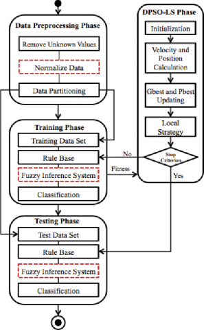
Some of the assumptions in Discrete Particle Swarm Optimization (DPSO) are spawning a swarm, favourable adaptation, and negative adaptation. Simple ideas are executed in a way that resembles natural selection. When a swarm is exposed to a longer time of survival, it has a higher chance of producing progeny. The swarm's life span will be prolonged if it finds a more suitable state, such as favourable adaptation, but it will be reduced if it fails to find a suitable state, such as unfavourable adaptation. The fuzzy c-means clustering algorithm is effective, however the random selection of centre points reduces the iterative process to the local best answer. Researchers have proposed numerous changes to the FCM in order to improve its convergence during the previous few years. The ideas of Fuzzy and DPSO are combined to create Fuzzy DPSO which is illustrated in Figure 1. The algorithm's junction rate is managed using fractional calculus, and swarms of Fuzzy DPSOs compete using Darwin's survival-of-the-fittest principles. By applying these concepts, the particles are spared the challenge of local minima. At the same time, several PSO algorithms are run.
Figure 1. Fuzzy DPSO process
Swarm Intelligence Algorithm
Fuzzy DPSO Procedure
3.3 Dropout CNN classifier
To segment lung images, the Dropout CNN classifier is employed, and the CLAHE algorithm is used to improve the lung picture. The CLAHE approach separates an original image into non-overlapping contextual sub-images, tiles, or blocks. Block Size (BS) and Clip Limit (CL) are the two most important elements of the CLAHE (CL). These two properties are significantly responsible for the improvement in image quality [32]. Increased CL brightens the image and flattens the histogram because the input image has a low intensity. The image is of dynamic range, as well as its contrast, increases as the BS increases [33]. The two parameters determined at the location with the biggest entropy curvature, using the image's entropy, produce subjectively good image quality.
Procedure
The CT image is converted from RGB to YIQ colour space via linear transformation, and subsequently to HSI colour space via nonlinear transformation in this method. In the YIQ and HSI colour spaces, chromatic and brightness information are independent. Second, the brightness information is used to improve contrast while Rayleigh CLAHE is used to keep the chromatic information. Dropout is implemented per layer in a neural network. It works with a variety of layers, including dense fully connected layers, convolutional layers, and recurrent layers like the long short-term memory network layer. Dropout can be utilized on the visible or input layer, as well as any or all of the network's hidden layers. It isn't used on the output layer. The likelihood of the layer's outputs being dropped out or maintained is controlled by a new hyper parameter. We trained dropout neural networks for classification challenges on data sets from a variety of domains. We discovered that using dropout improved generalization p when compared to neural networks that did not use it.
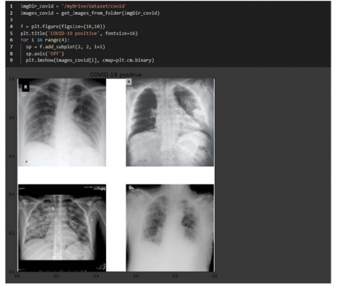
The Kaggle dataset was provided by the Radiological Society of North America (RSNA). Hundreds of pictures of healthy and pneumonia-infected people remain in this collection, which are useful for this study. To generate a training dataset, images from those two datasets must first be downloaded. The desired photographs are then picked and saved to the appropriate location. Images are then loaded and preprocessed before being sent into the training process by being turned into numpy arrays of the necessary size. It is vital to set up the study's working environment before collecting data. Colab (Google Colaboratory) is a free cloud-based Jupyter notebook environment that requires no installation. Colaboratory allows users to build and execute code as well as access advanced computational resources directly from their browser. Colab, in particular, generously provides GPU, which significantly accelerates the computationally intensive training process. For these reasons, Colab has become highly popular among Deep Learning and Data Science enthusiasts who may not have access to a PC with expensive GPUs. To begin, we show that our method works on a COVID-CT-Dataset, which contains COVID-19-positive CT scans as well as ground-truth lesions that a radiology specialist manually recognizes. Figure 2 depicts the sample Covid-19 lung images. Second, we examined newly released CT images from a nearby hospital that contained individuals who had tested positive for the coronavirus. Finally, the COVID-19 lesion and its repercussions over patient's lungs are depicted in three dimensions. The performance metrics compared in Table 1.
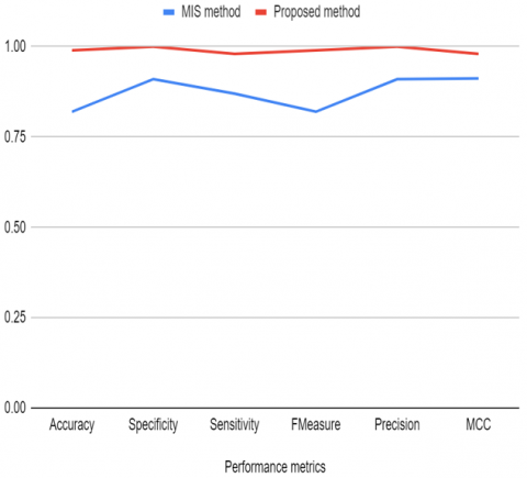
To analyze and find out the result of the recommended segmentation approach, the segmented COVID-19 lesion statistical values are matched to the results of the Medical Image Segmentation (MIS) Methodology as shown in Table 2 and Figure 3. The performance of the provided algorithms was evaluated using generally established assessment scores. One of our work's primary characteristics is the capacity to assess the COVID-19 lesion, show the polluted region, and to trace disease changes in real time. Furthermore, the proposed method may detect aberrant regions despite the low-intensity contrast among healthy tissues and lesions. Even if our suggestions were effective, there are still disadvantages to be aware of. We also want to improve the algorithms so that lesions like ground-glass opacity, crazy paving, and consolidation can be identified. To improve COVID-19 detection, diagnosis, and assessment, we want to merge imaging data with clinical markers and laboratory testing results. COVID-19 continues to spread over the world in an uncontrollable and unpredictable manner.
Table 1. Performance metrics comparison
|
Performance metrics |
MIS method |
Proposed method |
|
Accuracy |
0.843 |
0.99 |
|
Specificity |
0.967 |
1 |
|
Sensitivity |
0.854 |
0.99 |
|
Fmeasure |
0.812 |
0.12 |
|
Precision |
0.914 |
1 |
|
MCC |
0.917 |
0.97 |
|
DICE |
0.923 |
0.981 |
|
JACCARD |
0.891 |
0.960 |
Table 2. Summary of quantitative segmentation statistics
|
Lung (Vx) |
In (cm) |
Lesion |
In(cm) |
Ratio |
|
109530 |
2897 |
75149 |
1988.3 |
68.61 |
|
240827 |
6371.8 |
43752 |
1157.6 |
18.167 |
|
94919 |
2511.3 |
10578 |
279.87 |
11.14 |
|
38726 |
1024.6 |
20734 |
548.58 |
53.54 |
|
37637 |
995.8 |
30477 |
806.3 |
80.97 |
|
231634 |
6128.6 |
42215 |
1116.9 |
18.22 |
|
38372 |
1015.2 |
8920 |
236 |
23.24 |
|
151116 |
3998.2 |
7632 |
201.9 |
5.05 |
|
232166 |
6142.7 |
36725 |
971.6 |
15.81 |
|
44204 |
1169.5 |
7892 |
208.8 |
17.85 |
Figure 2. COVID-19 chest X-ray images
Figure 3. MIS Vs HFDPSO method
Until date, CT-scan imaging has been a widely used, low-cost, comprehensive screening method that efficiently aids in the visualization and rapid assessment of COVID-19 lesion severity. We assessed the efficacy of an automated technique for COVID-19 lung infection segmentation and quantification using chest CT data. The goal of this project is to build and analyze automated COVID-19 Lung Infection segmentation and quantification using Hybrid Swarm Intelligence and Fuzzy DPSO for chest CT images. According to extensive computer simulations, our persistent learning strategy for CT image segmentation using image enhancement is more efficient and adaptive than the Medical Image Segmentation (MIS) method. In future, the enormous dataset can be considered to approve our proposed model on it. Such strategies may be sought for further research to demonstrate their real case implementation.
[1] Barstugan, M., Ozkaya, U., Ozturk, S. (2020). Coronavirus (COVID-19) classification using CT images by machine learning methods. arXiv preprint arXiv:2003.09424.
[2] Mahajan, A., Somaraj, K., Sameer, M. (2021). Adopting artificial intelligence powered convnet to detect epileptic seizures. 2020 IEEE-EMBS Conference on Biomedical Engineering and Sciences (IECBES), Langkawi Island, Malaysia, pp. 427-432. https://doi.org/10.1109/IECBES48179.2021.9398832
[3] Nader, I. W., Zeilinger, E., Jomar, D., Zauchner, C. (2020). Analysing the effect of containment and mitigation measures on COVID-19 infection rates using machine learning on data of 95 countries: An observational study. Available at SSRN 3590467.
[4] Purohit, K., Kesarwani, A., Kisku, D.R., Dalui, M. (2020). COVID-19 detection on chest X-ray and CT scan images using multi-image augmented deep learning model. BioRxiv.
[5] Cifci, M.A. (2020). Deep learning model for diagnosis of corona virus disease from CT images. International Journal of Scientific & Engineering Research, 11(4): 273-278.
[6] Nabavi, S., Ejmalian, A., Moghaddam, M.E., Abin, A.A., Frangi, A.F., Mohammadi, M., Rad, H.S. (2021). Medical imaging and computational image analysis in COVID-19 diagnosis: A review. Computers in Biology and Medicine, 104605. https://doi.org/10.1016/j.compbiomed.2021.104605
[7] Farid, A.A., Selim, G.I., Khater, H.A.A. (2020). A novel approach of CT images feature analysis and prediction to screen for corona virus disease (COVID-19). International Journal of Scientific & Engineering Research, 11(3). http://dx.doi.org/10.14299/ijser.2020.03.02
[8] Zheng, C., Deng, X., Fu, Q., Zhou, Q., Feng, J., Ma, H., ..., Wang, X. (2020). Deep learning-based detection for COVID-19 from chest CT using weak label. MedRxiv. https://doi.org/10.1101/2020.03.12.20027185
[9] Chen, J., Wu, L., Zhang, J., Zhang, L., Gong, D., Zhao, Y., ..., Yu, H. (2020). Deep learning-based model for detecting 2019 novel coronavirus pneumonia on high-resolution computed tomography. Scientific reports, 10(1): 19196. https://doi.org/10.1038/s41598-020-76282-0
[10] Sethi, R., Mehrotra, M., Sethi, D. (2020). Deep learning based diagnosis recommendation for COVID-19 using chest x-rays images. 2020 Second International Conference on Inventive Research in Computing Applications (ICIRCA), Coimbatore, India, pp. 1-4. https://doi.org/10.1109/ICIRCA48905.2020.9183278
[11] Song, Y., Zheng, S., Li, L., Zhang, X., Zhang, X., Huang, Z., ..., Yang, Y. (2021). Deep learning enables accurate diagnosis of novel coronavirus (COVID-19) with CT images. IEEE/ACM Transactions on Computational Biology and Bioinformatics. https://doi.org/10.1109/TCBB.2021.3065361
[12] Shah, F.M., Joy, S.K.S., Ahmed, F., Hossain, T., Humaira, M., Ami, A.S., Paul, S., Jim, M.A.R.K., Ahmed, S. (2021). A comprehensive survey of COVID-19 detection using medical images. SN Computer Science, 2(6): 434. https://doi.org/10.1007/s42979-021-00823-1
[13] Sameer, M., Gupta, A.K., Chakraborty, C., Gupta, B. (2020). ROC analysis for detection of epileptical seizures using haralick features of gamma band. 2020 National Conference on Communications (NCC), pp. 1-5. https://doi.org/10.1109/NCC48643.2020.9056027
[14] Elaziz, M.A., Hosny, K.M., Salah, A., Darwish, M.M., Lu, S., Sahlol, A.T. (2020). New machine learning method for image-based diagnosis of COVID-19. Plos One, 15(6): e0235187. https://doi.org/10.1371/journal.pone.0235187
[15] Moutounet-Cartan, P.G. (2020). Deep convolutional neural networks to diagnose COVID-19 and other pneumonia diseases from posteroanterior chest x-rays. arXiv preprint arXiv:2005.00845.
[16] Satapathy, S.C., Hemanth, D.J., Kadry, S., Manogaran, G., Hannon, N.M., Rajinikanth, V. (2020). Segmentation and evaluation of COVID-19 lesion from CT scan slices-A study with Kapur/Otsu function and Cuckoo Search Algorithm. Research Square. https://doi.org/10.21203/rs.3.rs-40148/v1
[17] Gozes, O., Frid-Adar, M., Greenspan, H., Browning, P.D., Zhang, H., Ji, W., Beinheim, A., Siegel, E. (2020). Rapid ai development cycle for the coronavirus (COVID-19) pandemic: Initial results for automated detection & patient monitoring using deep learning CT image analysis. arXiv preprint arXiv:2003.05037.
[18] Herath, H., Karunasena, G., Ariyathunge, S., Priyankara, H., Madhusanka, B., Nimanthi, U. (2021). Deep Learning approach to recognition of novel COVID-19 using CT scans and digital image processing. Research Square. https://doi.org/10.21203/rs.3.rs-646890/v1
[19] King, B., Barve, S., Ford, A., Jha, R. (2020). Unsupervised clustering of COVID-19 chest X-ray images with a self-organizing feature map. 2020 IEEE 63rd International Midwest Symposium on Circuits and Systems (MWSCAS), Springfield, MA, USA, pp. 395-398. https://doi.org/10.1109/MWSCAS48704.2020.9184493
[20] Hu, S., Gao, Y., Niu, Z., Jiang, Y., Li, L., Xiao, X., Wang, M., Fang, E.F., Menpes-Smith, W., Xia, J., Yang, G. (2020). Weakly supervised deep learning for COVID-19 infection detection and classification from CT images. IEEE Access, 8: 118869-118883. https://doi.org/10.1109/ACCESS.2020.3005510
[21] Sangeetha, S.K.B., Dhaya, R., Shah, D.T., Dharanidharan, R., Reddy, K.P.S. (2021). An empirical analysis of machine learning frameworks for digital pathology in medical science. Journal of Physics: Conference Series, 1767: 012031. https://doi.org/10.1088/1742-6596/1767/1/012031
[22] Tello-Mijares, S., Woo, L. (2021). Computed tomography image processing analysis in COVID-19 patient follow-up assessment. Journal of Healthcare Engineering. https://doi.org/10.1155/2021/8869372
[23] El Asnaoui, K., Chawki, Y., Idri, A. (2021). Automated methods for detection and classification pneumonia based on x-ray images using deep learning. In Artificial Intelligence and Blockchain for Future Cybersecurity Applications, pp. 257-284. https://doi.org/10.1007/978-3-030-74575-2_14
[24] Oh, Y., Park, S., Ye, J.C. (2020). Deep learning COVID-19 features on CXR using limited training data sets. IEEE Transactions on Medical Imaging, 39(8): 2688-2700. https://doi.org/10.1109/TMI.2020.2993291
[25] Sameer, M., Gupta, B. (2021). Time–frequency statistical features of delta band for detection of epileptic seizures. Wireless Personal Communications. https://doi.org/10.1007/s11277-021-08909-y
[26] Gupta, S., Sameer, M., Mohan, N. (2021). Detection of epileptic seizures using convolutional neural network. 2021 International Conference on Emerging Smart Computing and Informatics (ESCI), Pune, India, pp. 786-790. https://doi.org/10.1109/ESCI50559.2021.9396983
[27] Beeraka, S.M., Kumar, A., Sameer, M., Ghosh, S., Gupta, B. (2021). Accuracy enhancement of epileptic seizure detection: A deep learning approach with hardware realization of STFT. Circuits, Systems, and Signal Processing. https://doi.org/10.1007/s00034-021-01789-4
[28] Sameer, M., Gupta, B. (2021). ROC analysis of EEG subbands for epileptic seizure detection using naïve bayes classifier. Journal of Mobile Multimedia, 17(1-3): 299-310. https://doi.org/10.13052/jmm1550-4646.171315
[29] Ghoshal, B., Tucker, A. (2020). Estimating uncertainty and interpretability in deep learning for coronavirus (COVID-19) detection. arXiv preprint arXiv:2003.10769.
[30] Apostolopoulos, I.D., Mpesiana, T.A. (2020). COVID-19: Automatic detection from x-ray images utilizing transfer learning with convolutional neural networks. Physical and Engineering Sciences in Medicine, 43(2): 635-640. https://doi.org/10.1007/s13246-020-00865-4
[31] Al-Timemy, A.H., Khushaba, R.N., Mosa, Z.M., Escudero, J. (2021). An efficient mixture of deep and machine learning models for COVID-19 and tuberculosis detection using X-ray images in resource limited settings. In Artificial Intelligence for COVID-19, pp. 77-100. https://doi.org/10.1007/978-3-030-69744-0_6
[32] Abdullah Farid, A., Khater, H., Selim, G. (2020). A CNN classification model for diagnosis COVID-19. Preprints. https://doi.org/10.20944/preprints202007.0591.v1.
[33] Amo-Boateng, M. (2020). Tracking and classifying global COVID-19 cases by using 1d deep convolution neural network. medRxiv. https://doi.org/10.1101/2020.06.09.20126565