Lihui Zheng* | Yiming Sun | Guolin Jing | Tianyi Wang
© 2021 IIETA. This article is published by IIETA and is licensed under the CC BY 4.0 license (http://creativecommons.org/licenses/by/4.0/).
OPEN ACCESS
Heptamethine cyanine dyes (HCDs) feature a large molar extinction coefficient, a high fluorescence quantum yield, and an adjustable structure, with maximum absorption and emission wavelengths located in the near-infrared region. Thanks to these excellent features, the HCDs have been widely used in biological imaging, ion detection, new energy, and many other fields. However, the unique structure of the HCDs brings some defects, such as poor hydrophilicity and low photostability. Many scholars have been striving to expand the application of the HCDs in biological analysis by improving properties like spectral properties, hydrophilicity, photostability, and fluorescence quantum yield. This paper thoroughly reviews the advances of structural modification in the sites of terminal heterocycles (indole, benzothiazole, benzoxazole and benzoselenide) and polymethine chains (straight chain or rigid bridged ring), and introduces the influences of functional materials over the features of the HCDs. Finally, the authors predicted the development trend of the HCDs.
Heptamethine cyanine dyes (HCDs), structure modification, spectral properties, functional materials
Fluorescence detection, being a technology with contamination-free, fast responding, highly sensitive, and easily operable technology, has been widely applied in ion detection [1-3], molecular recognition [4-9], cell labeling [10-12], deoxyribonucleic acid (DNA) sequencing [13], and photodynamic therapy [14-16]. Accompanying with the development of life sciences, the fluorescence technology demonstrates incomparable advantages in the analysis and detection of ultra-trace substances in the life process.
Many fluorescent probes have been developed, including porphyrins [17, 18], coumarins [19-21], rhodamines [22, 23], boron dipyrrole methylene (BODIPY) [24-28], and cyanine dyes [29-31]. Among them, cyanine dyes connect the electron donor section (D) and electron acceptor section (A) through a polymethine chain. The absorption and emission spectra, which are affected by the length of the methine chain and the substituents, fully cover the visible light region and the near-infrared region. As a result, cyanine dyes have been widely adopted for biological analysis [32]. In particular, heptamethine cyanine dyes (HCDs), with seven carbon atoms in the polymethine chain, have a maximum emission wavelength in the biological blank window region around 800nm. In biological analysis, the HCDs can effectively avoid the interference of biological background fluorescence, and improve the detection accuracy.
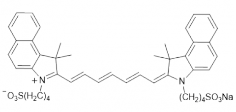
In 2000, the United States Food and Drug Administration (FDA) approved an HCD named indocyanine green (ICG) (Figure 1) for clinical use, kicking off further research on theHCDs. Nevertheless, the ICG and other HCDs face a poor photostability, because the electron donor section (D) and electron acceptor section (A) are connected via a linear methine chain. However, there are many other problems with the HCDs, including small Stokes shift, low fluorescence quantum yield, and propensity to fluorescence quenching. These problems must be solved urgently for the further application of the HCDs.
Figure 1. Structure of the ICG
To make up for the defects of the HCDs in actual applications, this paper probes into the molecular structure of the HCDs, and optimizes molecular properties through structural modification. The research results would provide an important guide for developing cyanine dyes with diverse functions.Structure of the HCDs.
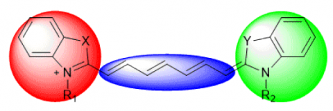
The HCDs are prepared through the condensation reaction of basic nitrogen heterocycles (indole, thiazole, oxazole, etc.), which contain active methylene groups, with dialdehydes compounds. Two basic nitrogen heterocycles are connected by a polymethine chain containing seven carbon atoms, forming a conjugated system with a certain electron push-pull effect (Figure 2). The whole molecule is a larger conjugated system with good coplanarity. That is the reason that the HCDs boast excellent fluorescence properties.
Figure 2. Structure of the HCDs
When the basic nitrogen heterocycles at the end of the molecule have the same structure (X=Y, and R1=R2), the molecule is highly symmetric, and the HCDs are symmetric. When the basic nitrogen heterocycles do not have the same structure (X≠Y, or R1≠R2), the molecule is no longer symmetric, and the HCDs are asymmetric. The electron push-pull effect and electron cloud density of the polymethine chain are greatly affected by the type of nitrogen heterocycles in the molecule, substituents on heterocyclic nitrogen, substituents on benzene rings, types of conjugated chains, and substituents on conjugated chains. They all play important roles in the photostability and spectral properties of the dyes.
2.1 Effects of the heterocycle type to the characteristics of HCDs
With the increasing basicity of the terminal nitrogen heterocycle, the absorption spectra of the HCDs will be red-shifted. For instance, Yao et al. [33] developed the HCDs whose terminal heterocycles are oxazole, indole, thiazole, and selenazole, respectively (Figure 3). In ethanol solution, the maximum absorption wavelengths (λab) of the four types of HCDs are 734.4 nm, 789.8 nm, 803.6 nm and 809.4 nm (R=H), respectively. In addition, the introduction of a heteroatom at the X position causes the two methyl groups on the original carbon atoms to be replaced, thereby reducing intermolecular hydrogen bonds. Then, the aggregation of dye molecules in protic solvents is weakened, which reduces the fluorescence quenching caused by molecular aggregation, and increases the fluorescence quantum yield of the molecule.
Nevertheless, the introduction of the heteroatom X in the molecular terminal heterocycle dampens the photostability of the dye. The greater the radius of the substituted heteroatom X, the worse the photostability of the dye. Through comparison, Yao et al. [33], Liu and Bao [34] agreed that, the best photostability is achieved with indole as the terminal nitrogen heterocycle (X = C) when the HCDs have the same polymethine chain structure. After the introduction of the substituted heteroatom into the heterocycle, the dyes can be ranked in descending order of photostability as oxazocyanines (X = O) > thiazocyanines (X = S) > selenazocyanines (X = Se). As a result, heptamethine indocyanine dyes are frequently studied in the follow-up research. There are occasional reports on heptamethine oxazocyanines and heptamethine thiazocyanines. For example, Vus et al. [35] and Zhytniakivska et al. [36] applied heptamethine thiazocyanines for the diagnosis of Alzheimer's disease and other illnesses. However, selenazole HCDs are rarely reported.
Figure 3. HCDs with different terminal heterocycles
2.2 Effects of the substituent on the N of heterocycle to the characteristics of HCDs
The N of the heterocyclic ring is generally connected to the corresponding substituent via a methylene group. Since the substituent is not directly connected to the heptamethine conjugated chain of the molecule, the substituent at this position does not significantly affect the spectral properties of the molecule. The limited effect is primarily the impacts on molecular stability and aggregation, which arise from electronic effect and spatial effect.
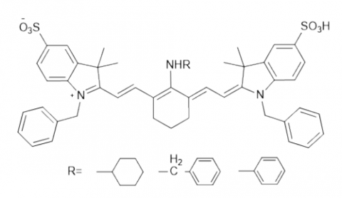
The introduction of substituents at this position can expand the application field of the molecule. For example, the introduction of hydrophilic substituents like -COOH [37], and -SO3H [38, 39] on the N of heterocycle (Figure 4) can greatly enhance the water solubility and fluorescence quantum yield of the dye [40], facilitating the application of the HCDs in biological analysis.
Ryu's research team [41] introduced a pyridyl group on the N of heterocycle (Figure 4), which enhances the water solubility of the molecule, improves the generation efficiency of singlet oxygen O·, and elevates the efficiency of dyes in photodynamic therapy. Chen et al. [42] found that the dye has a good photostability, when the substituent on N of heterocycle is a sterically hindered benzyl group. Chen et al. [43] introduced six substituents (carboxypentyl, benzyl, 4-methylbenzyl, 4-fluorobenzyl, 4-carboxybenzyl, and 4-nitrobenzyl) with different electron donating abilities to the N of heterocycle of heptamethine indocyanine, and discovered that: their λab values were all around 770 nm, and the photostability of the dye increased with the electron donating ability of the N substituent on the indole ring.
Figure 4. Substituents on the N of terminal heterocycle
Liu et al. [44] and Shi et al. [45] introduced glycine or carboxyl group on the N of heterocycle of heptamethine indocyanine, respectively, and managed to boost the cell permeability, strengthen the binding ability to cancer cells, and lower the biological toxicity of the dye. Khairutdinov and Serpone [46] demonstrated that, with the growing length of the substituted alkyl chain on the N of heterocycle of the HCDs, the molecular aggregation weakened, and the fluorescence quantum yield gradually increased. Yadav et al. [47] also found that, the molecule had the best cancer cell penetration ability, when there were six carbon atoms in the alkyl chain of the substituent on the N of heterocycle of heptamethine indocyanine.
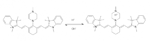
Achilefu's research team [48] developed an unsubstituted pH probe at the N of the indole ring, drawing on the effect of the protonation of the N atom of the indole ring on the push-pull of the polymethine chain (Figure 5a). The probe has no fluorescence in neutral and alkaline environments, but emits a strong fluorescence after contacting H+ in the weakly acidic environment of pH 4.00-6.00. After binding to cRGD, the probe can be used for early diagnosis of various cancers (e.g., breast cancer). Following this principle, Hou et al. [49] designed a water-soluble fluorescence-enhanced probe containing quaternary ammonium salts (Figure 5b) for pH detection in a weakly acidic environment. To generate molecular fluorescence, the probe uses H+ in the environment to positively charge the N of heterocycle of the dye, and enhance the push-pull of the polymethine chain. This generation method has been widely adopted to develop weakly acidic fluorescent probes.
Figure 5. The HCDs with different substituents on the N of heterocycle and their applications as pH probes
2.3 Effects of the substituents on the terminal benzene to the characteristics of HCDs
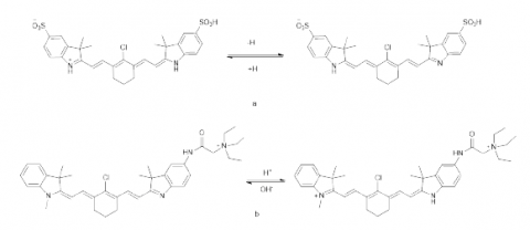
The substituents on the benzene ring, which are not directly connected to the polymethine chain of the molecule, do not significantly affect the spectral properties of the molecule, and are mostly adopted to expand the application field of cyanine dyes. For instance, Hubner et al. [50] introduced a carboxyl group (-COOH) at the 5 and 5' positions of the heptamethine cyanine indole ring, respectively, and realized the quantitative fluorescent labeling of dye molecules in the field of biological analysis through the amide condensation of the carboxyl group and the amine group. Zheng et al. research team [51] increased the water solubility of the dye by introducing sulfonic acid groups (-SO3H) at the 5 and 5' positions of heptamethineindocyanine, which greatly simplified the operation of the HCDs in biological analysis.
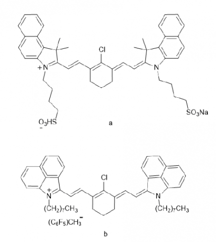
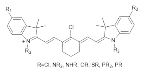
The substituents on the benzene ring do not directly participate in the conjugation of the molecule. But they can cause the red shift of the absorption spectrum of the dye, by regulating the degree of conjugation of the polymethine chain through lone pair electrons or σ electrons. The electron withdrawing group can decrease the electron cloud density of the conjugated system, thus enhancing the photostability of the dye. By contrast, the electron donating group can increase electron cloud density of the conjugated system, thus reducing the photostability of the dye. For example, after -Cl and -OCH3 were introduced on the terminal benzene ring of the HCDs (Figure 3), the λab values in ethanol solvent was red-shifted from 803.6 nm to 810 nm or 823 nm, respectively. The introduction of -Cl enhanced the photostability of the dye, while the introduction of -OCH3 reduced the photostability of the dye [33]. Wang et al. [40] further investigated the effect of substituents on the benzene ring on the fluorescence properties of the dye, and discovered that the introduction of -Cl on the benzene ring could reduce the fluorescence quantum yield of the dye from 6.5 to 1.2, owing to the heavy atom effect. Since the electron-withdrawing effect of the benzene ring can effectively improve the photostability of the dye, the HCDs (Figure 6) with a dibenzindole structure at the terminal position have also been well developed [52-54].
Figure 6. Structure of the HCDs with a dibenzindole structure at the terminal positions
The poor photostability of the HCDs is attributable to the fact that the long polymethine chains loss much energy in chemical bond rotation, and are easily attacked by oxygen or reactive radicals. The photostability is mostly improved through the direct modification of the polymethine chains of HCDs. The specific measures are changing the electron cloud density, increasing molecular rigidity, and adjusting steric hindrance. These structural modifications have a great impact on Stokes shift, fluorescence quantum yield, and application fields.
3.1 Effects of rigid bridge ring to the characteristics of HCDs
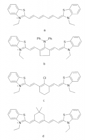
The introduction of a rigid bridged ring at the mid- position of the HCD molecule can enhance the coplanarity of the molecule, reduce the energy loss due to chemical bond rotation, lower the energy required for electronic transitions, and red-shift the molecular absorption spectrum. In addition, the molecular photostability can be improved at the expense of the fluorescence quantum yield of the dye molecule. When the mid-bridged ring is a five-membered ring, the red-shift of the absorption spectrum and the photostability of the molecule are better than those of the six-membered ring, thanks to the limited loss of energy. Yao et al. [55] prepared the HCDs with different polymethine chains (Figure 7), and measured the λab values of A, B, C, and D in ethanol as 760 nm, 803.6 nm, 799.8 nm (methanol) and 763.4 nm (methanol), respectively. Further investigation shows that C is more stable than A, while D is slightly less stable than A. Li et al. [56] also found through experiments that the photostability of the dye was greatly improved after the introduction of a chlorine-containing rigid bridged ring on the polymethine chain of the heptamethine indocyanine molecule.
Figure 7. The HCDs with different polymethine chains
Figure 8. The HCDs with different number of carbon atoms on the rigid bridged ring
Figure 9. The HCDs with substituents on the para-methylene group
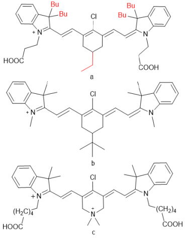
Henary's research team [57] synthesized rigid bridged cyclic cyanine dyes with different number of carbon atoms in the polymethine chain (Figure 8). It is also found that, when the rigid bridged ring is a five-membered ring, the dye absorption wavelength is the largest (803 nm), the molar absorptivity is the greatest, and the photostability is the best; when the rigid bridged ring is a six-membered ring or a seven-membered ring, the maximum absorption wavelengths of the dye are basically the same (λab= 780 nm or 781 nm). Moreover, when the bridged ring is a seven-membered ring, the dye has no fluorescence, the molar absorption coefficient is the lowest, and the photostability is the worst.
Apart from the rigid bridged ring, the introduction of substituents on the para-methylene group of the bridged ring will also affect the properties of the dye. If an alkyl chain is introduced at this position, the aggregation degree of the dye in solution will be reduced due to the large steric hinderance, yet enhanced in aqueous solution under the action of hydrogen bonding. Otsuka et al. [58] discovered that the aggregation degree of dye molecules can be effectively suppressed by introducing alkyl chains on the heterocyclic and bridged rings of the HCDs (Figure 9a). Henary's research team [57] noticed that the introduction of a tert-butyl group on the para-methylene group of the six-membered ring would increase the dye aggregation degree in the phosphate-buffered saline (PBS) buffer solution, and lower the fluorescence quantum yield. Thavornpradit [59] used dimethylamine to replace the para-methylene group on the six-membered ring (Figure 9c), which increases the molar absorption and fluorescence quantum yield of the dye by 1.7 and 3 times, respectively, compared with ICG. The replacement also enhances the photostability of dye molecules and their solubility in aqueous solutions, reducing their biological toxicity.
3.2 Effects of meso-substituents to the characteristics of HCDs
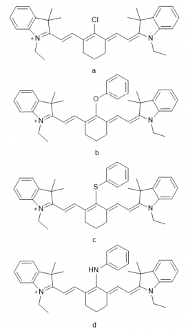
After the rigid bridged ring was introduced into the HCD molecule, a new functional group was added to allow further structural modifications. For example, chlorine or bromine can be incorporated to the middle position of the molecule, while the six-membered rigid bridged ring is introduced. The new element is easily substituted by nucleophilic reagents like amine, thiol, phenol, phosphide, and carbonyl, to form corresponding derivatives [60, 61] (Figure 10). The C-C bonds (hydrocarbyl or phenyl) can also be introduced into the middle position of polymethine chains by palladium-catalyzed Suzuki coupling or cross-coupling reaction [62-66]. The introduction of new functional groups on the substituents, coupled with the further derivatization of functional groups, greatly expands the application field of the dye.
Figure 10. The HCDs with different substituents at the middle position
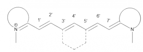
The median substituent of the polymethine chain, which is directly connected with the conjugated system of the dye, directly affects the spectral properties, photostability, and fluorescence quantum yield of the dye. Stackova et al. [67] examined the influence of substituents on the methine chain over dye properties. When the substituents are at the middle position C4’ (Figure 11), the electron-withdrawing and electron-donating groups cause the absorption spectrum of the dye molecule to redshift and blueshift, respectively. As the electron-withdrawing ability of the substituents weakens, the red-shift of the dye absorption spectrum becomes larger. As the electron-donating ability of the substituents enhances, the blue-shift of the dye absorption spectrum becomes larger. Lei et al. [68] also believed that the 1’-position substitution with either electron drawing group or electron donating group can cause the red shift of the absorption peak of dye molecules, without exerting much effect on the spectrum.
Figure 11. Number of carbon atoms in the polymethine chain of the HCDs
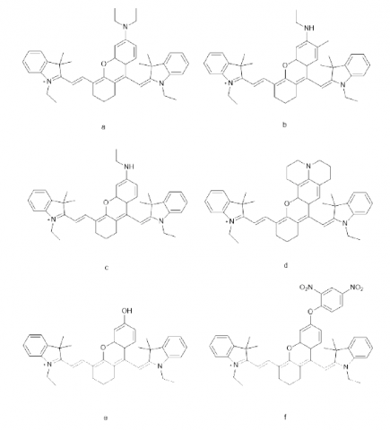
König and Krämer [65] introduced a C-C bond to the heptamethine indocyanine molecule through a coupling reaction, and obtained multiple fluorescent probes for Hela cell labeling. The test results show that, compared with the case of no substituent at the middle position (R=H), the absorption spectrum of the molecule is red-shifted when the middle position substituent is an electron withdrawing group like alkynyl, chlorine, and phenyl; that spectrum is blue-shifted when the middle position substituent is an anilino group. Among all the middle position substitution derivatives, the molecule whose middle position is substituted with phenyl boasts the highest fluorescence quantum yield and the best photostability. But the values are lower than those of the molecule with no substituent at the middle position (R=H) (Figure 12). Su et al. [69] compared the effects of middle position phenoxy (Figure 13a) and phenyl (Figure 13b) substitutions on molecular properties, revealing that molecule 13b has better photostability and fluorescence quantum yield than molecule 13a, due to its relatively good coplanarity. The fluorescence quantum yield of molecule 13b is about 3.4 times higher than that of ICG.
Figure 12. The HCDs with different substituents at the middle position
Figure 13. The HCDs with phenyl and phenoxy substituents at the middle position
Peng et al. [70] and Song et al. [71] research team prepared heptamethine indocyanine derivatives substituted with sulfur and secondary amine at the middle position (Figure 14), and made the following conclusions: The sulfur substituent at the middle position causes photo-induced electron transfer (PET) of heptamethine indocyanine. It not only red-shifts the λab and λem of the dye, but also greatly reduces the fluorescence quantum yield of the dye. The middle position substitution of secondary amine causes intramolecular electron transfer (ICT) of heptamethine indocyanine. It not only largely blue-shifts the λab and λem of the dye, but also boosts the Stokes shift of the molecule (greater than 140 nm), compared with the parent molecule. These findings lay the basis for developing bioluminescence probes with low self-absorption and high sensitivity.
Figure 14. The HCDs with sulfur or secondary amine at the middle position
Further research found that the conjugation effect between the middle position substituent and the molecular polymethine chain also affects the molecular properties. The stronger the conjugation effect, the greater the red-shift of the absorption spectrum. For example, Song et al. [71] discovered that, when the middle position substituents of the heptamethine indocyanine molecule were cyclohexylamino, benzylamino and aniline (Figure 15), the λab values in aqueous solution were 602 nm, 617 nm, and 713nm, respectively.
Figure 15. The HCDs with different conjugating substituents at the middle position
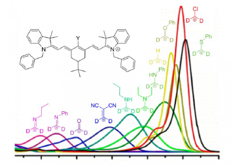
Nano's research team developed a series of fluorescent probes for middle position-substituted heptamethine indocyanines, and analyzed their properties. The experimental results show that, when the middle position substituent is phenoxy (Figure 16b), the photostability and fluorescence quantum yield of the heptamethine indocyanine molecule are greater than those of the parent molecule; when the middle position substituent is thiophenyl (Figure 16c) or anilino (Figure 16d), the photostability and fluorescence quantum yield of the heptamethine indocyanine molecule are smaller than those of the parent molecule. Drawing on the influence of the middle position substituents, the research team developed the fluorescence probes for detecting H+ in the environment (Figure 17).
Levitz et al. [57] synthesized heptamethine indocyanines with bromine, chlorine, methyl, and phenyl as the middle position substituents, and measured the maximum absorption wavelengths λab: 781nm, 780nm, 768nm, and 759nm. Pascal et al. [72] combined the time-dependent density functional theory (TD-DFT) with experiments to analyze the influence of the substituents on the six-membered bridged ring on the absorption spectra of dyes, and made the following discovery: With the growing electron-donating ability of the substituents, the charge distribution on the polymethine chain gradually moves from uniform state to centralized state. By the λab value, the HCDs with different middle position substituents can be ranked as C-S>C-Cl>C-OR>C-H>C-NHPh>C-NR2>C-NHR>C=CR2>C=O>C=NPh>C=NR (Figure 18).
Figure 16. The HCDs with different substituents at the middle position
Figure 17. The HCDs as pH sensor
Figure 18. Relationship of middle position substituents with absorption spectra of the HCDs [72]
The middle position substituents not only directly affect the properties of HCD molecules, but also regulate their photostability and fluorescence properties by combining with the conjugated chains. Chen et al. [73] used phenolic hydroxyl to replace the middle position chlorine of heptamethine indocyanine, and obtained several fluorescent probes of the structure "xanthene-cyanine" (Figure 19a-e). Compared with that of the parent molecule, the absorption spectrum of this molecule has a blue shift of about 60-80 nm, with a small shoulder peak at 500-600 nm, and a significant improvement of photostability and fluorescence quantum yield. In addition, Chen et al. [73] derived a H2S fluorescence-enhanced fluorescent probe from 19e (19f).
Figure 19. The HCDs with the structure "xanthene-cyanine"
Since the middle position substitution has a major impact on HCD molecules, and plays an important role in expanding molecular applications, the derivatization of dye molecules by middle position substitution can realize the fluorescence detection for small molecules and ions like hypochlorous acid, palladium, and promote the application of the HCDs in biological analysis. Liu et al. research team [77] introduced an acyl group to the middle carbonyl of the polymethine chain, and managed to determine the mitophagy in biological cells. Zhao et al. [78] introduced tetraphenylene at the middle position of heptamethine indocyanine, which successfully transforms heptamethanine molecules as photothermal conversion materials in cancer treatment, for the introduction effectively avoids the aggregated fluorescence quenching of dye molecules, and improves the photothermal conversion efficiency.
The application of the HCDs is limited by their water solubility, photostability, and fluorescence quantum yield. To expand the application scope, it is necessary to improve the aggregation degree, fluorescence properties and photostability by combining the HCDs with functional materials (such as metal oxide sols, polymers, surfactants, etc.).
Zheng et al. research team [79] combined cyanine dyes with metal oxides (TiO2, and SiO2) or cetrimonium bromide (CTAB), and observed a large increase in the photostability of the dyes, as well as an increment of the pH response range, compared with the pure dye solutions. Their research sheds new light on how to expand the use scope and application of the dyes.
The HCDs are also complexed with polyethylene glycol (PEG) to increase their water solubility, and then applied to the photodynamic therapy of cancer cells [80, 81]. Pais-Silva et al. [82] loaded IR780 into PEG-succinate vitamin E micelles, which increases its solubility in water by nearly 10 times, reduces its aggregation in aqueous solution, and greatly lowers the clinical dose for caner cell removal. Zhang et al. [83] and St-Lorenz et al. [84] loaded the HCDs onto nanoparticles like SiO2, and PEG-polycaprolactone (PCL), and observed marked improvements to dispersibility, photostability and photothermal conversion efficiency. The combined materials can be used clinically as photothermal conversion materials in cancer therapy.
Since the approval of the ICG by the FDA, more and more researchers turned their attention to the application of the HCDs in biological analysis. There is ample room to improve the HCD performance, owing to their defects in water solubility, photostability, and fluorescence quantum yield, as well as the many modifiable sites. In the field of biological analysis, several HCDs with excellent performance have been developed for biological imaging, photothermal conversion, and photoacoustic conversion, thanks to the HCDs’ low biological toxicity, lack of obvious side effects, and good targeting and clearing ability of cancer cells. In addition, the HCDs have been applied to photoelectric conversion tasks (e.g., Dye-Sensitized Solar Cells), because their wavelength is adjustable.
The further research on the structural modification of the HCDs will surely make up for their structural defects, and the modified HCDs will achieve better application results in biological analysis, photoelectric conversion, and other fields.
This work is funded by the Youth Science Fund, Northeast Petroleum University, Fundamental Research Funds for Undergraduate Universities in Heilongjiang Province, under the project “Fluorescence probes based on heptamethine indocyanine and their application in metal ion detection” (Grant No.: 2018QNQ-03).
[1] Zhong, S.L., Zhang L.L., Liu J., Liu, X.J., Chang T.J., Shangguan, L.H. (2022). Carbon quantum dots-based mesoporous nanomaterials for detection of copper ion. Chinese Journal of Analytical Chemistry, 50(1): 47-53. https://doi.org/10.19756/j.issn.0253-3820.211101
[2] Chailek, N., Kaewnok, N., Petdum, A., Sirirak, J., Chaneam, S., Kamkaew, A., Girdthep, S., Wanichacheva, N. (2021). Near infrared and colorimetric fluorescence sensor for ultra-selective detection of Cu2+ level with applications in diverse water samples, brain tumor cell and flow injection analysis. Journal of Photochemistry and Photobiology A-Chemistry, 421:113533. https://doi.org/10.1016/j.jphotochem.2021.113533
[3] Zhao, X.J., Chen, Y., Niu, G.Y., Gu, D.N., Wang, J.N., Cao, Y.M., Yin, Y.M., Li, X.G., Ding, D., Xi, R.M., Meng, M. (2019). Photostable pH-sensitive near- infrared aggregation-induced emission luminogen for long-term mitochondrial tracking. Acs Applied Materials and Interfaces, 11(14): 13134-13139. https://doi.org/10.1021/acsami.9b02228
[4] Fei, X.N., Zheng, Y.J., Gu, Y.C., Li, G.Y., Zhao, H.B., Zhang, B.L. (2021). Fluorescence imaging and chiral specific biological recognition. Spectroscopy and Spectral Analysis, 41(12): 3802-3807. https://doi.org/10.3964/j.issn.1000-0593(2021)12- 3802-06
[5] Huang, Z.K., Liu, C., Fu, Q.Q., Li, J., Zou, J.M., Xie S.T., Qiu, L.P. (2018). Aptamer-based fluorescence probe for bioanalysis and bioimaging. Chinese Journal of Applied Chemistry, 35(1): 28-39. https://doi.org/10.11944/j.issn.1000- 0518.2018.01.170363
[6] Maiya, B.G. (2000). Molecular recognition. Journal of Porphyrins and Phthalocyanines, 4(4): 393-397. https://doi.org/10.1002/(SICI)1099- 1409(200006/07)4:4<393::AID-JPP227>3.0.CO;2-B
[7] Cao, Z.Q., Liu, X.Y., Qu, D.H. (2017). The application of fluorescence molecular probe in saccharin sodium determination. Imaging Science and Photochemistry, 35(3): 316-321. https://doi.org/10.7517/j.issn.1674- 0475.2017.03.316
[8] Chen, X., Wang, F., Hyun, J.Y., Wei, T., Qiang, J., Ren, X., Shin, I., Yoon, J. (2016). Recent progress in the development of fluorescent, luminescent and colorimetric probes for detection of reactive oxygen and nitrogen species. Chemical Society Reviews, 45(10): 2976-3016. https://doi.org/10.1039/C6CS00192K
[9] Ge, Y.R., Yang, J., Li, Y.Y., Xu, Y.G. (2020). Advances of near-infrared fluorescent probes for detection of Alzheimer’s disease. Journal of China Pharmaceutical University, 51(2): 138-151.https://doi.org/10.11665/j.issn.1000-5048.20200203
[10] Li, L.P., Ren, X.F., Bai, P.R., Liu, Y., Xu, W.Y., Xie,J., Zhang, R.P. (2021). Near-infrared emission carbon dots for bio-imaging applications. New Carbon Materials, 36(3): 632-638. https://doi.org/10.1016/S1872- 5805(21)60041-0
[11] Wang, Y., Liao, X.Y., Sun, J.G., Yi, B., Luo, S.L., Liu, T., Tan, X., Liu, D.Q., Chen, Z.L., Wang, X., Shi, C.M. (2018). Characterization of HIF-1 alpha/glycolysis hyperactive cell population via small-molecule-based imaging of mitochondrial transporter activity. Advanced Science, 5(3): 1700392. https://doi.org/10.1002/advs.201700392
[12] Xu, Y.X., Liu, R.F., Xu, K., Dai, Z.F. (2021). Fluorescent probes for intraoperative navigation. Progress in Chemistry, 33(1): 52-65. https://doi.org/10.7536/PC201014
[13] Garrido-Cardenas, J.A., Garcia-Maroto, F., Alvarez- Bermejo, J.A., Manzano-Agugliaro, F. (2017). DNA Sequencing Sensors: An Overview. Sensors (Basel), 17(3): 588. https://doi.org/10.3390/s17030588
[14] Chen, H.W., Ren, X.Q., Paholak, H. L. J., Burnett, J., Ni, F., Fang, X.L., Sun, D.X. (2015). Facile fabrication of near-infrared-resonant and magnetic resonance imaging capable nanomediators for photothermal therapy. ACS Applied Materials & Interfaces, 7(23): 12814-12823. https://doi.org/10.1021/acsami.5b01991
[15] Yoo, Y., Jo, G., Jung, J.S., Yang, D.H., Hyun, H. (2020). Multivalent sorbitol probes for near infrared photothermal cancer therapy. Particle & Particle Systems Characterization, 37(2): 1900490. https://doi.org/10.1002/ppsc.201900490
[16] Shi, C.H., Wu, J.B., Pan, D.F. (2016). Review on near- infrared heptamethine cyanine dyes as theranostic agents for tumor imaging, targeting, and photodynamic therapy. Journal of Biomedical Optics, 21(5): 50901. https://doi.org/10.1117/1.JBO.21.5.050901
[17] Parsa, Z., Tahay, P., Safari, N. (2020). Co-sensitization of porphyrin and metal-free dye for panchromatic dye- sensitized solar cells. Journal of the Iranian Chemical Society, 17(2): 453-459. https://doi.org/10.1007/s13738-019-01782-4
[18] Gu, C.Z., Meng, S.X., Feng, Y.Q. (2015). Progress of porphyrin sensitizers for dye-sensitized solar cells. Chinese Journal of Organic Chemistry, 35(6): 1229- 1237. https://doi.org/10.6023/ cjoc201412018
[19] Dandriyal, J., Singla, R., Kumar, M., Jaitak, V. (2016). Recent developments of C-4 substituted coumarin derivatives as anticancer agents. European Journal of Medicinal Chemistry, 119: 141-168. https://doi.org/10.1016/j.ejmech.2016.03.087
[20] Erande, Y., Kothavale, S., Sreenath, M.C., Chitrambalam, S., Joe, I.H., Sekar, N. (2018). Triphenylamine derived 3-acetyl and 3-benzothiazolyl bis and tris coumarins: synthesis, photophysical and DFT assisted hyperpolarizability study. Journal of Electronic Materials, 47(2): 1431-1446. https://doi.org/10.1007/s11664-017-5925-7
[21] Keri, R.S., Sasidhar, B.S., Nagaraja, B.M., Santos, M.A. (2015). Recent progress in the drug development of coumarin derivatives as potent antituberculosis agents. European Journal of Medicinal Chemistry, 100: 257-269. https://doi.org/10.1016/j.ejmech.2015.06.017
[22] Alamry, K.A., Georgiev, N.I., El-Daly, S.A., Taib, L.A., Bojinov, V.B. (2015). A ratiometric rhodamine– naphthalimide pH selective probe built on the basis of a PAMAM light-harvesting architecture. Journal of Luminescence, 158: 50-59. https://doi.org/10.1016/j.jlumin.2014.09.014
[23] Yu, H.B., Li, G.L., Zhang, B., Zhang, X.F., Xiao, Y., Wang, J.Q., Song, Y.T. (2016). A neutral pH probe of rhodamine derivatives inspired by effect of hydrogen bond on pKa and its organelle-targetable fluorescent imaging. Dyes and Pigments, 133: 93-99. https://doi.org/10.1016/j.dyepig.2016.05.028
[24] Xia, S., Fang, M.X., Wang, J.B., Bi, J.H., Mazi, W., Zhang, Y.B., Luck, R.L., Liu, H.Y. (2019). Near- infrared fluorescent probes with BODIPY donors and rhodamine and merocyanine acceptors for ratiometric determination of lysosomal pH variance. Sensors and Actuators B-Chemical, 294: 1-13. https://doi.org/10.1016/j.snb.2019.05.005
[25] Zuo, J.H., Pan, H.F., Zhang, Y.J., Chen, Y.F., Wang, H.C., Ren, X.K., Chen, Z.J. (2020). Near-infrared fluorescent amphiphilic aza-BODIPY dye: Synthesis, solvatochromic properties, and selective detection of Cu2+. Dyes and Pigments, 183: 108714. https://doi.org/10.1016/j.dyepig.2020.108714
[26] Liu, W.W., Yu, H.K., Hu, R.R., Xu, T., Lun, Y.P., Gan, J.L., Xu, S.H., Yang, Z.M., Tang, B.Z. (2020). Microlasers from AIE-active BODIPY derivative. Small, 16(8): 1907074. https://doi.org/10.1002/smll.201907074
[27] Li, Z.S., Li, L.J., Sun, T.T., Liu, L.M., Xie, Z.G. (2016). Benzimidazole-BODIPY as optical and fluorometric pH sensor. Dyes and Pigments, 128: 165-169. https://doi.org/10.1016/j.dyepig.2016.01.029
[28] Singh, P. K., Majumdar, P., Singh, S.P. (2021). Advances in BODIPY photocleavable protecting groups. Coordination Chemistry Reviews, 449: 214193. https://doi.org/10.1016/j.ccr.2021.214193
[29] Sun, W., Guo, S.G., Hu, C., Fan, J.L., Peng, X.J. (2016). Recent development of chemosensors based on cyanine platforms. Chemical Reviews, 116(14): 7768-7817. https://doi.org/10.1021/acs.chemrev.6b00001
[30] Gopika, G.S., Prasad, P.M.H., Lekshmi, A.G., Lekshmypriya, S., Sreesaila, S., Arunima, C., Kumar, M.S., Anil, A., Sreekumar, A., Pillai, Z.S. (2021). Chemistry of cyanine dyes-A review. Materials Today: Proceedings, 46: 3102-3108. https://doi.org/10.1016/j.matpr.2021.02.622
[31] Dereje, D.M., Pontremoli, C., Moran Plata, M.J., Visentin, S., Barbero, N. (2022). Polymethine dyes for PDT: Recent advances and perspectives to drive future applications. Photochemical & Photobiological Sciences, pp. 1-23. https://doi.org/10.1007/s43630- 022-00175-6
[32] Yu, X., Bao, Q.Q., Jiang, C., Zhang, S.X., Zhang, F.Q., Chen, C., Tao, Y.N., Zhang, Y.W. (2019). Applied research progress on cyanine-based near- infrared fluorescent probe in biological detection. Chemical Industry and Engineering Progress, 38(12): 5492-5503. https://doi.org/10.16085/j.issn.1000- 6613.2019-0442
[33] Yao, Z.G., Fang, X., Zhao, F.X., Zhu, Z.H. (1991). Study on tricarbocyanine dyes. Chinese Journal of Applied Chemistry, 8(6): 82-84. https://doi.org/cnki:sun:yyhx.0.1991-06-018
[34] Liu, Z.H., Bao, L.H. (2019). Synthesis and spectral properties of four near-infrared cyanine dyes. Journal of Beijing Institute of Clothing Technology, 39(1): 33-38. https://doi.org/0.16454/j.cnki.issn.1001- 0564.2019.01.006
[35] Vus, K., Tarabara, U., Kurutos, A., Ryzhova, O., Gorbenko, G., Trusova, V., Gadjev, N., Deligeorgiev, T. (2017). Aggregation behavior of novel heptamethine cyanine dyes upon their binding to native and fibrillar lysozyme. Molecular Biosystems, 13(5): 970-980. https://doi.org/10.1039/c7mb00185a
[36] Zhytniakivska, O., Kurutos, A., Tarabara, U., Vus, K., Trusova, V., Gorbenko, G., Gadjev, N., Deligeorgiev, T. (2020). Probing the amyloid protein aggregates with unsymmetrical monocationic trimethine cyanine dyes. Journal of Molecular Liquids, 311: 113287. https://doi.org/10.1016/j.molliq.2020.113287
[37] Leitao, M.M., de Melo-Diogo, D., Alves, C.G., Lima- Sousa, R., Correia, I.J. (2020). Prototypic heptamethine cyanine incorporating nanomaterials for cancer phototheragnostic. Advanced Healthcare Materials, Lihui Zheng, Yiming Sun, Guolin Jing, Tianyi Wang / J. New Mat. Electrochem. Systems, 9(6): 1901665. https://doi.org/10.1002/adhm.201901665
[38] Williams, R.J., Lipowska, M., Patonay, G., Strekowski, L. (1993). Comparison of covalent and noncovalent labeling with near-infrared dyes for the high- performance liquid chromatographic determination of human serum albumin. Analytical Chemistry, 65(5): 601-605. https://doi.org/10.1021/ac00053a019
[39] Duong, T., Li, X., Yang, B., Schumann, C., Albarqi, H.A., Taratula, O., Taratula, O. (2017). Phototheranostic nanoplatform based on a single cyanine dye for image guided combinatorial phototherapy. Nanomedicine : Nanotechnology, Biology, and Medicine, 13(3): 955-963. https://doi.org/10.1016/j.nano.2016.11.005
[40] Wang, L., Jin, J., Chen, X., Fan, H.-H., Li, B.K.F., Cheah, K.W., Ding, N., Ju, S., Wong, W.T., Li, C. (2012). A cyanine based fluorophore emitting both single photon near infrared fluorescence and two photon deep red fluorescence in aqueous solution. Organic & Biomolecular Chemistry, 10(28): 5366- 5370. https://doi.org/10.1039/C2OB25619C
[41] Thomas, A.P., Palanikumar, L., Jeena, M.T., Kim, K., Ryu, J.H. (2017). Cancer mitochondria targeted photodynamic therapy with supramolecular assembly of HA and a water soluble NIR cyanine dye. Chemical Science, 8(12): 8351-8356. https://doi.org/10.1039/C7SC03169F
[42] Chen, X., Yao, Z.G. (1996). Synthesis and properties of N-benzyindotricarbocyanine dyes. Chemical Journal of Chinese Universities, 17(10): 1613-1616. http://www.cjcu.jlu.edu.cn/CN/Y1996/V17/I10/1613
[43] Chen, X.Y., Peng, X.J., Cui, A.J., Wang, B.S., Wang, L., Zhang, R. (2006). Photostabilities of novel heptamethine 3H-indolenine cyanine dyes with different N-substituents. Journal of Photochemistry and Photobiology A-Chemistry, 181(1): 79-85. https://doi.org/10.1016/j.jphotochem.2005.11.004
[44] Liu, T., Luo, S.L., Wang, Y., Tan, X., Qi, Q.R., Shi, C.M. (2014). Synthesis and characterization of a glycine-modified heptamethine indocyanine dye for in vivo cancer-targeted near-infrared imaging. Drug Design Development and Therapy, (8): 1287-1297. https://doi.org/10.2147/Dddt.S6569
[45] Shi, C.H., Wu, J.B., Chu, G.C., Li, Q.L., Wang, R.X., Zhang, C.Q., Zhang, Y., Kim, H.L., Wang, J., Zhau, H.E., Pan, D.F., Chung, L.W. (2014). Heptamethine carbocyanine dye mediated near infrared imaging of canine and human cancers through the HIF-1 alpha/OATPs signaling axis. Oncotarget, 5(20): 10114- 10126. https://doi.org/10.18632/oncotarget.2464
[46] Khairutdinov, R.F., Serpone, N. (1997). Photophysics of cyanine dyes: Subnanosecond relaxation dynamics in monomers, dimers, and H- and J-aggregates in solution. The Journal of Physical Chemistry B, 101(14): 2602- 2610. https://doi.org/10.1021/jp9621134
[47] Yadav, Y., Levitz, A., Dharma, S., Aneja, R., Henary, (2017). Effects of heterocyclic N-alkyl chain length on cancer cell uptake of near infrared heptamethine cyanine dyes. Dyes and Pigments, 145: 307-314. https://doi.org/10.1016/j.dyepig.2017.06.016
[48] Zhang, Z.R., Achilefu, S. (2005). Design, synthesis and evaluation of near-infrared fluorescent pH indicators in a physiologically relevant range. Chemical Communications, 37(47): 5887-5889. https://doi.org/10.1039/b512315a
[49] Hou, J.R., Jin, D., Chen, B., Si, L.L., Jin, Y.H., Chen, L.G., Yan, X.L., Wang, B.W., Li, Y. (2017). Two near infrared highly sensitive cyanine fluorescent probes for pH monitoring. Chinese Chemical Letters, 28(8): 1681- 1687. https://doi.org/ 10.1016/j.cclet.2017.03.037
[50] Hübner, R., Paretzki, A., von Kiedrowski, V., Maspero, M., Cheng, X., Davarci, G., Braun, D., Damerow, H., Judmann, B., Filippou, V., Dallanoce, C., Schirrmacher, R., Wängler, B., Wängler, C. (2021). PESIN conjugates for multimodal imaging: can multimerization compensate charge influences on cell binding properties? A case study. Pharmaceuticals, 14(6): 531. https://doi.org/10.3390/ph14060531
[51] Zheng, L.H., Wang, L.Q., Wang, P.J., Sun, Q., Liu, X.L., Zhang, X.B., Qiu, S.B. (2016). Substitution nitrogen for chlorine of heptamethine cyanines for large Stokes shift fluorescent probes. Tetrahedron Letters, 57(8): 932-936. http://dx.doi.org/10.1016/j.tetlet.2016.01.057
[52] Funabiki, K., Yanagawa, R., Kubota, Y., Inuzuka, T. (2019). Thermo- and photo-stable symmetrical benzo[cd]indolenyl-substituted heptamethine cyanine dye carrying a tetrakis(pentafluorophenyl)borate that absorbs only near-infrared light over 1000 nm. New Journal of Chemistry, 43(19): 7491-7501. https://doi.org/10.1039/C9NJ00867E
[53] Lee, H., Berezin, M.Y., Tang, R., Zhegalova, N., Achilefu, S. (2013). Pyrazole substituted near infrared cyanine dyes exhibit pH-dependent fluorescence lifetime properties. Photochemistry and Photobiology, 89(2): 326-331. https://doi.org/10.1111/php.12009
[54] Sun, C.L., Du, W., Wang, B.Q., Dong, B., Wang, B.G. (2020). Research progress of near infrared fluorescence probes based on indole heptamethine cyanine dyes in vivo and in vitro. Bmc Chemistry, 14(1): 21. https://doi.org/10.1186/s13065-020-00677-3
[55] Yao.Z.G., Pan, W.X., Zhu, Z.H. (1993). Synthesis of thiatricarbocyanine dyes with chloro substitute and their properties. Chemical Research and Application, 5(3): 59-62. https://doi.org/ CNKI:SUN:HXYJ.0.1993- 03-009
[56] Li, J., Chen, P. Zhao, J., Zheng, D.S., Tsuneki, O. Masaaki, H. (1997). The influence of molecular chain structureon stability against photooxidation of near- infrared absorbing cyanine dyes. Imaging Science and Photochemistry, 15(4): 343-350. http://www.yxkxyghx.org/CN/10.7517/j.issn.1674- 0475.1997.04.343
[57] Levitz, A., Marmarchi, F., Henary, M. (2018). Introduction of various substitutions to the methine bridge of heptamethine cyanine dyes via substituted dianil linkers. Photochemical and Photobiological Sciences, 17(10): 1409-1416. https://doi.org/10.1039/C8PP00218E
[58] Otsuka, A., Funabiki, K., Sugiyama, N., Mase, H., Yoshida, T., Minoura, H., Matsui, M. (2008). Design and synthesis of near infrared active heptamethine cyanine dyes to suppress aggregation in a dye sensitized porous zinc oxide solar cell. Chemistry Letters, 37(2): 176-177. https://doi.org/10.1246/Cl.2008.176
[59] Thavornpradit, S., Usama, S.M., Park, G.K., Shrestha, J.P., Nomura, S., Baek, Y., Choi, H.S., Burgess, K. (2019). QuatCy: A heptamethine cyanine modification with improved characteristics. Theranostics, 9(10): 2856-2867. https://doi.org/10.7150/thno.33595
[60] Ma, X., Laramie, M., Henary, M. (2018). Synthesis, optical properties and cytotoxicity of meso-heteroatom substituted IR-786 analogs. Bioorganic and Medicinal Chemistry Letters, 28(3): 509-514. https://doi.org/10.1016/j.bmcl.2017.12.001
[61] Lee, H., Mason, J.C., Achilefu, S. (2008). Synthesis and spectral properties of near infrared aminophenyl-, hydroxyphenyl-, and phenyl-substituted heptamethine cyanines. Journal of Organic Chemistry, 73(2): 723- 725. https://doi.org/10.1021/Jo701793h
[62] Liu, Z.H., Bao, L.H. (2020). Synthesis and spectral properties of dihydroxy heptamethine cyanine dyes. Fine Chemicals, 37(3): 609-614. https://doi.org/10.13550/j.jxhg.20190499
[63] Hyun, H., Owens, E.A., Narayana, L., Wada, H., Gravier, J., Bao, K., Frangioni, J.V., Choi, H.S., Henary, M. (2014). Central C-C bonding increases optical and chemical stability of NIR fluorophores. RSC Advances, 4(102): 58762-58768. https://doi.org/10.1039/c4ra11225c
[64] Lee, H., Mason, J.C., Achilefu, S. (2006). Heptamethine cyanine dyes with a robust C-C bond at the central position of the chromophore. Journal of Organic Chemistry, 71(20): 7862-7865. https://doi.org/10.1021/Jo061284u
[65] König, S.G., Krämer, R. (2017). Accessing structurally diverse near-infrared cyanine dyes for folate receptor- targeted cancer cell staining. Chemistry– A European Journal, 23(39): 9306-9312. https://doi.org/10.1002/chem.201700026
[66] Li, D.H., Schreiber, C.L., Smith, B.D. (2020). Sterically shielded heptamethine cyanine dyes for bioconjugation and high performance near-infrared fluorescence imaging. Angewandte Chemie-International Edition, 59(29): 12154-12161. https://doi.org/10.1002/anie.202004449
[67] Stackova, L., Stacko, P., Klan, P. (2019). Approach to a substituted heptamethine cyanine chain by the ring opening of zincke salts. Journal of the American Chemical Society, 141(17): 7155-7162. https://doi.org/10.1021/jacs.9b02537
[68] Lei, Y.L., Huo, J.C., Duan, X.H., Ye, X., Sun, C.C. (2007). The relations between carbocyanine structures and properties and its quantum chemical theoretic calculation. Progress in Chemistry, 19(6): 878-883. https://doi.org/10.3321/j.issn:1005-281X.2007.06.005
[69] Su, D.D.,Teoh, C.L., Samanta, A., Kang, N.Y., Parka, S.J., Chang, Y.T. (2015). The development of a highly photostable and chemically stable zwitterionic near- infrared dye for imaging applications. Chemical Communications, 51(19): 3989-3992. https://doi.org/10.1039/c4cc08814j
[70] Peng, X.J., Song, F.L., Lu, E.H., Wang, Y.N., Zhou, W., Fan, J.L., Gao, Y.L. (2005). Heptamethine cyanine dyes with a large stokes shift and strong fluorescence: A paradigm for excited-state intramolecular charge transfer. Journal of the American Chemical Society, 127(12): 4170-4171. https://doi.org/10.1021/ja043413z
[71] Song, F.L., Peng, X.J., Lu, E.H., Wang, Y.N,, Zhou, W., Fan, J.L. (2005). Tuning the photoinduced electron transfer in near-infrared heptamethine cyanine dyes. Tetrahedron Letters, 46(28): 4817-4820. https://doi.org/10.1016/j.tetlet.2005.04.089
[72] Pascal, S., Haefele, A., Monnereau, C., Charaf-Eddin, A., Jacquemin, D., Le Guennic, B., Andraud, C., Maury, O. (2014). Expanding the polymethine paradigm: Evidence for the contribution of a bis-dipolar electronic structure. Journal of Physical Chemistry A, 118(23): 4038-4047. https://doi.org/10.1021/jp501358q
[73] Chen, H., Lin, W.Y., Cui, H.J., Jiang, W.Q. (2015). Development of unique xanthene–cyanine fused near- infrared fluorescent fluorophores with superior chemical stability for biological fluorescence imaging. Chemistry -A European Journal, 21(2): 733-745. https://doi.org/10.1002/chem.201404718
[74] Su, W., Gu, B., Hu, X.J., Duan, X.L., Zhang, Y.Y., Li, H.T., Yao, S.Z. (2017). A near-infrared and colorimetric fluorescent probe for palladium detection and bioimaging. Dyes and Pigments, 137: 293-298. https://doi.org/10.1016/j.dyepig.2016.10.052
[75] Cheng, G.H., Fan, J.L., Sun, W., Cao, J.F., Hu, C., Peng, X.J. (2014). A near-infrared fluorescent probe for selective detection of HClO based on Se-sensitized aggregation of heptamethine cyanine dye. Chemical communications, 50(8): 1018-1020. https://doi.org/10.1039/c3cc47864e
[76] Wang, J., Song, F., Wang, J., Peng, X.J. (2013). A near- infrared and ratiometric fluorescent chemosensor for palladium. Analyst, 138(13): 3667-3672. https://doi.org/10.1039/c3an00616f
[77] Liu, Y., Zhou, J., Wang, L.L., Hu, X.X., Liu, X.J., Liu, M.R., Cao, Z.H., Shangguan, D.H., Tan, W.H. (2016). A cyanine dye to probe mitophagy: simultaneous detection of mitochondria and autolysosomes in live cells. Journal of the American Chemical Society, 138(38): 12368-12374. https://doi.org/10.1021/jacs.6b04048
[78] Zhao, X.J., Fan, Z.W., Qiao, Y.Q., Chen, Y., Wang, S., Yue, X.M., Shen, T.L., Liu, W.T., Yang, J., Gao, H.Q., Zhan, X.L., Shang, L.Q., Yin, Y.M., Zhao, W., Ding, D., Xi, R.M., Meng, M. (2020). AIEgens conjugation improves photothermal efficacy and NIR imaging of heptamethine cyanine IR-780. ACS Applied Materials and Interfaces, 12(14): 16114-16124. https://doi.org/10.1021/acsami.0c01715
[79] Zheng, L.H., Wang, L.Q., Liu, Y., Guo, C.X., Hou, Y.J., Liu, X.L., Zhang, X.B. (2018). The pH response of near infrared 5,5’-bisulfonic heptamethine indocyanines in water, CTAB solution and metal oxide- based sol under extremely acidic and basic conditions. Colloids and Surfaces A: Physicochemical and Engineering Aspects, 546: 83-90. https://doi.org/10.1016/j.colsurfa.2018.03.007
[80] Xu, Q., Shen, Y.J., Zhang, Y.C., Shao, X.S. (2019). PEGylated cyanine dye nanoparticles as photothermal agents for mosquito and cancer cell control. Bioorganic and Medicinal Chemistry Letters, 29(16): 2398-2404. https://doi.org/10.1016/j.bmcl.2019.05.057
[81] Patel, N.J., Manivannan, E., Joshi, P., Ohulchanskyy, T.J., Nani, R.R., Schnermann, M.J., Pandey, R.K. (2015). Impact of substituents in tumor uptake and fluorescence imaging ability of near-infrared cyanine- like dyes. Photochemistry and Photobiology, 91(5): 1219-1230. https://doi.org/10.1111/php.12482
[82] Pais-Silva, C., de Melo-Diogo, D., Correia, I.J. (2017). IR780-loaded TPGS-TOS micelles for breast cancer photodynamic therapy. European Journal of Pharmaceutics and Biopharmaceutics, 113: 108-117. https://doi.org/10.1016/j.ejpb.2017.01.002
[83] Zhang, Y.Y., Ang, C.Y., Li, M.H., Tan, S.Y., Qu, Q.Y., Zhao, Y.L. (2016). Polymeric prodrug grafted hollow mesoporous silica nanoparticles encapsulating near infrared absorbing dye for potent combined photothermal-chemotherapy. Acs Applied Materials and Interfaces, 8(11): 6869-6879. https://doi.org/10.1021/acsami.6b00376
[84] St-Lorenz, A., Buabeng, E.R., Taratula, O., Taratula, O., Henary, M. (2021). Near infrared heptamethine cyanine dyes for nanoparticle based photoacoustic imaging and photothermal therapy. Journal of Medicinal Chemistry, 64(12): 8798-8805. https://doi.org/10.1021/acs.jmedchem.1c00771