Shaga Anoosha*![]() | B. Seetharamulu
| B. Seetharamulu![]()
© 2025 The authors. This article is published by IIETA and is licensed under the CC BY 4.0 license (http://creativecommons.org/licenses/by/4.0/).
OPEN ACCESS
This study proposes a federated learning framework for brain tumor classification. The diagnostic AI system operates by using ResNet-18 models for scanning MRI images which subsequently enables glioma and meningioma and pituitary tumor and normal brain category recognition. The system supports three user roles including administrator and patient and doctor and allows functions for image upload and distributed training with diagnosis viewing capabilities and appointment scheduling. The system operates under administrator control for managing core functionalities and establishing training sessions and dealing with feedback data to enhance performance. The safe transfer of Brain MRI scans to medical centres through a system which uses AI recommendations allows for enhanced clinical decision processes. Through this platform physicians achieve better report controls which facilitates diagnostic speed and enables active healthcare delivery to patients. The model achieves 98% accuracy while ensuring data privacy, demonstrating clinical potential.
brain tumor classification, federated learning, ResNet18, FedAvg, privacy preserving AI, MRI segmentation, deep learning
Medical image with help of image processing gets enhanced and adjust to make important issues prominent using some editing and computer-based techniques to make it easy for the doctor to detect key diagnostic features. The diagnostic analysis of brain MRI scans to detect tumours along with tumor classification represents the most important application because brain tumors are potentially life- threatening while brain structures make identification challenging. The application of artificial intelligence through deep learning architecture ResNet delivers exceptional success when determining brain tumors. Federated learning extends collaborative model training through various medical facilities to protect patient data privacy which eliminates healthcare institutions' doubts about sharing medical information. The paper follows this format: Section 2 describes the problem while Section 3 defines the goals and Section 4 examines existing research. Section 5 details the dataset and methodology with the model design. Section 6 presents result alongside evaluations. Section 7 summaries the findings. The comparison section displays performance evaluation. A performance evaluation of the models exists to affirm the validity of the proposed method.
Despite recent progress in deep learning for medical imaging, limited studies have investigated the generalization ability of federated learning (FL) in multi-center MRI data settings. Most centralized models often fail to adapt well across diverse healthcare institutions with varying data distributions, making them less effective in real-world applications. Addressing this research gap is crucial for building robust diagnostic systems that can perform reliably across different hospitals and patient populations.
Furthermore, this research emphasizes clinical relevance, particularly in addressing rural healthcare disparities. By reducing reliance on centralized infrastructure and specialized radiologists, the proposed FL-based system ensures equitable access to diagnostic support for underserved regions. This approach contributes to fairer healthcare delivery and timely tumor detection, thereby improving survival rates.
A modern deep learning-based brain tumor classification system seeks to establish an identification tool which categories brain tumors when they are still in their early stages. Using this approach would substantially improves both the accuracy of Neural diagnosis and treatment and their accessibility while streamlining their timeliness steps. This objective is supported by the following specific goals:
(1) The research investigates how CNNs specifically ResNet - 18 functions in MRI scan tumor classification, as illustrated in Figure 1. A federated learning training of this model will be evaluated to determine its ability in diagnostic accuracy like expert radiologists for glioma meningioma and pituitary tumor detection.
(2) A user-focused platform will be designed for clinicians and researchers to work with easily without confusion. The interface provides secure features to upload MRI files together with metadata and generates readable output results to enable urgent clinical decision-making.
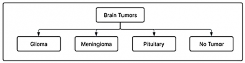
Figure 1. Brain tumor types
(3) The system will establish capability to facilitate language options beginning with English and Telugu as part of enhancing user reach among diverse linguistic populations. The inclusive tool proves its value in regional areas together with underserved communities, so it helps overcome language barriers with digital healthcare systems.
(4) A complete assessment of the AI system should measure its effects on three critical clinical aspects including diagnosis precision and system operating speed and examination volume management capabilities. The supplementary evaluations will investigate both patient satisfaction metrics and how the tool performs for triage purposes by identifying urgent cases which reduces radiologist workloads and optimizes medical resource distribution.
(5) The system aims to enhance medical results through rapid brain tumor diagnosis which requires less centralized data processing and manual reading. General medical diagnostic uses of AI become more widespread because this method enhances healthcare delivery efficiency and fairness.
The classification of brain tumors through deep learning techniques uses MRI image analysis of shape measurements along with texture analysis and intensity characteristics. Research shows that Convolutional Neural Networks (CNNs) especially ResNet successfully identify the tumors glioma and meningioma and pituitary tumors. Full-resolution neural networks serve segmentation operations to achieve better results. The security model training method known as federated learning enables institutions to work together through collaborative processes without revealing sensitive information.
A brain tumor classification system was developed by Lu et al. [1] through MRI scans under federated learning with EfficientNet-B0 and FedAvg to mitigate security risks for non-IID data and tumor variability protection. The developed framework used patient age together with tumor location and scan modality parameters to reach 80.17% accuracy for BraTS dataset evaluation and outperformed standard CNNs and ResNet models in distributed operations. Our initiative coincides with the project's objective to establish ResNet- based federated learning which safeguards medical information privacy and maintains accurate prediction results. The medical images benefit from deep feature extraction capabilities of ResNet in our approach but Efficient Net provides computational efficiency in the researchers' model. The study results demonstrate how FL brings highly effective healthcare solutions by enabling diagnostic systems to secure patient information through collaborative operations.
The research conducted by Neamah et al. [2] presents a privacy-preserved brain tumor classification framework through federated learning (FL) to prevent central data accumulation [3]. The training of parameters for Multiple Convolutional Neural Network (CNN) models occurred locally using distributed networks on MRI datasets for medical image privacy protection. The ensemble classification system selected three models as its leading performers. FL allowed ensemble classifiers to grab parameter information from various local network devices for additional training purposes. The system allowed various institutions to work together for collaborative learning by preventing the transmission of original patient data from their separate devices. The method execution reached an accuracy rate of 92.68% which demonstrates that integrating various models achieves success in distributed learning systems.
Neamah et al. [2] conducted a systematic review of deep learning models that studied brain tumor MRI recognition during the period from 2019 to 2022. The research demonstrated that DNNs achieved high performance accuracy exceeding 95% scenarios. The authors highlighted data sensitivity issues together with small data quantities as major obstacles in their work. The presented research promotes using high-quality datasets to build robust models yet pushes future development of highly reliable diagnostic instruments with general applicability.
The research conducted by Saleh et al. [4] demonstrated the use of Deep Neural Networks (DNNs) for brain tumor classification which produced results of 96.97% accuracy. The analysis capabilities of DNNs for healthcare image patterns made it possible for them to handle brain tumor MRI images for classification purposes. Deep learning models demonstrate strong capabilities for improving brain tumor detection diagnostics because they handle effective analysis procedures effectively. The complete data evaluation at a central data centre makes healthcare decision systems vulnerable to medical data privacy risks. The need for standard privacy protection methods in patient data requires attention because medical imaging necessitates secure procedures to protect sensitive patient information. Research activities generate essential knowledge to build federated learning methods that protect safe data systems and ensure patient privacy integrity.
Zhang et al. [5] developed a brain tumor classification method through a combination of SVMs and CNNs for distributive systems. The brain MRI analyses through CNN- SVM method reached 90.5% successful classification results. Although the presented model exhibited promising performance the researcher's recognized challenges with diagnosing rare tumor cases. Further development of the model remained necessary to adequately categorize different types of tumors. Research established both performance- improving potentials of hybrid approaches and solution methods for present model generalization issues.
Through their research work by Mumtaz Zahoor et al. [6] constructed an advanced deep learning system that merged transfer learning approaches for brain tumor MRI scan classification. The research group achieved 96.08% accuracy on Kaggle data through model fine-tuning of pre-trained Exception along with ResNet50, VGG16, InceptionV3, and MobileNet models. This study demonstrates that transfer learning achieves successful diagnosis performance outcomes and handles restricted medical data effectively.
Amin et al. [7] studied how deep learning algorithms sort brain tumors when using MRI images. The study performed analytical evaluation of AlexNet and GoogLeNet and ResNet50 during their research on a labelled.
Kaggle dataset. ResNet50 proved to deliver the most accurate results because it produced an 85.71% accuracy rate in testing trials. The research showed present models had limitations which made it necessary to develop sophisticated CNN-based approaches to boost classification results. These researchers add value to continuous research into medical imaging tumor detection during early stages through deep learning techniques.
Using CNNs successfully detected brain tumors through a proposed network which generated 92.4% accuracy when processing data from the Kaggle Brain Tumor MRI dataset. Operationally the model demonstrated good detection outcomes but its ability to function was adversely affected by inconsistent MRI picture formats along with processing sensitivity issues. The authors emphasized through their research that better data processing methods together with augmentation technologies allow developers to build reliable models that function effectively on real-world datasets.
The researchers employed deep neural networks (DNNs) for brain tumor classification which demonstrated 89.6% accuracy through analysis of a publicly accessible brain MRI dataset [8, 9]. Although promising research outcomes were obtained the authors recognized two primary areas for enhancement: class imbalance resolution and usage of additional training datasets that better represent diversity. Further advancements in the model design may enhance its generalized performance and stability during the analysis of multiple medical image types.
Zhou et al. [10] developed a multiscale convolutional neural network (CNN) for automatic brain tumor identification as well as segmentation work. The model works with MRI images across different spatial levels to duplicate human vision operations. On a 3,064-slice dataset containing 233 patients, the proposed classification system attained 97.3% accuracy, which proved superior to alternative methods working on the same dataset.
Research by Zhou et al. [11] developed a lightweight ensemble deep learning solution for detecting brain tumors along with their categorization from MRI images. A total of 285 MRI scans from glioma patients in the BraTS 2020 dataset received the model implementation. Performance results from the authors reached substantial levels with accuracy at 93.0% and precision at 0.94, while recall reached 0.93, and the F1 score obtained 0.94, and AUC-ROC achieved 0.984. The model's performance emerges as very effective in medical imaging, thus establishing capabilities for early brain tumor detection.
Islam et al. [12] developed a brain tumor detection system for MRI images through the integration of convolutional deep learning algorithms and particular machine learning approaches. The authors developed a classification system for brain tumors in MRI pictures using a 2D Convolutional Neural Network (CNN) alongside an autoencoder network. A deep learning model reached an outstanding 96.47% accuracy level when tested on the dataset, which emphasized the ability of deep learning methods to perform medical image analysis.
Other works have investigated deep learning with MRI based on tumor detection, which achieved excellent performance while paying attention to the robustness of models [13].
Hybrid and ensemble techniques have also been suggested for improved tumor diagnosis in MRI [14, 15].
CNN-based methods adapted are still yielding improvements regarding the classification accuracy from images obtained from MRI scans [16].
Optimized CNN architectures have been applied to robust brain tumor detection as well [9].
Classical CNN designs such as VGG-16 have also been evaluated in tumor detection with promising success [17-21].
CNN-based MRI classification has also been shown by other works to have competitive accuracies.
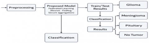
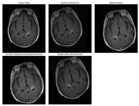
The following report details the complete approach of the proposed brain tumor classification system. The publicly accessible database provided brain MRI scan images, which are used for classification (Figure 2). A set of preprocessing operations is applied to establish consistency while increasing the input quality level. Process interventions start with a 224×224 pixels image resize, during which images become grayscale before receiving pixel normalization to stabilize the training process. Examples of augmented MRI images are provided in Figure 3. Data augmentation techniques that apply random rotation and horizontal flipping and scaling help enlarge dataset size while improving model generality. Image preprocessing completes before deep learning model application with the deployment of ResNet-18 architecture. The model operates with grayscale MRI data focusing on glioma and meningioma and pituitary tumor and no tumor classifications for training purposes. This phase instructs the model to detect patterned visuals that correspond to tumor classification, while validation tracks multiple epoch performance developments. The federated learning framework allows the development of an entire system to facilitate secure model training between simulated client nodes.
Figure 2. Flowchart for proposed methodology
Figure 3. Sample of augmented images
3.1 Dataset
The Brain Tumor MRI dataset from Nampalle et al. [20] is used in this study which consists of 7023 MRI images classified into four classes: glioma, meningioma, pituitary tumor and no tumor, as shown in Table 1. It includes the training and testing folders, preorganized into the dataset, and no further splitting is needed here. No metadata accompanies each of these images, and it is the single input feature. They resized images to 224×224, passed them to tensors and normalized, augmented images to ensure consistency and model performance.
As shown in the images above, the first image represents a glioma tumor, the second image shows a meningioma, the third image is a pituitary tumor, and the fourth image displays a case with no tumor. These MRI scans highlight the structural and visual differences among the tumor types, which are used by the model for accurate classification.
Data Preprocessing- The collected MRI scans are pre- processed to enhance their consistency and quality. In this step we do the grayscale image conversion to an RGB format, equalize pixel values, enhance contrast and these images are rescaled. The identifiers of the patients are anonymized to ensure privacy and follow ethical standards.
The MRI images were preprocessed by converting grayscale scans into three channels and resized to 224×224 pixels. Data augmentation included random cropping, flipping (p = 0.5), rotation (±15°), color jitter, and sharpness adjustment to enhance variability. Finally, images were converted to tensors and normalized using ImageNet mean [0.485, 0.456, 0.406] and std [0.229, 0.224, 0.225].
Table 1. Dataset used for training and testing
|
S. No. |
Train/Test |
Classification |
Images |
|
1. |
training |
Glioma Meningioma Pituitary No Tumor |
6592 |
|
2. |
testing |
Glioma Meningioma Pituitary No Tumor |
1311 |
|
|
total |
|
7023 |
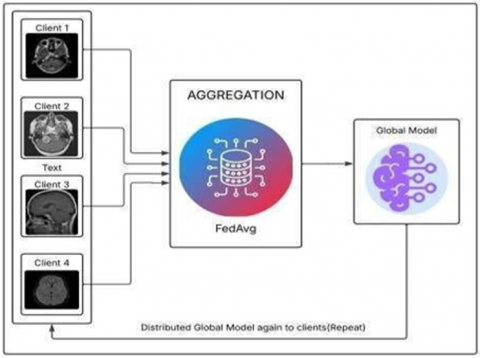
In Figure 4, the proposed federated learning-based system consists of a decentralized architecture with multiple clients (organizations), aggregation server and global model. Each participating organization, e.g. hospitals or research centres, is a client that has its own local set of MRI brain scans. What we do is these institutions train their models for the region using the information they hold in their database without sharing it among themselves. The federated learning starts out with learning a global model and distributing it to all clients for training. This model is trained by each client using its local data and is only sent the updated weights of the model to a centralized server, rather than the actual data. With the aggregated averaging (FedAvg) algorithm, the aggregation server takes the updates and aggregates them into a new global model. The above model is then updated and redistributed to all clients for further training. By using this approach, the privacy of data is maintained, whereas we enable collaborative learning across several institutions. As the entire federated system is overseen by the Admin/Owner, it is the Admin/Owner who maintains all plugins and maintains the entire backend. They are responsible for managing the training pipeline, monitoring model performance across clients, updating the global model with newer versions and resolving feedback from different organizations where the global model is used. The admin makes sure the system is improved and secured. The platform allows for those organizations to upload MRI scans, receive the tumor classification predictions, and analyze trends over time. And they will also have the system incorporated in their diagnostic workflows, including reviewing with radiology experts and using AI-generated insights as clinical decision support. By ensuring continuity of interaction between institutional use cases and the machine learning infrastructure, it guarantees continuity.
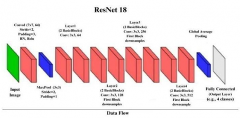
Figure 4. ResNet-18 architecture showing convolutional blocks, residual connections, and final classification layer used in this study
Activation Function - The ResNet-18 has each convolutional layer then followed by the ReLU (Rectified Linear Unit) activation function. The models benefit from the introduction of ReLU to provide the model with non-linearity that facilitates the capturing of complex MRI data patterns. It is easy to compute and prevents vanishing gradient problem. Mathematically, it is expressed as:
f(x)=max(0,x)
It propagates only positive values forward.
3.2 Max pooling
Max pooling is activated after convolutional and activation layers to reduce the spatial dimensions of the feature maps. This step will reduce the computational overhead with retain the most important features. Down sampling the input helps it avoid overfitting and accelerate training.
3.3 Flattening and fully connected layers
The output feature maps are passed through an adaptive average pooling layer and have the shape (512, 1, 1) after the convolutional and pooling stages. It is then flattened into one dimensional vector, and a fully connected layer maps on it into four brain tumor categories. The model can flatten the spatial features into the format that is suitable for classification.
3.4 Loss function
To train the model well, we opted for categorical cross- entropy as the loss function. For multi class classification tasks with one hot encoded label, it is well suited. It measures the dissimilarity between the predicted probability distribution and the actual distribution and is the function to optimize to improve prediction accuracy.
3.5 Model training and validation
In the federated learning approach, model trains in a collaborative manner across multiple clients such as hospitals or medical institutions without sharing raw patient data. However, each client gives the model access to its own brain tumor dataset while training it locally to preserve data privacy and security. The only thing sent to the central server is the model updates (e.g., weights and gradients). Then these updates are aggregated via the Federated Averaging (FedAvg) algorithm to obtain a global model benefiting from diversity of the data of all clients.
During training, the model of each client evaluates its performance based on a local validation set to prevent overfitting It will also perform local validation which helps demonstrate that the model can learn generalizability patterns and not overfit the datasets. During simultaneous communication rounds, the global model evolves to high accuracy and robustness on a diverse set of tumor types including glioma, meningioma, and pituitary.
3.6 Proposed models
3.6.1 Convolutional Neural Networks (CNN)
Being of that they had excelled at the image classification tasks(Convolutional Neural Networks) and were capable of learning complex visual features automatically, Convolutional Neural Networks (CNNs) were used as a core model for this project. In the context of brain MRI analysis, CNNs are specially well suited since they are able to provide both low level and high-level spatial features directly from raw image data. In early convolutional layers, edges, textures, and contrasts are captured; the deeper the layer, the more abstract the found pattern: tumor boundary, shape, and location within the brain. Being able to distinguish amongst tumor types with similar appearances is crucial because it is hierarchical feature learning. CNNs shed manual feature engineering and build an efficient, scalable base on which deep learning based medical diagnostics can be carried out.
3.7 ResNet-18
To make the model deeper and more powerful to learning, we adopted ResNet-18, an already popular convolutional neural network via the residual learning way. Deep neural networks when trained traditionally do suffer from vanishing gradient problem and it is increasingly difficult to train deep neural networks. To tackle it, ResNet utilizes shortcut connections to learn the residual mappings and preserve meaningful features during the consecutive layers. In classifications of brain tumors, ResNet-18 provides a tradeoff between model complexity and computational efficiency for deep feature extraction maintaining stability of the model. The ResNet-18 architecture is shown in Figure 5. Being proven success on many medical imaging tasks, it is very suitable for classifying similar tumor types such as glioma, meningioma, and pituitary tumors, and to identify non tumor cases.
Figure 5. Federated learning
3.7.1 Federated learning (FedAvg)
The choice to use the FedAvg algorithm in federated learning (FL) is because centralizing data in one location may cause serious ethical, legal, and practical trouble. In this approach, different organizations or devices individually train the model with their own MRI brain scan dataset and only transfer model updates (i.e., the model weights or gradients) to a central server. Since there is never raw data leaving the local system, sensitive patient information is always protected and compliant with data privacy legislation such as HIPAA and GDPR.
After which, the central server using the Federated Averaging (FedAvg) algorithm therefore aggregates these updates to create a global model that harnesses the combined intelligence of all the nodes participating. It sets up this way and decreases the probability of the data breach happening even as the model learns from as many different sources of data, the more it learns and the more successful it is.
Specifically, the impact of this method is especially high for healthcare, as medical data is commonly divided between different hospitals, research centres and diagnostic labs, which each work with strict privacy regulations.
Our approach aligns with prior federated frameworks for brain tumor segmentation that preserve patient data privacy [13].
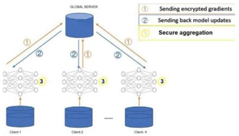
3.7.2 Secure aggregation
We secured aggregation during communication, to facilitate trust and confidentiality reinforcement, in the federated learning system. Secure Aggregation makes sure that every client update is locked in an encrypted way and combined in a way that the server can only obtain the result but not obtain any of the inputs. In this way, no data leakage or inference attack can be performed even if the server were compromised or is trusted. This process is depicted in Figure 6. Secure aggregation serves as another critical security layer for proxying patient data sensitivity when cooperation was needed on a medical setting, allowing institutions to develop collaborative AI without compromising on the data ownership or patient privacy. Such considerations also improve adoption potential since they address important issues in federated AI governance.
Figure 6. Secure aggregation
Each client is trained with its own private data across 10 FL rounds. It was trained with 30 epochs and with batch size 32. The proposed model federated learning approach using ResNet-18 with FedAvg algorithm, which has a train accuracy of 99.93%, validation accuracy of 96.07% and test accuracy of 98%, on classifying brain tumors between four categories.
4.1 Experimental setup
The experimental setup was carried out on a machine running Windows 11 (64-bit) with an Intel Core i5-1135G7 CPU @ 2.40 GHz and 8 GB RAM. This project was implemented using Python 3.10 with the important libraries like PyTorch, NumPy, Matplotlib, Seaborn, and OS. ResNet- 18 is chosen as the base model architecture, and the federated learning setup is applied using FedAvg which is a secure model aggregation algorithm. To train model, it was performed over the artificial clients in local environment and evaluated using pre-split Kaggle MRI data set without any external deployment tools and centralized database.
Federated learning employed the FedAvg algorithm, with aggregation performed at the end of each communication round and clients randomly sampled to participate, reflecting real-world multi-center variability.
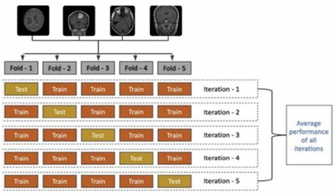
Finally, according to the above model proposed, experiments are conducted to discuss the performance of using deep learning method under the FL framework for classifying different types of brain tumors. Figure 7 demonstrates our experiments. A rigorous type of the brain tumour classification is carried out using K-Fold cross validation. The accompanying medical image dataset of the brain MRI scan data is separated into 5 folds, as shown in Tables 2 to 6. The model is locally trained on four folds for each iteration (four to start learning the model in different client nodes and one to evaluate the model at the central server). However, in this way we make sure that each fold never becomes test one more time and at last we can have a integrated evaluation of the generality of our model on various subsets.getParent. Using this, we also computed the mean accuracy, precision, recall and f1-score to assess the success and efficacy of our proposed disease stage classification method as well as its robustness were calculated across all 5 folds.
Figure 7. K-Fold classification
4.2 Evaluation metrics
In order to study the performance of the brain tumor classification system, the methods based on the standard measures of accuracy, precision, recall and F1-score were applied. These metrics were computed using the predictions of the final federated ResNet-18 model on the test dataset. The evaluation helps assess the model’s ability to correctly classify the four tumor classes: glioma, meningioma, pituitary tumor, and no tumor.
The formulas used for the key metrics are
Accuracy $=T P+T N$
Precision $=\frac{T P}{T P+F P}$
Recall $=\frac{T P}{T P+F N}$
F1-Score $=2 \times \frac{\text { Precision } \times \text { Recall }}{\text { Precision }+ \text { Recall }}$
where, TP = True Positive, TN = True Negative, FP = False Positive, FN = False Negative.
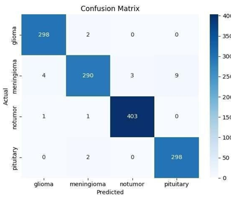
Figure 8. Confusion matrix
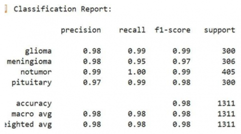
Finally, a test accuracy of 98% was achieved using the final federated ResNet-18 model. The Confusion Matrix is presented in Figure 8. Visualization of the classification results and misclassifications across all tumor classes was also offered through generation of a confusion matrix, classification report, bar graphs. The detailed classification report is shown in Figure 9.
Figure 9. Classification report
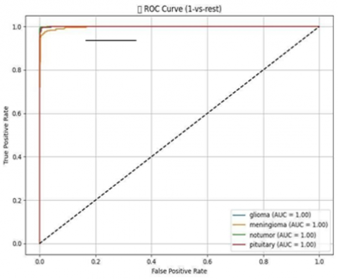
Figure 10. ROC curve
The ROC curves are illustrated in Figure 10. AUC values are reported for each class to complement ROC-based performance visualization.
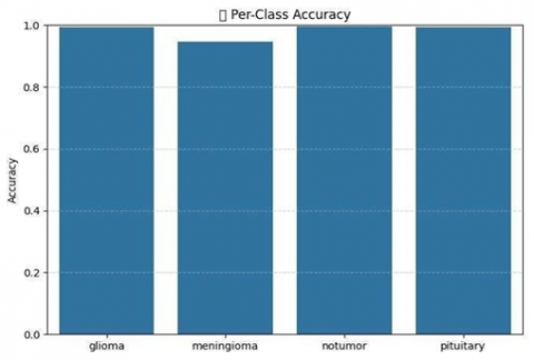
The per-class accuracy values are summarized and visualized in Figure 11, which demonstrates that the proposed federated ResNet-18 model maintains stable classification performance across all tumor categories, confirming the model’s generalization capability.
Figure 11. Per class accuracy
The following is the summary of the results achieved for all the folds:
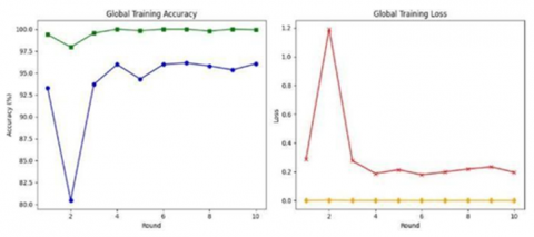
Training and validation curves are shown in Figures 12-15. Using the dataset, our federated learning model’s performance for each fold of the 5 folds cross validation is shown in Tables 2-6. This method guarantees the model is tested on different subsets of the data for the real evaluation of its generalization capability. The global model’s performance is illustrated in Figures 16-17. The model is trained on 4 partitions and validated for each fold using the 1 partition on which the model has never seen before to minimize any bias and variance of the performance prediction.
Table 2. Performance results of FOLD-1
|
Class |
Precison |
Recall |
F1-Score |
Support |
|
Glioma |
0.951613 |
0.967213 |
0.959350 |
61.0 |
|
Meningioma |
0.947368 |
0.900000 |
0.923077 |
60.0 |
|
No tumor |
1.000000 |
0.987500 |
0.993711 |
80.0 |
|
Pituitary |
0.953846 |
1.000000 |
0.976378 |
62.0 |
Table 3. Performance results of FOLD-2
|
Class |
Precison |
Recall |
F1-Score |
Support |
|
Glioma |
0.982143 |
1.000000 |
0.990991 |
55.0 |
|
Meningioma |
0.986111 |
0.986111 |
0.986111 |
72.0 |
|
No tumor |
1.000000 |
0.986486 |
0.993197 |
74.0 |
|
Pituitary |
0.983607 |
0.983607 |
0.983697 |
61.0 |
Table 4. Performance results of FOLD-3
|
Class |
Precison |
Recall |
F1-Score |
Support |
|
Glioma |
1.000000 |
1.000000 |
1.000000 |
67.0 |
|
Meningioma |
1.000000 |
0.938462 |
0.968254 |
65.0 |
|
No tumor |
0.987342 |
1.000000 |
0.993631 |
78.0 |
|
Pituitary |
0.945455 |
1.000000 |
0.971963 |
52.0 |
Table 5. Performance results of FOLD-4
|
Class |
Precison |
Recall |
F1-Score |
Support |
|
Glioma |
0.983051 |
1.000000 |
0.991453 |
58.0 |
|
Meningioma |
0.981818 |
0.964286 |
0.972973 |
56.0 |
|
No tumor |
1.000000 |
1.000000 |
1.000000 |
96.0 |
|
Pituitary |
0.980769 |
0.980769 |
0.980769 |
52.0 |
Table 6. Performance results of FOLD-5
|
Class |
Precison |
Recall |
F1-Score |
Support |
|
Glioma |
1.000000 |
1.000000 |
1.000000 |
59.0 |
|
Meningioma |
1.000000 |
0.943396 |
0.970874 |
53.0 |
|
No tumor |
0.974684 |
1.000000 |
0.987179 |
77.0 |
|
Pituitary |
0.986486 |
1.000000 |
0.993197 |
73.0 |
For each fold the model has been reported to follow metrics like Accuracy, Precision, Recall, F1-score and AUC, all of them are stable showing consistent performance of the model for each data split.
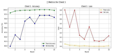
Figure 12. Client-1 training and validation accuracy/loss
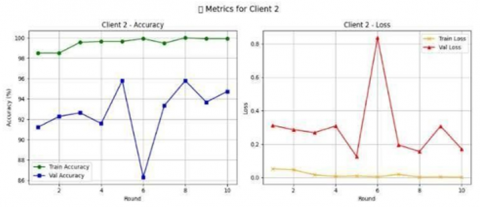
Figure 13. Client-2 training and validation accuracy/loss
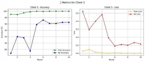
Figure 14. Client-3 training and validation accuracy/loss
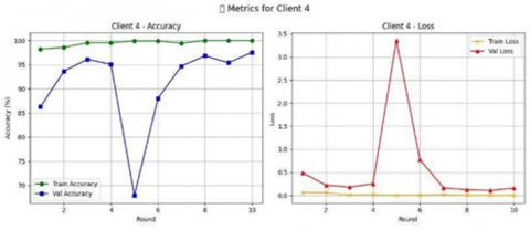
Figure 15. Client-4 training and validation accuracy and loss
Above are the training and validation accuracy and loss curves for federated ResNet-18 model of 4 clients.
Figure 16. Final global model training and validation accuracy/loss
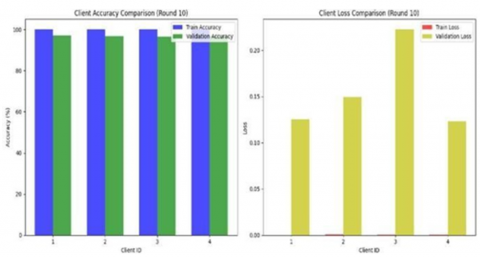
Figure 17. Bar graph of training and validation accuracy/loss of four clients
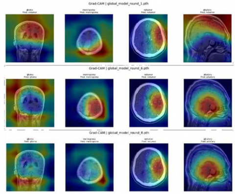
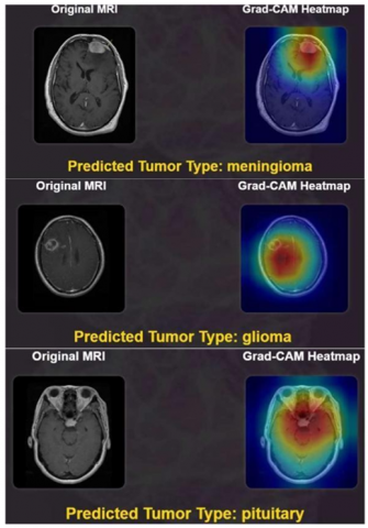
Our project has developed a user interface, which is a simple and intuitive web-based system to interact with our brain tumor model easily. Users can straight forwardly upload MRI images through a streamlined upload section. Grad-CAM visualizations are provided in Figures 18-19. After the image is uploaded, the model runs it in the back end, by using the ResNet-18 architecture that was trained using the federated learning framework on the uploaded image. The result contains a original input image and the Grad-CAM mapped for its prediction.
Figure 18. Grad-CAM visualization across federated learning rounds
Figure 19. Grad-CAM visualization of tumor images
In this study, a federated learning based deep learning approach to brain tumor classification incorporating privacy is presented. The ResNet-18 architecture was used to develop the system, and it was trained on the MRI images from the Kaggle Brain Tumor MRI dataset. The FedAvg algorithm was used for secure aggregation of model updates from multiple clients so as to ensure data privacy and decentralized learning. The proposed model was able to achieve a test accuracy of 98% in classifying four types of brain tumors: glioma, meningioma, pituitary tumor and no tumor. The results also indicate the feasibility of leveraging CNN-based architectures in combination with federated learning for application in the context of real-time, secure diagnostic tools for medical imaging.
More diverse MRI data from different institutions used for training in point 1 gives the system an opportunity to be generalised. A further step could be the application of the federated setup in real-world scenarios and adopt explainable AI as an add-on to enhance the transparency of the prediction and clinical reliability. Further studies could exploit federated frameworks, that is built-in in the management models addressed to tumor classification from MRI [18]. Combining simultaneous segmentation and classification techniques could lead to even more usable clinical tools [21].
[1] Lu, N.H., Huang, Y.H., Liu, K.Y., Chen, T.B. (2025). Deep learning-driven brain tumor classification and segmentation using non-contrast MRI. Scientific Reports, 15(1): 27831. https://doi.org/10.1038/s41598-025-13591-2
[2] Neamah, K., Mohamed, F., Adnan, M.M., Saba, T., et al. (2023). Brain tumor classification and detection based DL models: A systematic review. IEEE Access, 12: 2517-2542. https://doi.org/10.1109/ACCESS.2023.3347545
[3] Abdusalomov, A.B., Mukhiddinov, M., Whangbo, T.K. (2023). Brain tumor detection based on deep learning approaches and magnetic resonance imaging. Cancers, 15(16), 4172. https://doi.org/10.3390/cancers15164172
[4] Saleh, A., Sukaik, R., Abu-Naser, S.S. (2020). Brain tumor classification using deep learning. In 2020 International Conference on Assistive and Rehabilitation Technologies (iCareTech), Gaza, Palestine, pp. 131-136. https://doi.org/10.1109/iCareTech49914.2020.00032.
[5] Zhang, W., Jin, W., Rho, S., Jiang, F., Yang, C.F. (2024). A federated learning framework for brain tumor segmentation without sharing patient data. International Journal of Imaging Systems and Technology, 34(4): e23147. https://doi.org/10.1002/ima.23147
[6] Mumtaz Zahoor, M., Qureshi, S.A., Hussain Khan, S., Khan, A. (2022). A new deep hybrid boosted and ensemble learning-based brain tumor analysis using MRI. arXiv e-prints, arXiv-2201. https://ui.adsabs.harvard.edu/link_gateway/2022arXiv220105373M/doi:10.48550/arXiv.2201.05373
[7] Amin, A., Hasan, K., Zein-Sabatto, S., Chimba, D., et al. (2024). Empowering healthcare through privacy-preserving MRI analysis. In Southeast Con 2024, Atlanta, GA, USA, pp. 1534-1539. https://doi.org/10.1109/ACCESS.2024.1234567
[8] Yue, G., Wei, P., Zhou, T., Song, Y., Zhao, C., Wang, T., Lei, B. (2023). Specificity-aware federated learning with dynamic feature fusion network for imbalanced medical image classification. IEEE Journal of Biomedical and Health Informatics. https://doi.org/10.1109/JBHI.2023.3319516
[9] Yan, R., Qu, L., Wei, Q., Huang, S.C., Shen, L., Rubin, D.L., Xing, L., Zhou, Y. (2023). Label-efficient self-supervised federated learning for tackling data heterogeneity in medical imaging. IEEE Transactions on Medical Imaging, 42(7): 1932-1943. https://doi.org/10.1109/TMI.2022.3233574
[10] Zhou, Z., Luo, G., Chen, M., Weng, Z., Zhu, Y. (2025). Federated learning for medical image classification: A comprehensive benchmark. arXiv preprint arXiv:2504.05238. https://doi.org/10.48550/arXiv.2504.05238
[11] Zhou, L., Wang, M., Zhou, N. (2024). Distributed federated learning-based deep learning model for privacy mri brain tumor detection. arXiv preprint arXiv:2404.10026. https://doi.org/10.48550/arXiv.2404.10026
[12] Islam, M., Reza, M.T., Kaosar, M., Parvez, M.Z. (2023). Effectiveness of federated learning and CNN ensemble architectures for identifying brain tumors using MRI images. Neural Processing Letters, 55(4): 3779-3809. https://doi.org/10.1007/s11063-022-11014-1
[13] Wen, J., Li, X., Ye, X., Li, X., Mao, H. (2025). A highly generalized federated learning algorithm for brain tumor segmentation. Scientific Reports, 15(1): 21053. https://doi.org/10.1038/s41598-025-05297-2
[14] Alphonse, S., Mathew, F., Dhanush, K., Dinesh, V. (2025). Federated learning with integrated attention multiscale model for brain tumor segmentation. Scientific Reports, 15(1): 11889. https://doi.org/10.1038/s41598-025-96416-6
[15] Islam, M.M., Barua, P., Rahman, M., Ahammed, T., Akter, L., Uddin, J. (2023). Transfer learning architectures with fine-tuning for brain tumor classification using magnetic resonance imaging. Healthcare Analytics, 4: 100270. https://doi.org/10.1016/j.health.2023.100270
[16] Manthe, M., Duffner, S., Lartizien, C. (2024). Federated brain tumor segmentation: An extensive benchmark. Medical Image Analysis, 97: 103270. https://doi.org/10.1016/j.media.2024.103270
[17] Gade, V.S.R., Cherian, R.K., Rajarao, B., Kumar, M.A. (2024). BMO based improved lite swin transformer for brain tumor detection using MRI images. Biomedical Signal Processing and Control, 92: 106091. https://doi.org/10.1016/j.bspc.2024.106091
[18] Mathivanan, S.K., Sonaimuthu, S., Murugesan, S., Rajadurai, H., Shivahare, B.D., Shah, M.A. (2024). Employing deep learning and transfer learning for accurate brain tumor detection. Scientific Reports, 14(1): 7232. https://doi.org/10.1038/s41598-024-57970-7
[19] Fares, M.H., Saad, A.M.S.E. (2024). Towards privacy-preserving medical imaging: Federated learning with differential privacy and secure aggregation using a modified resnet architecture. arXiv preprint arXiv:2412.00687. https://doi.org/10.48550/arXiv.2412.00687
[20] Nampalle, K.B., Singh, P., Narayan, U.V., Raman, B. (2023). Vision through the veil: Differential privacy in federated learning for medical image classification. arXiv preprint arXiv:2306.17794. https://doi.org/10.48550/arXiv.2306.17794
[21] Rodriguez-Aguilar, R., Marmolejo-Saucedo, J.A., Köse, U. (2025). Federated learning based on an Internet of medical things framework for a secure brain tumor diagnostic system: A capsule networks application. Mathematics, 13(15): 2393. https://doi.org/10.3390/math13152393