Jamasri![]() | Ferriawan Yudhanto*
| Ferriawan Yudhanto*![]() | Venditias Yudha
| Venditias Yudha![]() | Edi Syafri
| Edi Syafri![]()
© 2023 IIETA. This article is published by IIETA and is licensed under the CC BY 4.0 license (http://creativecommons.org/licenses/by/4.0/).
OPEN ACCESS
PVA (polyvinyl alcohol) and chitosan polymers are two widely used polymers in bioplastic due to their biodegradable properties. This research aims to characterize the mechanical, physical, and thermal properties of combined PVA 3 wt.% and chitosan by 0.5, 1, and 2 wt.% as bioplastic films. It was tested using tensile, X-ray diffraction, Fourier transforms infrared, scanning electron microscopy, and thermogravimetry analysis. Adding 1 wt.% chitosan increases the bioplastic film's tensile strength and thermal stability by 60% and 49℃, respectively. In addition, adding this chitosan also increases the bioplastic film's crystallinity index. However, adding 2 wt.% chitosan affected stiffness and brittle, which indicates a decrease in the elongation at break by 27%.
PVA, chitosan, tensile strength, physical, thermal stability
PVA (polyvinyl alcohol) and chitosan polymers are two types of polymers that are widely used in plastic packaging due to their biodegradable properties. Chitosan with a chemical formula (C6H11NO4)n is a linear polysaccharide resulting from the deacetylation of chitin. Chitosan can be dissolved easily in weak acids. High hydrophilicity based on the presence of amino and hydroxyl in the chitosan chemical functional group [1-3]. Chitosan is deacetylated from chitin which has an amine group (-NH2) that can inhibit bacteria and microbes. The properties of chitosan have made many researchers use it in the biomedical field, including to prevent and treat infections [3, 4], manufacture wound healing dressings [5] and drug release [6]. However, some of the weaknesses of chitosan are weak mechanical properties, brittle, and can only be dissolved in acidic solutions. Several modifications have been made through various techniques to solve the weakness, including adding and blending of myofibrillar protein, cellulose nanocrystals, polymers similar to PLA (polylatic acid), PVA, which can improve biocompatibility, thereby increasing mechanical strength, elongation at break and elasticity of bioplastic materials [7-10]. PVA is a synthetic polymer widely used in biomedical applications due to having prime properties like hydrophilicity and biodegradable material [11]. According to Jiang et al. [12], PVA is a potential biomaterial for vascular cell culture processes. Ahmed et al. [13] have successfully used hydrogels made from PVA and PEG (polyethylene glycol) for wound-healing dressings. PVA is biocompatible and suitable for natural tissue simulation. Basically, PVA has good oxygen permeability, is not immunogenic, good film formation, emulsifier and it is can be moisturized [14]. The mixing of the two types of polymers has been investigated by Vo et al. [15], who made bioplastic films using PVA, chitosan, and anthocyanin extracts from red cabbage. Bioplastic films have been synthesized from hydrogels prepared by mixing 1% PVA and 1% chitosan with anthocyanin. In particular, anthocyanins extracted from red cabbage are used as pH indicators and crosslinking agents to provide good mechanical properties. Vimala et al. [16] explores the fabrication of chitosan-polyvinyl alcohol-silver nanoparticles (AgNPs) films were increased applications as anti-microbial packaging, wound dressings, and antibacterial agents. Silver ions were extracted into silver nanoparticles (AgNPs) and mixed in a solution of chitosan and PVA so that their functional chemical groups (-OH, -COOH, -NH2) bind together. As a result, silver-chitosan-PVA nanoparticle film has inhibited microbial and fungal activity. This has been proven by experiments on E. coli, Pseudomonas, Staphylococcus, Micrococcus, C. albicans, and P. aeruginosa. Yudhanto [17] indicates the high crystallinity was affected the good mechanical properties of the materials. The crystallinity was observed by X-ray diffraction (XRD) test to describe amount of crystalline structure on materials and fourier transform infrared (FTIR) to characterized the chemical bond properties of materials. This research characterizes the mechanical, physical, and thermal properties of the composition between a mixture of PVA and chitosan polymers without any plasticizer binders.
PVA (Polyvinyl alcohol) with molecular weights (MW) 89,000-98,000, 99% hydrolyzed. Table 1 shows the product specification of PVA from Sigma Aldrich with CAS number 9002-89-5.
The Table 2 shows product specification of chitosan (Deacetylated chitin, Poly (D-glucosamine) from Sigma Aldrich with CAS number 9012-76-4.
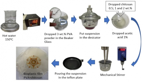
Figure 1 shows the scheme of bioplastic film process, A weight concentration of 3 wt.% PVA dropped in the purified water on a Beaker glass and heated at 150℃, stirrer by 350 rpm for 30 minutes. The PVA suspension was then placed in a desiccator for 24 hours until the bubbles on the surfaces disappeared. Chitosan as a filler was included in a 3 wt.% PVA suspension with concentrations of 0.5, 1, and 2 wt.%. Acetic acid (CH3COOH) 1% dropped into PVA and chitosan was done using a mechanical stirrer with 520 rpm rotation at room temperature (27℃) for 20 minutes to get the homogeneous suspension. PVA+chitosan suspension was poured 30 ml into a Teflon plate which has 150 mm in diameter. It is taken off from the Teflon plate after 24 hours and produces a bioplastic film.
2.1 Tensile test
The tensile test of bioplastic films according ASTM D882 for thin plastic sheets, including thickness film 0-1 mm could be seen on Figure 2. The thin plastic sheet specimen is 80 mm in overall length and 25 mm in width. For the attaching thin plastic sheet into paper 20 mm in gage length and 5 mm in gage width. The universal testing machine (UTM) Pearson Panke Ltd. has a maximum load of 200 N and a set crosshead rate of 2 mm/minutes.
Table 1. Specification of PVA CAS number. 9002-89-5
|
No |
Physical and Chemical Properties of PVA |
Specification |
|
1 |
Appearance (form) |
Crystalline (powder) |
|
2 |
Appearance (color) |
White |
|
3 |
Formula |
(C2H4O) n |
|
4 |
Melting Point |
250℃ |
|
5 |
Relative density |
1.19-1.31 g/cm3 |
|
6 |
Water solubility |
96℃ |
|
7 |
pH |
5-7 |
Table 2. Specification of chitosan CAS number. 9012-76-4
|
No |
Physical and Chemical Properties of Chitosan |
Specification |
|
1 |
Appearance (form) |
powder |
|
2 |
Appearance (color) |
Yellow |
|
3 |
Formula |
(C12H24N2O9) |
|
4 |
Melting Point |
102.5℃ |
|
5 |
Relative density |
1 g/cm3 |
Figure 1. Scheme of mixing process between PVA and chitosan to produce bioplastic film
Figure 2. Tensile test specimen according ASTM D882
2.2 FTIR test
FTIR (Fourier Transform Infrared) analysis finds the range of wavelengths in the infrared region absorbed by the bioplastic film. Infrared radiation (IR) is applied to a sample of a material. The ability of a selection to absorb infrared light energy at a wavelength of 4000-400 cm-1 is measured to determine the type of vibration and the molecular structure of the bioplastic film. Thin pellets were prepared with KBr (potassium bromide), and the samples were measured with a Shimadzu 8400S.
2.3 XRD test
The X-ray diffraction (XRD) analysis of the bioplastic films using a Rigaku Miniflex 600 Benchtop. The instrument was operated at 40kV, and 30mA. X-ray radiation filtered by CuKα radiation (λ=1.54060Ȧ). It was a of 2θ scanning range 5 to 40, with a step increment of 0.02° s-1 at room temperature. Increasing the incident beam peak energy would give greater penetration through the sample test it the range. This technique is applicable for films with thickness between 1-100 μm.
2.4 TGA test
Bioplastic film thermal stability was founded using thermogravimetry analysis (TGA). The tool used has the STA 7300 originating from Switzerland. The size of specimen weighed about 1-3 mg. Sample in the PAN Al2O3 is heated at a rate of 10 degrees per minute in the range of 30-800℃, using a Nitrogen flow of 20 ml per minute.
2.5 Morphology
The surfaces of bioplastic film have been identified by Scanning Electron Microscopy (SEM), JSM-6510, LA-JEOL type, and the changes of characters of materials from PVA to PVA+chitosan bioplastic films. SEM was set on the voltage by 40 kV; the sample was coated with Au using a sputtering method. The bioplastic film sample test by 10×10 mm. To be imaged, the specimen is put in a high vacuum because it needs a conductive surface.
3.1 Tensile strength
The bioplastic film from blended PVA and chitosan approximately have a thickness of 60-80 μm. Figure 3 shows the tensile strength values of PVA film and PVA film with adding chitosan. The pure PVA film gives tensile strength and elongation at the break by 27.3 MPa and 110%, respectively. Adding 1 wt.% chitosan into PVA film increases tensile strength by 60% (43.7 MPa) but decreases the elongation slightly by 7% (95%). Adding 2 wt.% chitosan in the PVA film reduces tensile and elongation properties by 17% and 27%, respectively. That causes the bioplastic film to be more brittle than pure PVA film. Generally, a substance added to produce flexibility and reduce brittleness is a plasticizer.
The addition of plasticizers by adding several polymers, such as glycerol and sorbitol, has improved elongation properties. The plasticizers have been developed by Cerqueira et al. [18], who examined the effect of adding glycerol/sorbitol to chitosan. With an optimal concentration of 1.5 wt.% chitosan and adding 0.5% glycerol/sorbitol, it produces a bioplastic film with a tensile strength of 12.8 MPa and an elongation break of 81.9%. Adding more glycerol (2.0%) increases elongation at a break of 109.9%, but the tensile strength decreased to 8.5 MPa. The use of PVA and chitosan as packaging for strawberries has been investigated by Liu et al. [19]. The variation made was the comparison of the weight concentrations of PVA and chitosan. Suspension 3 wt.% PVA was added 0, 0.60, 0.75, and 0.90 wt.%. Tensile testing on pure PVA resulted in a tensile strength value of 25.1 MPa and an elongation of 82.9%. The optimum addition at the addition of 0.75 wt.% PVA was 35.4 MPa, and the elongation decreased to 75.3%. Choo et al. [20] conducted a study by adding chitosan every 25, 50, and 100 wt.% weight of chitosan into PVA. The highest tensile strength by adding chitosan in the PVA by a ratio of 50:50 there is 29 MPa. The elongation at break decreases from 220 to 95%. Habibie et al. [21] examined the use of chitosan in a paper to improve the paper's mechanical properties. Adding 1 wt.% chitosan increased the strength of the mechanical paper so that it would not tear easily. The addition of 2% chitosan makes the paper material more rigid so that when it is rolled during production, the paper is easily torn.
Figure 3. Mechanical properties of PVA and PVA+chitosan bioplastic films
3.2 Crystallinity analysis
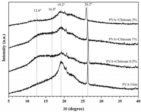
The results of the XRD test showed that the combination of PVA and chitosan showed good inter and intra molecular bonds. It is shown by Figure 4, the peak at 2θ=19.2°, showing the (220) plane, which indicates a crystal form I type lattice [22, 23]. The amorphous region is at 2θ=16.8°, the (100) plane [22]. At the addition of 2 wt.% chitosan into PVA films, there is a change in the shape of the peak at 2θ=12.6°. shows the crystal form type II lattice, the (010) plane [24].
Figure 4. x-ray diffraction patterns of PVA and PVA+chitosan bioplastic films
The presence of an inhomogeneous mixture can cause the intensity of wave absorption in that area to decrease. Adding 2 wt.% chitosan to PVA also causes the value of two thetas to shift from 26.2° to 24.8. The value of 26.2° indicates the reflection of the α waveform caused by the chitin content in chitosan. The pure PVA film crystallinity value is 44.1%. Therefore, mixing PVA and chitosan resulted in a change the crystallinity value of the bioplastic films.
The character of chitosan from crab and shrimp shells has a brittle character with a crystallinity value of 31.9-38.8%, according to Narudin et al. [25]. Mixing with PVA and chitosan improved the mechanical properties even better, and the elongation was relatively good, it is still above 75%. Figure 4 shows the adding of chitosan 0.5, 1, and 2 wt.% increased the crystallinity values 16.8, 25.5, and 34.2%, sequentially. this indicated compatibility between PVA and chitosan in terms of inter and intra-molecular bonds. They crystallinity were resulted 33.4, 43.7, and 36.2 MPA. Adding 2 wt.%, the bioplastic film was increased crystallinity but partially chitosan agglomerates so it changes structure more brittleness.
3.3 FTIR analysis
Figure 5 shows the Fourier transform infrared (FTIR) spectra of wave absorption of bioplastic films. The spectrum of pure PVA film at 3450 cm−1, confirms the stretching vibration of the hydroxyl O-H group bonds in PVA. The highest intensity on the wave 3450 cm−1 shows the pure PVA film characteristic. The vibration band observed at 2928 cm-1 presence of intramolecular bond of C-H from the alkyl group [26-28]. Increasing the chitosan content in the PVA film reduces the intensity of the wave peaks. It is caused by forming of new chemical bonds in the bioplastic film, which contains amide groups [29]. The disappearance of the sharp peak at 2928 cm-1 absorption indicated a chemical bond change from the pure PVA group to PVA/chitosan. This absorption suggests the existence of an intermolecular bending vibration bond between the hydroxyl group (-OH) and the amine group (-NH2) [30, 31]. The waves 1561 cm-1 and 1651 cm-1 showed N-H and C=O bending stretching bonds, which indicates the presence of amide II and amide I, commonly found in chitosan [32].
Figure 5. Fourier transform infrared spectra of PVA and PVA+chitosan bioplastic films
Table 3. FTIR characteristic bands of PVA and PVA+chitosan films
|
Sample |
C-C |
C-O Stretch |
C-H |
N-H Amide II |
C=O Amide I |
C-H |
O-H |
|
PVA |
840 cm-1 |
1100 cm-1 |
1261 cm-1 |
1561 cm-1 |
1651 cm-1 |
2928 cm-1 |
3450 cm-1 |
|
PVA+chitosan 0.5% |
840 cm-1 |
1100 cm-1 |
1261 cm-1 |
1561 cm-1 |
1651 cm-1 |
2928 cm-1 |
3450 cm-1 |
|
PVA+chitosan 1% |
840 cm-1 |
1100 cm-1 |
1261 cm-1 |
1561 cm-1 |
1651 cm-1 |
2928 cm-1 |
3450 cm-1 |
|
PVA+chitosan 2% |
- |
1100 cm-1 |
- |
1561 cm-1 |
1651 cm-1 |
- |
3450 cm-1 |
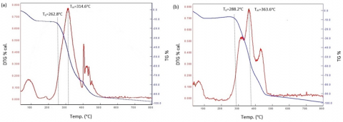
Figure 6. Thermogravimetry analysis (TGA) and derivative thermogravimetry (DTG) curve; (a) PVA film and (b) PVA+chitosan 1%
The change in intensity at the absorption of 1100 cm-1 indicates stretching vibrations, and the presence of C-O stretching bonds that show a saccharide structure indicates the chitosan properties. Due to the high chitosan concentration impact, the disappearing waves are 1261 cm-1 and 840 cm-1. This suggests that the chitosan chemical bond structure did not include C-H and C-C structures. The characteristic chemical bond in the bioplastic films can be seen in Table 3.
3.4 Thermal analysis
Thermal stability on proteins based on bioplastics tested through the Thermogravimetric analysis (TGA) and derivative thermogravimetry (DTG) which showed on Figure 6. Evaporation of the water contained in the bioplastic film at temperatures below 150℃ causes a decrease in the weight of the material [32, 33]. It is used to determine the hydrophilic character of the protein contained in chitin. The difference in water content in several bioplastics is almost the same in the initial range of degradation from 180o to 350℃. Heating can increase the movement of proteins that cause physical interactions between biopolymer chains to change. Mixing PVA 3 wt.% and chitosan 1 wt.% increases the initial degradation temperature (To). It occurred faster in PVA film is 262.8℃ (To) showed on Figure 6(a), while in bioplastic film PVA+Chitosan at 288.2℃ (To) showed on Figure 6(b). As a result, the weight loss occur is 10%, the same as the research conducted by Abraham et al. [32], a decrease in the initial degradation by 10% due to mixing the 5 wt.% PVA and 2 wt.% chitosan. Bio-composite PVA/chitosan film has To=289℃. Adding a plasticizer with a concentration of 25% glycerol on the bio-composite film reduces the weight loss at the 30%. The second degradation occurs at a temperature range of 350-500℃. It caused some of the material partition is starting to burn. Its condition is called the maximum temperature (Tm). The Tm increases from 314.6 to 363.6℃, due to adding chitosan 1 wt.% into PVA film.
3.5 Morphology
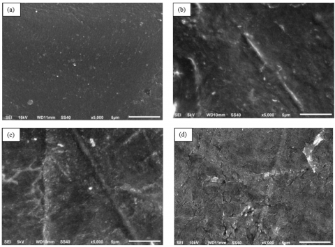
A bioplastic film morphology test was conducted using a scanning electron microscope (SEM). In general, SEM provides information about the surface character of bioplastic films. The test sample of SEM was observed by 20×20 mm, and chose the central location around on bioplastic film; it showed by image SEM with 5,000 x magnification (Figure 7). This test aims to identified the surface structure of bioplastics film. Figure 7(a) shows the appearance of pure PVA film. It is clearly from aggregates in the surfaces of a matrix and has excellent transparency. Figure 7(b) shows the arrival of the smooth wrinkle on the characters of bioplastic film. Figure 7(c) shows it homogeneous dispersion of the mixture of chitosan and PVA in the matrix. Nevertheless, it is effective in the tensile properties of bioplastic films. In addition, it blends indicates good intermolecular bonding between the amino and hydroxyl groups in chitosan and PVA. Finally, Figure 7(d) shows a brittle and rough surface. It has visual appearance slightly yellowish color in bioplastic film.
Figure 7. SEM image of bioplastic film (a) PVA film, (b) PVA+chitosan 0.5%, (c) PVA+chitosan 1%, and (d) PVA+chitosan 2%
Mixing PVA and chitosan with various weight concentrations produces bioplastic films. The bioplastic film PVA+chitosan 1 wt.% has resulted in high tensile strength and good thermal stability by 43.7 MPa and 363.6℃, respectively. XRD analysis shows that adding chitosan improves the crystallinity of bioplastic film. However, adding 2wt.% chitosan can cause the material to agglomerate and affects stiffness and brittle, which causes elongation at break decrease. FTIR analysis shows that when the two polymers are blended, it changes the spectra peaks' characteristics. It indicates the good chemical interaction between chitosan's amino and hydroxyl groups and PVA's hydroxyl groups.
The author is grateful for the financial support of the DTMI (Departemen Teknik Mesin dan Industri) Universitas Gadjah Mada through Independent Research Grant Program with contract No. 791803/UN1/FTK.1/III/PM/2022.
[1] Abraham, A., Soloman, P.A., Rejini, V.O. (2016). Preparation of chitosan-polyvinyl alcohol blends and studies on thermal and mechanical properties. Procedia Technology, 24: 741-748. https://doi.org/10.1016/j.protcy.2016.05.206
[2] Ramasamy, P., Shanmugam, A. (2015). Characterization and wound healing property of collagen–chitosan film from Sepia kobiensis (Hoyle, 1885). International Journal of Biological Macromolecules, 74: 93-102. https://doi.org/10.1016/j.ijbiomac.2014.11.034
[3] Lin, S., Chen, L.H., Huang, L.L., Cao, S.L., Luo, X.L., Liu, K. (2015). Novel antimicrobial chitosan–cellulose composite films bioconjugated with silver nanoparticles. Industrial Crops and Products, 70: 395-403. https://doi.org/10.1016/j.indcrop.2015.03.040
[4] Dai, T., Tanaka, M., Huang, Y.Y., Hamblin, M.R. (2011). Chitosan preparations for wounds and burns: antimicrobial and wound-healing effects. Expert Review of Anti-infective Therapy, 9(7): 857-879. https://doi.org/10.1586/eri.11.59
[5] Jayakumar, R., Prabaharan, M., Nair, S.V., Tamura, H. (2010). Novel chitin and chitosan nanofibers in biomedical applications. Biotechnology Advances, 28(1): 142-150. https://doi.org/10.1016/j.biotechadv.2009.11.001
[6] Yuan, Q., Shah, J., Hein, S., Misra, R.D.K. (2010). Controlled and extended drug release behavior of chitosan-based nanoparticle carrier. Acta Biomaterialia, 6(3): 1140-1148. https://doi.org/10.1016/j.actbio.2009.08.027
[7] Nie, X.H., Gong, Y.D., Wang, N.N., Meng, X.H. (2015). Preparation and characterization of edible myofibrillar protein-based film incorporated with grape seed procyanidins and green tea polyphenol. LWT-Food Science and Technology, 64(2): 1042-1046. https://doi.org/10.1016/j.lwt.2015.07.006
[8] Wang, D., Cheng, W.L., Yue, Y.Y., Xuan, L.H., Ni, X.H., Han, G.P. (2018). Electrospun cellulose nanocrystals/chitosan/polyvinyl alcohol nanofibrous films and their exploration to metal ions adsorption. Polymers, 10(10): 1046. https://doi.org/10.3390/polym10101046
[9] Claro, P.I.C., Neto, A.R.S., Bibbo, A.C.C., Mattoso, L.H.C., Bastos, M.S.R., Marconcini, J.M. (2016). Biodegradable blends with potential use in packaging: A comparison of PLA/chitosan and PLA/cellulose acetate films. Journal of Polymers and the Environment, 24: 363-371. https://doi.org/10.1007/s10924-016-0785-4
[10] Yu, Z., Li, B.Q., Chu, J.Y., Zhang, P.F. (2018). Silica in situ enhanced PVA/chitosan biodegradable films for food packages. Carbohydrate Polymers, 184: 214-220. https://doi.org/10.1016/j.carbpol.2017.12.043
[11] Paradossi, G., Cavalieri, F., Chiessi, E., Spagnoli, C., Cowman, M.K. (2003). Poly (vinyl alcohol) as versatile biomaterial for potential biomedical applications. Journal of Materials Science: Materials in Medicine, 14(8): 687-691. https://doi.org/10.1023/a:1024907615244
[12] Jiang, S., Liu, S., Feng, W.H. (2011). PVA hydrogel properties for biomedical application. Journal of the Mechanical Behavior of Biomedical Materials, 4(7): 1228-1233. https://doi.org/10.1016/j.jmbbm.2011.04.005
[13] Ahmed, A.S., Mandal, U.K., Taher, M., Susanti, D., Jaffri, J.M. (2018). PVA-PEG physically cross-linked hydrogel film as a wound dressing: experimental design and optimization. Pharmaceutical Development and Technology, 23(8): 751-760. https://doi.org/10.1080/10837450.2017.1295067
[14] Hayder, N., Hashim, A., Habeeb, M.A., Rabee, B.H., Hadi, A.G., Mohammed, M.K. (2022). Analysis of dielectric properties of PVA/PEG/In2O3 nanostructures for electronics devices. Revue des Composites et des Matériaux Avancés, 32(5). https://doi.org/10.18280/rcma.320507
[15] Vo, T.V., Dang, T.H., Chen, B.H. (2019). Synthesis of intelligent pH indicative films from chitosan/poly (vinyl alcohol)/anthocyanin extracted from red cabbage. Polymers, 11(7): 1088. https://doi.org/10.3390/polym11071088
[16] Vimala, K., Yallapu, M.M., Varaprasad, K., Reddy, N.N., Ravindra, S., Naidu, N.S., Raju, K.M. (2011). Fabrication of curcumin encapsulated chitosan-PVA silver nanocomposite films for improved antimicrobial activity. Journal of Biomaterials and Nanobiotechnology, 2(1): 55. https://doi.org/10.4236/jbnb.2011.21008
[17] Yudhanto, F. (2022). The effect of alkali treatment and addition of microcrystalline cellulose (MCC) on physical and tensile properties of ramie/polyester laminated composites. Revue des Composites et des Matériaux Avancés, 32(2). https://doi.org/10.18280/rcma.320204
[18] Cerqueira, M.A., Souza, B.W., Teixeira, J.A., Vicente, A.A. (2012). Effects of interactions between the constituents of chitosan-edible films on their physical properties. Food and Bioprocess Technology, 5: 3181-3192. https://doi.org/10.1007/s11947-011-0663-y
[19] Liu, Y.W., Wang, S.Y., Lan, W.T., Qin, W. (2017). Fabrication and testing of PVA/Chitosan bilayer films for strawberry packaging. Coatings, 7(8): 109. https://doi.org/10.3390/coatings7080109
[20] Choo, K., Ching, Y.C., Chuah, C.H., Julai, S., Liou, N.S. (2016). Preparation and characterization of polyvinyl alcohol-chitosan composite films reinforced with cellulose nanofiber. Materials, 9(8): 644. https://doi.org/10.3390/ma9080644
[21] Habibie, S., Hamzah, M., Anggaravidya, M., Kalembang, E. (2016). The effect of chitosan on physical and mechanical properties of paper. Journal of Chemical Engineering and Materials Science, 7(1): 1-10. https://doi.org/10.5897/JCEMS2015.0235
[22] Ioelovich, M. (2014). Crystallinity and hydrophility of chitin and chitosan. Journal of Chemistry, 3(3): 7-14.
[23] Kusmono, Wildan, M.W., Lubis, F.I. (2021). Fabrication and characterization of chitosan/cellulose nanocrystal/glycerol bio-composite films. Polymers, 13(7): 1096. https://doi.org/10.3390/polym13071096
[24] Sirotkin, N., Khlyustova, A., Costerin, D., Naumova, I., Kalazhokov, Z., Kalazhokov, K., Titov, V., Agafonov, A. (2022). Synthesis of chitosan/PVA/metal oxide nanocomposite using underwater discharge plasma: characterization and antibacterial activities. Polymer Bulletin, 1-20. https://doi.org/10.1007/s00289-022-04348-2
[25] Narudin, N.A.H., Mahadi, A.H., Kusrini, E., Usman, A. (2020). Chitin, chitosan and submicron-sized chitosan particles prepared from Scylla serrata shells. Materials International, 2(2): 139-149. https://doi.org/10.33263/Materials22.139149
[26] Yudhanto, F., Jamasri, J., Rochardjo, H.S.B. (2020). Physical and mechanical characterization of polyvinyl alcohol nanocomposite made from cellulose nanofibers. In Materials Science Forum, Trans Tech Publications Ltd, 988: 65-72. https://doi.org/10.4028/www.scientific.net/MSF.988.65
[27] Rochardjo, H.S., Fatkhurrohman, A.K., Yudhanto, F. (2021). Fabrication of nanofiltration membrane based on polyvinyl alcohol nanofibers reinforced with cellulose nanocrystal using electrospinning techniques. International Journal of Technology, 12(2): 329-338. https://doi.org/10.14716/ijtech.v12i2.4173
[28] Yudhanto, F., Jamasri, J., Rochardjo, H.S.B., Kusumaatmaja, A. (2021). Experimental study of polyvinyl alcohol nanocomposite film reinforced by cellulose nanofibers from agave cantala. International Journal of Engineering, 34(4): 987-998. https://doi.org/10.5829/ije.2021.34.04a.25
[29] Queiroz, M.F., Teodosio Melo, K.R., Sabry, D.A., Sassaki, G.L., Rocha, H.A.O. (2014). Does the use of chitosan contribute to oxalate kidney stone formation? Marine Drugs, 13(1): 141-158. https://doi.org/10.3390/md13010141
[30] Li, Q., Zhou, J.P., Zhang, L.N. (2009). Structure and properties of the nanocomposite films of chitosan reinforced with cellulose whiskers. Journal of Polymer Science Part B: Polymer Physics, 47(11): 1069-1077. https://doi.org/10.1002/polb.21711
[31] Yang, W., Owczarek, J.S., Fortunati, E., Kozanecki, M., Mazzaglia, A., Balestra, G.M., Kenny, J.M., Puglia, D. (2016). Antioxidant and antibacterial lignin nanoparticles in polyvinyl alcohol/chitosan films for active packaging. Industrial Crops and Products, 94: 800-811. https://doi.org/10.1016/j.indcrop.2016.09.061
[32] Abraham, A., Soloman, P.A., Rejini, V.O. (2016). Preparation of chitosan-polyvinyl alcohol blends and studies on thermal and mechanical properties. Procedia Technology, 24: 741-748. https://doi.org/10.1016/j.protcy.2016.05.206
[33] Terzioğlu, P., Güney, F., Parın, F.N., Şen, İ., Tuna, S. (2021). Biowaste orange peel incorporated chitosan/polyvinyl alcohol composite films for food packaging applications. Food Packaging and Shelf Life, 30: 100742. https://doi.org/10.1016/j.fpsl.2021.100742