Enhancement of an Inhibition of Plant Pathogen Alternaria alternata with ZnO Nanoparticles and Lactobacillus plantarum

Sraa Nsayef Muslim*![]() | Noor Riyadh Ibrahim
| Noor Riyadh Ibrahim![]() | Zainab Salim Hussin
| Zainab Salim Hussin![]() | Inas Sattar Abd
| Inas Sattar Abd![]()
© 2025 The authors. This article is published by IIETA and is licensed under the CC BY 4.0 license (http://creativecommons.org/licenses/by/4.0/).
OPEN ACCESS
The global fungal pathogen Alternaria alternata causes economically significant yield losses in various crop varieties through symptoms like leaf blight and seed rot. Because green synthesis of nanoparticles is safe, environmentally friendly, and cost-effective, it is preferred over other methods of synthesis. The purpose of this work was to evaluate the potential role of zinc oxide nanoparticles, or ZnO-NPs, and Lactobacillus plantarum crude extract in improving plant tolerance against A. alternata attack. ZnO nanoparticles were biosynthesized with water extract from dandelion roots and leaves. Atomic force microscopy (AFM) for ZnO nanoparticles were 87.62 nm in size, and scanning electron microscopy showed that they were spherical. FTIR spectroscopy for ZnO nanoparticles revealed functional groups which demonstrated formation of the ZnO structure and its purity, and UV-visible absorption spectrum showed a distinct peak at 355 nm. With a 76% inhibition rate, ZnO nanoparticles exhibited the highest antifungal activity against Alternaria alternata at a concentration of 200 mg/ml. Lactobacillus plantarum crude extract inhibited Alternaria alternata at 72%, but when ZnO nanoparticles and Lactobacillus plantarum crude extract were combined, the activity against Alternaria alternata increased to 93%. These findings support the use of that combination to combat a variety of plant-infecting pathogenic fungi.
Alternaria alternata, ZnO nanoparticles, Lactobacillus plantarum, green synthesis, antifungal activity
Alternaria is a widespread genus of microfungi that are found in soil as normal parts of the microflora. In the field and during the postharvest phase, they can infect a broad range of crops, leading to significant losses from fruit and vegetable rotting [1]. They are also responsible for the spoilage of these goods during refrigerated storage and transportation since they can proliferate even at low temperatures [2].
There are already around 70 secondary metabolites of Alternaria that are harmful to plants, but only a tiny percentage of these phytotoxins have been chemically identified and shown to act as mycotoxins in both humans and animals [3]. The most significant species of Alternaria fungus that produces mycotoxin is Alternaria alternata, which primarily infects fruits and vegetables [3, 4].
The existence of an endophyte alone or a specific strain of A. alternata posing concern to food safety must be determined by plant pathologists [5] due to the fact that the pathogen is present on numerous significant crop plants and coexists with many plants as an endophyte in asymptomatic symbiosis [4]. Due to the prevalence of endophytic A. alternata [2], conservationists also need to understand if changes in abiotic environment could lead to asymptomatic A. alternata infections developing into parasitism, further stressing delicate plant populations [4, 5].
There are many pharmacologically active compounds in dandelion. Flavonoids, luteolin, apigenin, and chlorogenic acid are all present in dandelion extract [6]. Vitamins A, C, K, and B complex, minerals, calcium, potassium, magnesium, iron, and zinc, and micronutrients are all found in dandelion roots and leaves [7].
There are three types of nanotechnology that are currently causing a lot of excitement: physical, chemical, and biogenic [8]. Environmental contamination, high temperatures, high pressures, and costly equipment are all associated with the chemical and physical processes [9]. Rather, green nanoparticle production is increasingly being done via biological methods [8]. They are environmentally friendly, economical, and typically have one-step protocols, which are just a few of their many benefits over other methods. Plant diseases are becoming more common and pose serious risks to social and economic stability worldwide. These plant pathogens include bacteria, fungus, viruses, nematodes, and parasitic plants [8, 9].
ZnO nanoparticles (ZnNP) are believed to be safe, non-toxic, and biocompatible. As a result, they have attracted special attention. ZnO nanoparticles also have UV filtering properties, optical, catalytic, electrical, and antibacterial properties [10]. Due to its involvement in numerous enzymatic and physiological processes, for plants to grow and flourish, metallic zinc is an essential mineral [11]. In metabolism of macromolecules, zinc functions as an enzyme component, a catalyst, or structural cofactor. Along with biosynthesis of chlorophyll and the production of energy, it also plays a role in the synthesis of proteins, carbohydrates, and nucleic acids [11].
Zinc increases the pace at which seeds germinate, encourages the quick growth of radicles, impacts the ability to transport and absorb water and protects against harmful effects of drought, high temperatures, and salt stress. Additionally, the production of plant hormones like auxins and gibberellins depends on zinc [12, 13]. Few researches have been done to examine the cytogenic effects in plants as well as the dual impacts of biosynthesized ZnO nanoparticles on various plant diseases. ZnO nanoparticles are among the most often created nanoparticles worldwide; however, they are not as common as silver NP, carbon nanotube, titanium dioxide NP, and gold nanoparticles [14].
In order to combat Alternaria alternata, an endophyte disease of numerous plants, this study aims to explore the inhibitory effect of Alternaria alternata through green synthesis of ZnO nanoparticles and combined with Lactobacillus plantarum extract, with a view to providing information for the control of plant pathogenic fungi.
2.1 Preparation of plant extract
In a homogenized blender, blend 200 milliliters of deionized water with 20 grams of fresh dandelion plant material (leaves and roots) for three minutesat 60℃. The extract was filtered via gauze and then filter paper after boiling for ten minutes. They removed the residue. Furthermore, the filtrate was used right away for the biosynthesis of nanoparticles.
2.2 Biosynthesis of ZnO nanoparticles
A flask containing 100 milliliters of produced plant extract and 2.5 milligrams per milliliter of zinc chloride reagent (ZnCl2) (a granulated zinc dissolved in a minimum amount of 2 M HCl, then the purified water was added) was placed on a magnetic stirrer hot plate set at 50 degrees Celsius and 500 revolutions per second for five hours. After letting the solutions cool to ambient temperature, the final product was filtered, deionized water was added to the nanoparticles, and centrifugation was performed many times for ten minutes at 3000 rpm. Following each centrifugation, after being washed with deionized water, the pellet was put in a hot air oven set at 40 degrees for eight hours, or until it was fully dry [15]. The dry weight of the powdered ZnO nanoparticles produced via biosynthesis was estimated.
2.3 Characterization of ZnO nanoparticles
2.3.1 AFM
The ZnO nanoparticles' size, surface, topography, and granularity volume distribution were all examined using atomic force spectroscopy. A thin layer of the sample was created by dropping 100 microliters of it onto a glass slide. Let it dry for five minutes. A scan of the slide was made [16].
2.3.2 SEM
ZnO nanoparticles were examined using a scanning electron microscope (SEM). Standard protocol was followed in the preparation of the SEM sample.
2.3.3 FTIR spectroscopy
ZnO nanoparticles' different functional groups were determined using the FTIR spectrum (4000-400 cm-1) was used to identify the functional groups, using the KBr technique.The peaks were compared with the standard ZnO nanoparticle.
2.3.4 UV-visible spectroscopy
ZnO nanoparticles' UV-vis absorption spectra were captured using a spectrophotometer and they have a resolution of 1 nm in the 200-800 nm range.
2.4 Preparation of Lactobacillus plantarum extracts
In addition to microscopic and biochemical analyses, the Lactobacillus plantarum isolate was obtained from a milk buffalo sample using Lactobacillus MRS-agar and was diagnosed based on the morphological characteristics of the colonies, such as their size, shape, color, texture, opacity, and margin [17]. The supernatant was obtained as crude extract after an overnight bacterial culture, containing up to 1.5 × 10⁸ CFU/ml, was centrifuged for 1 minute at 14000 × g.
2.5 Replication of Alternaria alternata fungus
A 1.5 cm diameter mycelium disc is inoculated in the middle of each Petri dish; Alternaria alternata isolate was duplicated and cultivated in potato dextrose agar. It was then cultured for seven days at 25℃ [15].
2.6 Evaluation of the ZnONP efficiency in the growth inhibition of Alternaria alternata
ZnO nanoparticles' antifungal properties against Alternaria alternata growth was assessed by adding varying concentrations of ZnONP (200, 20, 2, 0.2) mg/ml to the potato dextrose agar culture medium. Once the ZnO nanoparticles had been ultrasonically treated to guarantee their dispersion in medium, they were put into Petri dishes and left to harden. Four millimeters of fungal plugs were used to inoculate the middle of Petri dishes and incubated at 25℃ for 7 days. Using the following formula [18], the radials, colony growth, and percentage of mycelia growth inhibition were computed.
$Inhibition \,\,of\,\, Mycelial\,\, Growth \,\,(\%)=\frac{(C-T)}{C} \times 100$
where, C is diameter of colony in control plates and T is colony diameter in treated plate.
2.7 Evaluation of Lactobacillus plantarum crude extracts efficiency in the growth inhibition of Alternaria alternata
By adding various concentrations of Lactobacillus plantarum crude extract with 5%, 10%, and 15% to the culture medium potato dextrose agar, the antifungal activity of the extract against the growth of Alternaria alternata was assessed. The experiment was then repeated as previously described, and the percentage of fungal inhibition was noted.
2.8 Evaluation of the combination of ZnO nanoparticles and Lactobacillus plantarum crude extract efficiency in the growth inhibition of Alternaria alternate
After mixing culture media with 200 mg/ml of ZnO nanoparticles and 15% crude extract of Lactobacillus plantarum, the same procedure was carried out to ascertain the effectiveness of the growth inhibition of Alternaria alternata.
2.9 Statistical analysis
Three duplicates of the experiment were conducted. The mean ± standard deviation (SD) is used to record the results.
3.1 Biosynthesis of ZnO nanoparticles from plant extract
The ZnO nanoparticles were biosynthesised using the crude extract from dandelion (Taraxcum officinale) by reducing the ZnCl2 reagent with the extract. ZnO nanoparticle production and transformation were revealed by formation of white precipitate at the bottom of the universal tube. ZnO nanoparticles are made via biosynthesis using dandelion plant extracts, which is a straightforward, inexpensive, and an eco-friendly capping and lowering agent. 323 mg of ZnO nanoparticles were generated per 100 milliliters.
The hazardous and/or detrimental consequences often associated with wet chemical procedures are effectively eliminated by the green synthesis method [19]. ZnO nanoparticles, or biosynthesized zinc oxide nanoparticles, have grown in significance due to its numerous uses and environmental friendliness where by a one-pot process can produce ZnO nanoparticles without the need for an additional stabilizing and reducing agent [20]. Utilizing a variety of plant extracts, recent studies have examined the ecologically benign synthesis of zinc oxide nanoparticles (ZnO NPs). Royal jelly, Cassia fistula, Melia azadarach, and Limoniumbruinosum L. chaz are some of these extracts. Depending on the particular plant extract utilized, the produced zinc oxide nanoparticles (ZnO NPs) size and shape can vary [21].
3.2 Characterization of ZnO nanoparticles
3.2.1 AFM
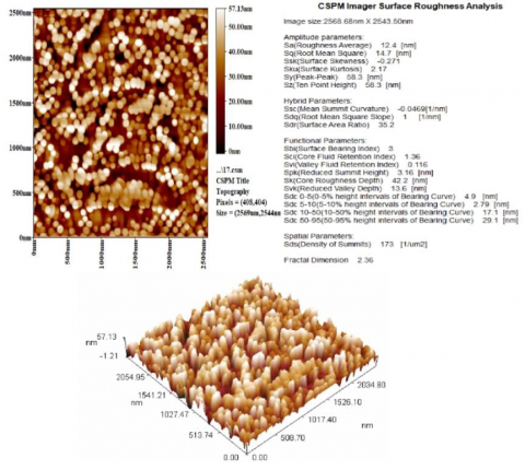
Atomic force microscopy analysis of the generated ZnO nanoparticles revealed that their average size was 87.62 nm. According to Figure 1, the average particle size and roughness rose as the density of ZnO nanoparticles increased.
3.2.2 SEM
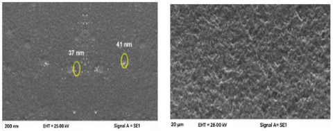
The spherical shape of the biosynthesized ZnO nanoparticles must be seen in the scanning electron microscope picture, as shown in Figure 2. The creation of smaller aggregations was shown in this image, and the structure confirms that the material is densely packed together, demonstrating good film adhesiveness.
Figure 1. Atomic force microscopy pattern for biosynthesized ZnO nanoparticles by dandelion extract
Figure 2. Scanning electron microscope image for biosynthesized ZnO nanoparticles by dandelion extract
3.2.3 FTIR spectroscopy
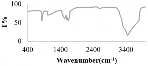
The spectroscopy method known as FTIR is based on how atoms in molecules vibrate. The primary Ir characteristics of pure ZnO biosynthesized by dandelion (Taraxcum officinale) extract were displayed in Figure 3. One feature of the hydroxyl functional group in the spectra is the strong, broad peak at 3444.27 cm−1. The carbonyl and ethylene groups are represented by the peaks at 1640.12 and 1588.14 cm−1, respectively. It is the protein amine bands that caused the peak at 1523 cm-1 in the sample. The detected band at 1020 cm−1 was caused by C-N stretching vibrations. These peaks, that show development and purity of ZnO structure, are caused by ZnO stretching mode.
Figure 3. FTIR spectroscopy analysis for biosynthesized ZnO nanoparticles by dandelion extract
Despite using organic substances to synthesize the ZnO nanoparticles, It is evident that the bending band at 1626 cm−1 contains water molecules, while band at 3482 cm-1 has hydroxyl groups, as well as that there are no bands that may be linked to the solid's organic phase [22].
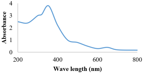
Figure 4. UV-visible absorption analysis for biosynthesized ZnO nanoparticles by dandelion extract
3.2.4 UV-visible spectroscopy
The generated nanoparticles' UV-visible absorption spectra validated the earlier ocular observation. They confirmed their synthesis with a distinct peak at 355 nm (Figure 4), which is a feature of ZnO nanoparticles [4].
3.3 Evaluation of the ZnO nanoparticles efficiency in the growth inhibition of Alternaria alternata
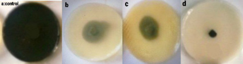
The findings showed that the Alternaria alternata fungus was inhibited in its growth as the concentration of ZnO nanoparticles increased, with the highest inhibition of 76% occurring at 200 mg/ml of ZnO nanoparticles (Figure 5(b) and Table 1).
Table 1. Effect of ZnO nanoparticles on mycelial growth of Alternaria alternataat different concentrations
|
Concentration of ZnO Nanoparticles (mg/ml) |
Diameter of A. Alternata Disc. (mm) |
Percentage of Mycelia Inhibition (%) |
|
200 |
16 |
76 |
|
20 |
22 |
62 |
|
2 |
37 |
52 |
|
0.2 |
44 |
37 |
Figure 5. Antifungal activity of (a) Control, (b) ZnO nanoparticles alone, (c) Lactobacillus plantarum crude extract alone, (d) Combination of ZnO nanoparticles and Lactobacillus plantarum crude extract against Alternaria alternata
For bacteria to be affected, ZnO nanoparticles nanofluid concentration may need to be lower. But, since the nanoparticles' modes of action on bacteria were more varied and numerous than those on fungi, it's also plausible that fungi require higher amounts to be suppressed [20].
3.4 Evaluation of Lactobacillus plantarum crude extracts efficiency in the growth inhibition of Alternaria alternata
The findings showed that employing varying concentrations of Lactobacillus plantarum cell-free supernatant (5%, 10%, and 15%) inhibited the growth of Alternaria alternata fungus progressed with increasing crude extract concentrations, reaching a maximum inhibition of 72% at concentrations of 15% in Figure 5(c) and Table 2.
Table 2. Inhibition of mycelial growth of Alternaria alternata by Lactobacillus plantarum crude extract at different concentrations
|
Concentration of Lactobacillus Plantarum Crude Extract (%) |
Diameter of A. Alternata Disc (mm) |
Percentage of Mycelia Inhibition (%) |
|
15 |
18 |
72 |
|
10 |
27 |
62 |
|
5 |
35 |
47 |
|
2.5 |
49 |
32 |
Numerous bacteriocins, lipopeptides, organic acids, diacetyl, proteases, lipases, hydrogen peroxide, amylases, ethanol, and other antibacterial compounds have been found to be abundant in Lactobacillus species. These substances are produced during the lactate fermentation of Lactobacillus species [23]. Lactic acid, the main byproduct of LAB, is made from glucose and substances that impede development such bacteriocins, hydrogen peroxide and diacyls that stop food spoilage bacteria and pathogens from growing [24]. Lactic acid generation, the primary end metabolic product of carbohydrate fermentation, is what distinguishes them [25].
3.5 Evaluation of the combination of ZnO nanoparticles and Lactobacillus plantarum crude extract efficiency in the growth inhibition of Alternaria alternata
The findings demonstrated that the diameter of Alternaria alternata fungus growth was reduced when cell free supernatant of Lactobacillus plantarum and ZnO nanoparticles were present together, as opposed to when ZnO nanoparticles and Lactobacillus plantarum cell free supernatant were present separately, as illustrated in Figure 5(d). The fungus growth diameter reached 9 mm, while the inhibition percentage was 93%.
Because ZnO nanoparticles make it easier for antibiotics to enter cells, boost their antibacterial effectiveness, and allow access to their target inside cells, they can have a synergistic or additive impact. The synergy between ZnO nanoparticles and the filtrate of Lactobacillus plantarum has been attributed to ZnO nanoparticles destabilizing the cell membrane, encouraging the cell to internalize the compounds in the filtrate while also increasing the microbicidal activity [15].
The cytoplasmic contents were liquefied by ZnO nanoparticles, which reduced the electron density of the cytoplasm and led the fungal cell wall to noticeably separate. When examining the mechanism of ZnO nanoparticles action on harmful, disease-causing fungus, these results should be further investigated because they might turn out to be typical [26].
The electrostatic interaction between metal ions and the microbial cell membrane may account for the prepared nanoparticles' mode of action. Subsequent steps increase the inhibitory activity of the nanoparticles by damaging the intracellular organelles and cell membrane [27, 28]. By interacting with the electron transport chain, breaking phosphate and hydrogen bonds in DNA, denaturing proteins by changing their tertiary structure, and causing mitochondrial death through oxidative stress, nanoparticle penetration into microbial cells promotes microbial inhibition [29]. Cell damage is caused by reactive oxygen species that are created as a result of interactions between inorganic metal and metal oxide nanoparticles [29, 30]. The treatment of fungal plant diseases is one of the many uses for environmentally produced inorganic metal and metal oxide nanoparticles in agriculture.
Over the past few decades, the research community has been eager to build green, economical, and environmentally acceptable nanoparticles. This is because green sources serve as both stabilizing and reducing agents, which helps create shape- and size-controlled nanoparticles that may be used to a variety of processes. In this study, we used dandelion (Taraxcumofficinale) to manufacture ZnO NPs. Utilizing AFM, FTIR, SEM, and UV-visible spectroscopy, the nanoparticles were described. The results of the investigation showed that the plant extract contained biomolecules that might have been essential to the development of zinc oxide nanoparticles. Higher inhibition against Alternaria alternata was shown by ZnO nanoparticles and Lactobacillus plantarum crude extract than by ZnO-NPs and Lactobacillus plantarum crude extract alone. In the field of agriculture, this combination may be utilized as a promising antifungal agent.
Authors introduce their thanks and gratefulness to Department of Microbiology, College of Science, Al-Karkh University of Science, Baghdad, Iraq for their help to complete this research.
[1] Dall, A.C., Cirlini, M., Falavigna, C. (2014). Chapter three-mycotoxins from Alternaria: Toxicological implications. Advances in Molecular Toxicology, 8: 107-121.
[2] Gupta, S., Saxena, S. (2023). Endophytes: Saviour of apples from post-harvest fungal pathogens. Biological Control, 182: 105234. https://doi.org/10.1016/j.biocontrol.2023.105234
[3] Ostry, V. (2008). Alternaria mycotoxins: An overview of chemical characterization, producers, toxicity, analysis and occurrence in foodstuffs. World Mycotoxin Journal, 1(2): 175-188. https://doi.org/10.3920/WMJ2008.x013
[4] Patriarca, A., da Cruz Cabral, L., Pavicich, M.A., Nielsen, K.F., Andersen, B. (2019). Secondary metabolite profiles of small-spored Alternaria support the new phylogenetic organization of the genus. International Journal of Food Microbiology, 291: 135-143. https://doi.org/10.1016/j.ijfoodmicro.2018.11.022
[5] DeMers, M., May, G. (2021). Habitat-scale heterogeneity maintains fungal endophyte diversity in two native prairie legumes. Mycologia, 113(1): 20-32. https://doi.org/10.1080/00275514.2020.1813487
[6] Fatima, T., Bashir, O., Naseer, B., Hussain, S.Z. (2018). Dandelion: Phytochemistry and clinical potential. Journal of Medicinal Plants Studies, 6(2): 198-202.
[7] Sigstedt, S.C., Hooten, C.J., Callewaert, M.C., Jenkins, A.R., Romero, A.E., Pullin, M.J., Steelant, W.F. (2008). Evaluation of aqueous extracts of Taraxacum officinale on growth and invasion of breast and prostate cancer cells. International Journal of Oncology, 32(5): 1085-1090. https://doi.org/10.3892/ijo.32.5.1085
[8] Al-Dhabi, N.A., Valan Arasu, M. (2018). Environmentally-friendly green approach for the production of zinc oxide nanoparticles and their anti-fungal, ovicidal, and larvicidal properties. Nanomaterials, 8(7): 500. https://doi.org/10.3390/nano8070500
[9] Taner, M., Sayar, N., Yulug, I.G., Suzer, S. (2011). Synthesis, characterization and antibacterial investigation of silver–copper nanoalloys. Journal of Materials Chemistry, 21(35): 13150-13154. https://doi.org/10.1039/C1JM11718A
[10] Rehman, S., Jermy, B.R., Akhtar, S., Borgio, J.F., Abdul Azeez, S., Ravinayagam, V., Gani, A. (2019). Isolation and characterization of a novel thermophile; Bacillus haynesii, applied for the green synthesis of ZnO nanoparticles. Artificial Cells, Nanomedicine, and Biotechnology, 47(1): 2072-2082. https://doi.org/10.1080/21691401.2019.1620254
[11] Rajput, V.D., Minkina, T., Fedorenko, A., Chernikova, N., Hassan, T., Mandzhieva, S., Burachevskaya, M. (2021). Effects of zinc oxide nanoparticles on physiological and anatomical indices in spring barley tissues. Nanomaterials, 11(7): 1722. https://doi.org/10.3390/nano11071722
[12] de la Rosa, G., López-Moreno, M.L., de Haro, D., Botez, C.E., Peralta-Videa, J.R., Gardea-Torresdey, J.L. (2013). Effects of ZnO nanoparticles in alfalfa, tomato, and cucumber at the germination stage: Root development and X-ray absorption spectroscopy studies. Pure and Applied Chemistry, 85(12): 2161-2174. https://doi.org/10.1351/pac-con-12-09-05
[13] Eisvand, H.R., Kamaei, H., Nazarian, F. (2018). Chlorophyll fluorescence, yield and yield components of bread wheat affected by phosphate bio-fertilizer, zinc and boron under late-season heat stress. Photosynthetica, 56: 1287-1296. https://doi.org/10.1007/s11099-018-0829-1
[14] Mohd Yusof, H., Abdul Rahman, N.A., Mohamad, R., Zaidan, U.H., Samsudin, A.A. (2020). Biosynthesis of zinc oxide nanoparticles by cell-biomass and supernatant of Lactobacillus plantarum TA4 and its antibacterial and biocompatibility properties. Scientific Reports, 10(1): 19996. https://doi.org/10.1038/s41598-020-76402-w
[15] Arciniegas-Grijalba, P.A., Patiño-Portela, M.C., Mosquera-Sánchez, L.P., Guerrero-Vargas, J.A., Rodríguez-Páez, J.E. (2017). ZnO nanoparticles (ZnO-NPs) and their antifungal activity against coffee fungus Erythricium salmonicolor. Applied Nanoscience, 7: 225-241. https://doi.org/10.1007/s13204-017-0561-3
[16] Hemath Naveen, K.S., Kumar, G., Karthik, L., Bhaskara Rao, K.V. (2010). Extracellular biosynthesis of silver nanoparticles using the filamentous fungus Penicillium sp. Archives of Applied Science Research, 2(6): 161-167.
[17] Kekuda, P.T.R., Vivek, M.N., Kambar, Y., Manasa, M. (2014). Biocontrol potential of Parmotrema species against Colletotrichum capsici isolated from anthracnose of chilli. Journal of Biological and Scientific Opinion, 2(2): 166-169.
[18] Syed Yaacob, S.N., Huyop, F., Kamarulzaman Raja Ibrahim, R., Wahab, R.A. (2018). Identification of Lactobacillus spp. and Fructobacillus spp. isolated from fresh Heterotrigona itama honey and their antagonistic activities against clinical pathogenic bacteria. Journal of Apicultural Research, 57(3): 395-405. https://doi.org/10.1080/00218839.2018.1428047
[19] Mustafa, S.M., Barzinjy, A.A., Hamad, A.H., Hamad, S.M. (2022). Green synthesis of Ni doped ZnO nanoparticles using dandelion leaf extract and its solar cell applications. Ceramics International, 48(19): 29257-29266. https://doi.org/10.1016/j.ceramint.2022.05.202
[20] Barzinjy, A.A., Hamad, S.M., Abdulrahman, A.F., Biro, S.J., Ghafor, A.A. (2020). Biosynthesis, characterization and mechanism of formation of ZnO nanoparticles using Petroselinum crispum leaf extract. Current Organic Synthesis, 17(7): 558-566. https://doi.org/10.2174/1570179417666200628140547
[21] Al-darwesh, M.Y., Ibrahim, S.S., Mohammed, M.A. (2024). A review on plant extract mediated green synthesis of zinc oxide nanoparticles and their biomedical applications. Results in Chemistry, 7: 101368. https://doi.org/10.1016/j.rechem.2024.101368
[22] Kalaba, M.H., Moghannem, S.A., El-Hawary, A.S., Radwan, A.A., Sharaf, M.H., Shaban, A.S. (2021). Green synthesized ZnO nanoparticles mediated by Streptomyces plicatus: Characterizations, antimicrobial and nematicidal activities and cytogenetic effects. Plants, 10(9): 1760.https://doi.org/10.3390/plants10091760
[23] Ruiz Rodríguez, L.G., Mohamed, F., Bleckwedel, J., Medina, R., De Vuyst, L., Hebert, E.M., Mozzi, F. (2019). Diversity and functional properties of lactic acid bacteria isolated from wild fruits and flowers present in Northern Argentina. Frontiers in Microbiology, 10: 1091. https://doi.org/10.3389/fmicb.2019.01091
[24] Mokoena, M.P. (2017). Lactic acid bacteria and their bacteriocins: classification, biosynthesis and applications against uropathogens: A mini-review. Molecules, 22(8): 1255. https://doi.org/10.3390/molecules22081255
[25] Montero-Zamora, J., Rojas-Vargas, M.D., Barboza, N., López-Gómez, J.P., Mora-Villalobos, J.A., Redondo-Solano, M. (2022). Potential of new bacterial strains for a multiproduct bioprocess application: A case study using isolates of lactic acid bacteria from pineapple silage of costa rican agro-industrial residues. Fermentation, 8(8): 361. https://doi.org/10.3390/fermentation8080361
[26] Kumar, H., Seth, S. (2011). Bacterial and fungal study of 100 cases of chronic suppurative otitis media. Journal of Clinical and Diagnostic Research, 5(6): 1224-1227.
[27] Nisar, P., Ali, N., Rahman, L., Ali, M., Shinwari, Z.K. (2019). Antimicrobial activities of biologically synthesized metal nanoparticles: An insight into the mechanism of action. JBIC Journal of Biological Inorganic Chemistry, 24: 929-941. https://doi.org/10.1007/s00775-019-01717-7
[28] Gold, K., Slay, B., Knackstedt, M., Gaharwar, A.K. (2018). Antimicrobial activity of metal and metal-oxide based nanoparticles. Advanced Therapeutics, 1(3): 1700033. https://doi.org/10.1002/adtp.201700033
[29] Elshafie, H.S., Osman, A., El-Saber, M.M., Camele, I., Abbas, E. (2023). Antifungal activity of green and chemically synthesized ZnO nanoparticles against Alternaria citri, the causal agent citrus black rot. The Plant Pathology Journal, 39(3): 265-274. https://doi.org/10.5423/PPJ.OA.02.2023.0035
[30] Abd-Ellatif, S., Ibrahim, A.A., Safhi, F.A., Abdel Razik, E.S., Kabeil, S.S.A., Aloufi, S., Alyamani, A.A., Basuoni, M.M., Alshamrani, S.M, Elshafie, H.S. (2022). Green synthesized of Thymus vulgaris chitosan nanoparticles induce relative WRKY-genes expression in Solanum lycopersicum against Fusarium solani, the causal agent of root rot disease. Plants, 11(22): 3129. https://doi.org/10.3390/plants11223129