Comparative Effects of Nd: YAG and Diode Laser Irradiation on Escherichia coli Clinical Isolates: A Molecular Analysis

Suha Maher Abed*![]() | Sahar Naji Rashid
| Sahar Naji Rashid![]() | Fatima Mustafa Al-Najar
| Fatima Mustafa Al-Najar![]() | Mohammed Ahmed Mustafa
| Mohammed Ahmed Mustafa![]()
© 2024 The authors. This article is published by IIETA and is licensed under the CC BY 4.0 license (http://creativecommons.org/licenses/by/4.0/).
OPEN ACCESS
This work aims to explore various stimulation by two laser types on three samples of Escherichia coli obtained from wound infected patients. Initially the bacterial isolates were collected and diagnosed at the species level. The experiments of bacterial exposure was performed using two laser types: the neodymium-doped yttrium aluminum garnet pulse Nd: YAG (energy: 800 mJ, distance: 20 cm, time: 40 sec. with 532 nm wave length) and the Diode laser (energy: 5 mw, distance: 20 cm, time: 40 sec. with 650 nm wave length). Viable bacterial count was investigated before and after exposure with laser and then the effect was evaluated at the molecular level. The molecular changes were assessed by Random Amplified Polymorphic DNA technique-PCR (RAPD-PCR) through using six oligomeric primers. Results revealed decreased number of viable bacterial colony after being exposed to the irradiation then sub-cultured and incubated at 37℃ for 24 hours the mean of decrease ratio with Diode was 53% while Nd: YAG recorded 86%, the electrophoretic profile results showed appearance of new bands and missing of others at certain molecular weights of treated samples comparing with controls. The oligomeric random primer OPB-07 scored highest number of bands while primer OPS-13 scored the least number. The laser effects on genomic DNA were characterized by distinctive mutant bands which alters gene expression of bacteria and, ultimately, inhibits bacterial growth and activity. The Nd: YAG laser scored high effects than the diode phenotypically. Thermal propagation caused more changes in the DNA. We can conclude that laser light has antibacterial activity and toxigenecity against the studied bacteria and eventually in healing wounds caused by bacteria also we conclude that the Random Amplified Polymorphic technique was effective in detecting laser changes at the molecular level as a simple low-cost method.
antibacterial, diode laser, E. coli, laser therapy, Nd: YAG laser, RAPD-PCR
Laser irradiation has been widely used in biology and medicine, and modern laser systems for diagnosis and treatment in major health centers and hospitals. After the invention of laser, many studies were conducted on the possible interaction between laser and tissue and on all types of lase [1]. The action of a laser begins with the absorption of light by specific chromophores. Lasers interact with chromophores throughout several mechanisms, e.g. photochemical and photothermal interaction, plasmainduced ablation, photoablation, and photodisruption, depending on the influence and pulse duration of the laser [2]. Lasers have become an indispensable tool for some specialties where it is heated [3]. In a few seconds, a protein when the temperature of the tissue increases by 60℃, the cells are destroyed and the coagulation occurs. If the temperature continues to rise to 100℃, the water content of the cells evaporates, causing the cells to shrink and disappear. The continued temperature increase leads to burning and burning of the cells. Time is the primary such as cosmetic surgery and had the ability to kill microorganisms such as bacteria and fungi, via thermal effects because of its high absorption by water molecules, where water occupies a high proportion of the living cell [4, 5]. There are many devices that produce laser with a wavelength range between X-rays and infrared (IR). Well-known lasers like CO2, Neodymium Doped Yttrium Aluminum Garnet (Nd: YAG), fiber laser, and lasers dye are usually applied in medicine. When laser-beam is shed, the power is converted to heat, some of which is absorbed by the treated tissue, evaporated and eliminated by evaporation of the water content in the cells. The remaining part is transferred to adjacent tissue factor in the thermal effect of laser [6]. The chemical effects of the laser in the reaction of chemical reactions in living molecules, inhibition of cell due to genetic alteration in the DNA of the living cell. The laser bacterial killing is related to several factors, including what is related to the interaction of laser with the medium, such as the optical properties of the medium, the extent of its reflection and absorption, thermal diffusion coefficient, thermal conductivity, laser wavelength, capacity and mode of operation are all important factors in the interaction of laser radiation with the medium containing bacteria Less important factor than other factors [7].
The depth of the laser penetration in tissues is the most land mark determining the suitability of the laser type for surgical operations. However, this penetrated depth is proportional to the absorption coefficient of laser radiation in tissues. The intensity of the laser beam is weakened by dispersion and attenuation across the tissue. When laser-beam falls on the biological tissue, several interaction mechanisms related to laser energy intensity and exposure time can be created. There are five types of interactions may produce plasma-included ablation, (photo-disruption, photo-ablation, thermal interaction, and photochemical interaction) [8]. Other interactions may involve light mechanical phenomena in the same temperature range, and the thermal reaction system is frequently important for medical applications. Depending on laser wavelength and the tissue nature, the absorbed part for lasers can give optical light or photochemical effects [9]. Physical and chemical conditions affect the growth and activity of bacteria, including heat, pressure, oxygen concentration and hydrogen ion concentration. These conditions transmit and emit energy through the physical medium that contains bacteria. This energy is known as radiation. The laser is used to sterilize water, milk and some nutrients. However, the effect of radiation requires a direct and increased effect by increasing the radiation dose. The energy density of the unit of area depends on the wavelength of the laser used and the laser capacity and the time required for irradiation [10].
Random Amplified Polymorphic DNA technique has extensively been used by many researchers to assess DNA damage. It is of great importance and widespread use, since It's relatively simple, cheap, fast and gives information on a large number of positions. RAPD pattern differences between control (non- exposed samples) and being exposed is represented as changes in intensity as well as loss or appearance of new bands [11]. The current work aimed to assess the antibacterial activity of Neodymium and Diode Laser in vitro based on viable count and RAPD PCR technique, where there was a need to use clean energy with a local effect, such as Nd: YAG and Diode lasers, this work was conducted to inhibit pathogenic bacteria and evaluate Nd: YAG and Diode effectiveness as a support or alternative to antibiotics and the possibility of developing this mechanism in the future.
2.1 Bacterial isolation
Wound swabs were used to collect the bacteria involved in this research; Escherichia coli was routinely identified based on cultural characteristics and biochemical tests including IMVC tests (Indole, Methyl red, Vogus proskauer and utilization of carbon as a sole source), sugar fermentation and growth on Eosine Methylene Blue media. E. coli isolates were selected to be tested on with the laser based on the number of viable colonies counts and on their genome.
2.2 Irradiation conditions
Three replicates of each isolate were involved; first tube was control, second and third were used for laser exposure experiments. A bacterial suspension prepared from nutrient broth was inoculated with E. coli. The laser device included a pulse duration of: neodymium-doped yttrium aluminum garnet pulse Nd: YAG with energy measured: 800 mJ, distance: 20 cm, time: 40 sec. with wave length 532 nm spectral range) and red diode laser radiation that operates at energy: 5 mw, distance: 20 cm, time: 40 sec. with 650 coulombs nm spectral range) at a repetition rate as fast as 2.0 Hz [12]. The use of lasers has been described as a therapeutic modality to help prevent bacterial growth from this subsequent stimulation and exposed to a single red laser irradiation. Effects tested using power density 0.54 W/cm2 laser.
2.3 Laser exposure conditions and cells viability
Viable bacterial count of the studied samples as control and after being affected by laser irradiation was measured using the Pour-plate method with serial dilution following procedure described in the study by Hussain et al. [13] which involves preparations decimal serial dilution (from 10-1 to 10-6) then adding 0.1ml of the bacterial suspension to the nutrient agar petri dishes. Nutrient agar medium was prepared following manufacturer's instructions and pouring the suspension onto the dishes. The dishes were stirred long enough to mix the sample with the medium and incubated at 37℃ for 24 hours and then counting the bacterial colonies developed on the dish depending on the formula: Total bacterial number (CFU/ml) = number of colonies × inverted dilution factor. Referring that dilution 10-6 was used for pre-treatment (control) and post-treatment including diode and Nd: YAG laser for viable colony counting. Each suspension prior to serial dilutions was adjusted with 0.5 McFarland tube [14].
2.4 Genomic DNA preparation
The phenol chloroform manual extraction was performed following procedure presented by Abed et al. [15] with minor modification. Initially, 1.5 ml of the bacterial culture was transferred into an Eppendorf tube and centrifuged at 14.000 rpm for 3 min in then washed with 200 μL TE buffer followed by addition of 10% sodium dodecyl sulfate (SDS) 30μL and 20 μL of proteinaseK (20 mg/ml) with incubation for 1 h at 37℃ after incubation 50μL of phenol: chloroform: isoamylalcohol (25:24:1) was added and mixed by gentle inversion followed by centrifugation at 14000 rpm for 5 minutes. Aqueous phase was decanted into a new tube and 0.1 ml of 3 M sodium acetate and double volume of -20℃ absolute ethanol, to precipitate DNA was added to it. The mixture was swirled slowly (gentle inversion) until DNA precipitated then centrifuged at 14000 rpm for 5 minutes. DNA pellet was washed with 70% ethanol (500 μL) and centrifuged 14000 rpm for 5 minutes. The supernatant was discarded and 100μL of distil water was added to re-suspended DNA. The stock DNA was kept frozen at- 20℃ until further use. The concentration and purity were determined using nanodrop at wavelength 260/280 nm, the DNA was stored at 4℃ for later usage [16].
2.5 Preparation of RAPD-PCR reactions
The RAPD-PCR reactions was carried out with six oligomeric arbitrary primers (Operon Tech., Inc USA) listed in (Table 1) using (AccuPower PCR premix Kit) supplied by (Bioneer Co., Korea), each tube contained (20 mM Tris-HCl, pH 8.4, 10 mM KCl, 1.5 mM MgCl2, 0.25 mM dNTP and 1U Taq DNA polymerase). The PCR amplifications were performed in 20 μL of reaction mixture containing 25 ng of DNA, 1 μL of 10 p.mol random primer and made of the volume to 20 μL with deionized distilled water. The PCR conditions were as follows: denaturing at 94℃ for 5 sec, annealing at 36℃ for 1min and extension at 72℃ for 1 min. The products (8 μL each), were loaded in 2% agarose gels and electrophoresed at 100V for 1h. The gels were stained with red safe dye prior electrophoresis and vidualized using gel documentation device camera [17, 18]. Polymorphism percentage bands were calculated using the formula:
Polymorphism bands (%) = [Total No. of a + b bands / Total No. of control bands] × 100
where, a: number of new appeared bands; b: number of disappeared bands [19].
Table 1. Name and nucleotide sequence of random primers
|
No. |
Primer Code |
Sequences (5’- 3’) |
|
1. |
OPA-01 |
CAGGCCCTTC |
|
2. |
OPB-07 |
GGTGACGCAG |
|
3. |
OPS-11 |
AGTCGGGTGG |
|
4. |
OPA-13 |
CAGCACCCAC |
|
5. |
OPB-08 |
GTCCACACGG |
|
6. |
OPB-10 |
CTGCTGGGAC |
Thirteen isolates of Escherichia coli out of fifty specimens were collected from wounds of both genders with different age groups of patients using cotton swabs. Those isolates were identified based on cultural characteristics and biochemical tests including IMVC tests, sugar fermentation and growth on Eosine Methylene Blue media. Three of it were selected to test the effect of laser on their genome. Viable bacterial count of the studied samples as control and after being affected by laser irradiation was measured using the Pour-plate method. Results of colony counts illustrated in Figure 1 and Table 2 show that Nd: YAG laser reduced number of colonies more than Diode laser.
Figure 1. Colony counts of control and irradiated sample
Bacterial inactivation due to laser exposure has shown that neodymium-doped yttrium aluminum garnet pulse laser with energy 800 mJ had higher inhibitory effects than Diode5 mw.
The current result is in accordance with Tabit [20]. Also agreed with Ebid et al. [21] who published that those lasers cause denaturation of protein, increase the permeability, vaporization, and thermal decomposition (high damage and/0r burst cell) that may occur at high temperature. Altaee et al. [22] and Zahra [23] mentioned that high pulsed intensity Nd: YAG showed significant decrease in colony count and mortality.
Table 2. Viable counts of E. coli pre (control) and post irradiation (Diode, Nd: YAG)
|
No. of Isolates |
Control |
Diode |
Decrease Ratio |
Nd: YAG |
Decrease Ratio |
|
1 |
46 |
12 |
-0.34 |
8 |
-0.82 |
|
2 |
29 |
15 |
-0.48 |
4 |
-0.86 |
|
3 |
89 |
18 |
-0.79 |
7 |
-0.92 |
|
|
|
Mean |
53% |
Mean |
86% |
Al-Timimi [24] had proposed several explanations for the basis of low-level laser effects including multiple waves shock, a temperature-induced proteins furthermore bubble formation would occurs due to the properties of laser-induced by light-absorbing and raising the critical temperature of the water to 300℃ meaning that the vapor bubbles disrupt and destruct the cell membrane [25]. Further assessment was performed at the molecular level using RAPD-PCR technique with five primers after extraction the genome manually.
The genetic variations among the three isolates regardless of the type of treatments showed that polymorphism in isolates number 3 were the highest using primer OPB-10 (Table 3, Figure 2) while the isolate number1 gave high polymorphism with primer OPB-07, OPA-01 OPS-11 OPB-08 and OPA-13 (Tables 4-8, and Figures 3-7).
Figure 2. RAPD-PCR results electrophoresed on 2% agarose using OPB-10 primer
M: lane marker, C: control, D: Diode and Ng: Neodymium
Figure 3. RAPD-PCR results electrophoresed on 2% agarose using OPB-08 primer
M: lane marker, C: control, D: Diode and Ng: Neodymium
All in all, data of the current work presented in Table 9 show the total number of bands were 233, number of polymorphic bands 179 and percent polymorphism were 76.8% between three groups of control and two type lasers treated samples using six different random primers. It can be observed that random primers produced polymorphic bands. Polymorphic bands number ranged from 21 to 66 whose molecular weights range from100 to more than 2000 base pair agreed with Babu et al. [26] who declared that sufficient distinguishable bands number ranged between 250 and 1,500 bp in the product amplified. Among the three clinical isolates of E. coli, isolates number one gave the highest ratio of polymorphisms were 52.17% concerning with primer OPB-07. Regarding with the appearance of new molecular weight bends or disappearance of the original ones, primer OPB-10 gave a clear vision of new bands in the treatments with Ng laser comparing with diode (Table 3).
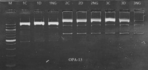
Figure 4. RAPD-PCR results electrophoresed on 2% agarose using OPA-13 primer
M: lane marker, C: control, D: Diode and Ng: Neodymium
Figure 5. RAPD-PCR results electrophoresed on 2% agarose using OPS-11 primer
M: lane marker, C: control, D: Diode and Ng
Figure 6. RAPD-PCR results electrophoresed on 2% agarose using OPB-07 primer
M: lane marker, C: control, D: Diode and Ng: Neodymium
Figure 7. RAPD-PCR results electrophoresed on 2% agarose using OPA-01 primer
M: lane marker, C: control, D: Diode and Ng: Neodymium
As obvious from RAPD electrophoretic figures and Table 3, OPB-07 scored highest number of bands while OPS-13 scored the least number. The dissimilarity of banding pattern and loci profile among control samples although they are of same species due to the fact, they are three distinct individuals that were collected from unrelated patients however six monomorphic bands with large size were detected agreed with Alipour and Mozafari [27]. Emaneini et al. [28] posted that RAPD markers exhibited to be efficient and useful marker system to detect the polymorphism and number of loci scored.
Table 3. Total and polymorphic bands of control and treated isolates (Diode and Neodymium) using OPB-10
|
Isolate |
Control Bands |
Treated 1 (D) |
Treated 2 (NG) |
Total Bands |
Polymorphic Bands |
Polymorphic of Each Primer |
||
|
A |
B |
A |
B |
|||||
|
1 |
4 |
- |
- |
- |
2000, 1500 |
13 |
2 |
15.38% |
|
2 |
4 |
- |
800 |
- |
2000 |
14 |
2 |
14.28% |
|
3 |
4 |
- |
800, 900 |
- |
800, 2000 |
16 |
4 |
25% |
|
Total |
12 |
0 |
3 |
0 |
5 |
43 |
8(18.60%) |
18.22% |
|
a+b |
3 |
5 |
- |
|||||
|
Polymorphism % |
25% |
41.66% |
- |
|||||
|
∑Polymorphism % |
33.33% |
|||||||
A: disappearance; B: appearance of new bands.
Table 4. Total and polymorphic bands of control and treated isolates (Diode and Neodymium) using OPB-08
|
Samples |
Control Bands |
Treated 1 (D) |
Treated 2 (NG) |
Total Bands |
Polymorphic Bands |
Polymorphic of Each Primer |
||
|
A |
B |
A |
B |
|||||
|
1 |
3 |
- |
- |
400 |
500 |
9 |
2 |
22.22% |
|
2 |
2 |
- |
- |
- |
- |
6 |
0 |
0% |
|
3 |
2 |
- |
- |
- |
- |
6 |
0 |
0% |
|
Total |
7 |
0 |
0 |
1 |
1 |
21 |
2(9.5%) |
7.4% |
|
a+b |
0 |
2 |
- |
|||||
|
Polymorphism % |
0% |
28.57% |
- |
|||||
|
∑Polymorphism % |
14.28% |
|||||||
A: disappearance; B: appearance of new bands.
Table 5. Total and polymorphic bands of control and treated isolates (Diode and Neodymium) using OPA-13
|
Samples |
Control Bands |
Treated 1 (D) |
Treated 2 (NG) |
Total Bands |
Polymorphic Bands |
Polymorphic of Each Primer |
||
|
A |
B |
A |
B |
|||||
|
1 |
3 |
- |
1500 |
- |
1500 |
11 |
2 |
18.18% |
|
2 |
3 |
- |
- |
- |
- |
9 |
0 |
0% |
|
3 |
3 |
- |
- |
- |
- |
9 |
0 |
0% |
|
Total |
9 |
0 |
1 |
0 |
1 |
29 |
2(6.89%) |
6.06% |
|
a+b |
1 |
1 |
- |
|||||
|
Polymorphism % |
11.11% |
11.11% |
- |
|||||
|
∑Polymorphism % |
11.11% |
|||||||
A: disappearance; B: appearance of new bands.
Table 6. Total and polymorphic bands of control and treated isolates (Diode and Neodymium) using OPS-11
|
Sample |
Co. |
Treated 1 (D) |
Treated 2 (NG) |
Total Bands |
Polymorphic Bands |
Polymorphic of Each Primer |
||
|
A |
B |
A |
B |
|||||
|
1 |
3 |
- |
- |
800 |
700, 1100 |
10 |
3 |
30% |
|
2 |
4 |
- |
- |
- |
- |
12 |
0 |
0% |
|
3 |
4 |
- |
1200 |
1100 |
- |
12 |
2 |
16.66% |
|
Total |
11 |
0 |
1 |
2 |
2 |
34 |
5(14.70%) |
15.55% |
|
a+b |
1 |
4 |
- |
|||||
|
Polymorphism % |
9.09% |
36.36% |
- |
|||||
|
∑Polymorphism % |
22.72% |
|||||||
A: disappearance; B: appearance of new bands.
Table 7. Total and Polymorphic bands of control and treated isolates (Diode and Neodymium) using OPB-07
|
Samples |
Control Bands |
Treated 1 (D) |
Treated 2 (NG) |
Total Bands |
Polymorphic Bands |
Polymorphic of Each Primer |
||
|
A |
B |
A |
B |
|||||
|
1 |
7 |
250, 300, 350 |
700, 900, 1500 |
250, 300, 350, |
700, 900, 1500 |
23 |
12 |
52.17% |
|
2 |
7 |
- |
- |
700, 900, 1200 |
200 |
19 |
4 |
21.05% |
|
3 |
8 |
- |
- |
- |
- |
24 |
0 |
0% |
|
Total |
22 |
3 |
3 |
6 |
4 |
66 |
16 (24.24%) |
36.61% |
|
a+b |
6 |
10 |
- |
|||||
|
Polymorphism % |
27.27% |
45.45% |
- |
|||||
|
∑Polymorphism % |
36.36% |
|||||||
A: disappearance; B: appearance of new bands.
Table 8. Total and polymorphic bands of control and treated isolates (Diode and Neodymium) using OPA-01
|
Samples |
Control Bands |
Treated 1 (D) |
Treated 2 (NG) |
Total Bands |
Polymorphic Bands |
Polymorphic of Each Primer |
||
|
A |
B |
A |
B |
|||||
|
1 |
5 |
100, 300 |
400 |
250 |
600, 700 |
15 |
6 |
40% |
|
2 |
5 |
1000 |
- |
1000 |
0 |
13 |
2 |
15.38% |
|
3 |
4 |
100 |
- |
600 |
250, 400, 700 |
13 |
5 |
38.46% |
|
Total |
14 |
4 |
1 |
3 |
5 |
41 |
13(31.70%) |
31.28% |
|
a+b |
5 |
8 |
- |
|||||
|
Polymorphism % |
35.71% |
57.14% |
- |
|||||
|
∑Polymorphism % |
46.42% |
|||||||
A: disappearance; B: appearance of new bands.
Table 9. Results of RAPD primer bands
|
Primer |
Molecular Size of Band (bp) |
No. of Polymorphic Bands |
No. of Monomorphic Band |
Total No. of Bands |
|
OPA-01 |
100-2000 |
40 |
0 |
40 |
|
OPB-07 |
˂200-˂1500 |
48 |
2 |
66 |
|
OPS-11 |
700-˂2000 |
16 |
2 |
34 |
|
OPA-13 |
˂ 400- ˂ 1500 |
21 |
0 |
21 |
|
OPB-10 |
400- ˂2000 |
25 |
2 |
43 |
|
OPB-08 |
(400-500)-2000 |
29 |
0 |
29 |
|
Total |
179 (76.8%) |
6 |
233 |
|
Numbers of E. coli isolates obtained from wound infection reflect a skin second stage infection including soft skin tissue and surgical intervention [29]. The Decreased number of the colony forming units is explained with laser photoexcitation of endogenous microbial porphyrin molecules existed in microorganisms, thereby evoking oxidative damage through reactive oxygen species (ROS), which have a high killing potential for bacteria also fungus, and viruses. The degree of destruction relay on the dose used, laser parameters and laser types, ranging from decreased cell growth to inhibition, loss of metabolic activity, and even damaging physical structural. Increasing the pulse energy or pulse rate, or irradiation time would create an extended diameter of the pyknotic cell zone [30].
The variation of amplified bands for both irradiated groups with respect to control (non treated E.coli isolates) indicates the occurrence of a mutation in a specific site. Results reveals that neodymium laser was more efficient phenotypically and genetically. The instability of the DNA from the lasers exposed cells comparing with the normal samples revealed that the rate of appearance of new bands were recorded in neodymium certainly in primer OPA- 01. DNA alterations and genetic changes can be detected by the random amplified polymorphic DNA (RAPD) these alterations include formation of DNA adducts, breaks, point mutations, large rearrangements, and/or others such as structural distortion induced by chemical or physical agents following direct or indirect interaction in the genomic DNA [31].
Many reasons could led to a lost bands such as: genetic material rearrangements or point mutations in oligonucleotide priming sites, damage of DNA in the primer binding sites; and interactions with DNA polymerase of tested organism [32].
The appearance of new bands at certain sites has after being exposed to a stimuli such as chemicals and physical factors or can be resulted in the deletion of a region of DNA [33]. Appearance of a new packages might be due to changes in complementary sites (Oligonucleotide priming) resulting for mutations; a new collision, large deletions or homologous recombinations [34]. Genetic influences like mutation, may not occur only because of an alteration in the sequence of nucleotides, since horizontal transfer of genetic material e.g. chromosome and transposon proteins in DNA can be inherited. Laser light could have been interacted with the DNA and caused damage to the genetic material more or less severe depending on the type of laser and its irradiation condition [33].
The occurrence of genetic mutations in the bacterial genome and the occurrence of genetic variation between control and treatment samples may reflect negatively or positively on the resistance and virulence characteristics of pathogenic bacteria for several reasons, including the occurrence of what is called an amber mutation, which is a change that occurs in the gene, which is the substitution of a genetic code for another. These mutations stimulate the production of additional copies of the gene through the duplication process, thus changing the trait or encouraging a mutant gene to revert to the wild type. The gene also changes due to mutation, so the gene product either stops or changes. It may affect stop codons, leading to irregular replication. It also affects silent genes, turning them into active mode.
Specific laser parameters can lead to contracting or shrinking of the bacterial cell and DNA, which alters gene expression of bacteria and, ultimately, inhibits bacterial growth and activity. Also, laser light stroke cell integrity directly after the application, including cell division inhibition and increasing of metabolically inactive cells [35].
Application of the effects of low-intensity laser therapy (LILT) to wounds, which provides long-term, multiple-use, broad-spectrum radiation in the range of 1-20 J/cm2, can influence bacterial production of great importance in wound healing. The results of this study may be useful LILT for infected wounds that are more reactive to wound-causing bacteria [36].
In this work, we conclude that laser light has antibacterial activity by lowering number of colony and toxigenecity against the studied bacteria and eventually in healing wounds caused by bacteria. The laser effects on genomic DNA were characterized by distinctive mutant bands those bands were considered a distinguishing characteristic and a diagnosis of these transactions, as they indicate the effect of either Nd: YAG or Diode on the genetic material. The appearance of new bands only in one treatment and not another. The RAPD PCR technique was effective in detecting laser changes at the molecular level as a simple low cost method. Also we conclude that Nd: YAG laser had affected more than diode laser. It can be used as an effective treatment against bacteria but further studies are needed to draw firmer findings such as gene expression of virulence factors and antibiotics resistance genes that is often facilitated by biofilm formation. Also this study suggest enhancement the antibacterial efficiency of antimicrobial drugs and nanoparticles using plasma-induced ablation.
[1] Lloyd, A.A., Graves, M.S., Ross, E.V. (2018). Laser-tissue interactions. Lasers in Dermatology and Medicine, 1-36. https://doi.org/10.1007/978-3-319-76118-3_1
[2] Bintanjoyo, L., Indramaya, D.M. (2023). Application of picosecond laser in dermatology. Laser, 35(2). https://doi.org/10.20473/bikk.V35.2.2023.158-162
[3] Tuchin, V.V. (2015). Tissue optics and photonics: Light-tissue interaction. Journal of Biomedical Photonics & Engineering, 1(2): 101-103. https://doi.org/10.18287/JBPE-2015-1-2-98
[4] Youssef, H.M., Alghamdi, N.A. (2018). High-order effect in two-temperature thermal lagging to thermal responses in biological tissue subjected to laser irradiation. Journal of Biomaterials and Tissue Engineering, 8(10): 1519-1526. https://doi.org/10.1166/jbt.2018.1897
[5] Mocan, L., Tabaran, F.A., Mocan, T., Pop, T., Mosteanu, O., Agoston-Coldea, L., Gonciar, D., Zdrehus, C., Iancu, C. (2017). Laser thermal ablation of multidrug-resistant bacteria using functionalized gold nanoparticles. International Journal of Nanomedicine, 12: 2255-2263. https://doi.org/10.2147/IJN.S124778
[6] Wang, L., Feng, Y., Wang, K., Liu, G. (2021). Solar water sterilization enabled by photothermal nanomaterials. Nano Energy, 87: 106158. https://doi.org/10.1016/j.nanoen.2021.106158
[7] Liu, P., Li, Y., Mao, B., Chen, M., Huang, Z., Wang, Q. (2021). Experimental study on thermal runaway and fire behaviors of large format lithium iron phosphate battery. Applied Thermal Engineering, 192: 116949. https://doi.org/10.1016/j.applthermaleng.2021.116949
[8] Keiser, G. (2016). Biophotonics: Concepts to Applications. Springer Singapore. https://doi.org/10.1007/978-981-19-3482-7
[9] Salman, A.M., Jaffar, A.F., Al-Taie, A.A.J. (2017). Studying of laser tissue interaction using biomedical tissue. Al-Nahrain Journal for Engineering Sciences, 20(4): 894-903.
[10] Kodeary, A.K. (2016). Study the effect of laser on bacteria (Enterococcus) Isolated from dairy products. Al-Bahir Journal for Engineering and Pure Sciences, 3(5+6).
[11] Marwan, A., Jasem, A.S. (2019). The influence of molecular effects on laser Nd: YAG and diode on trichophyton rubrum fungi using RAPD marker. Tikrit Journal of Pure Science, 24(6): 117-125. https://doi.org/10.25130/tjps.v24i6.446
[12] Noll, R., Fricke-Begemann, C., Connemann, S., Meinhardt, C., Sturm, V. (2018). LIBS analyses for industrial applications–An overview of developments from 2014 to 2018. Journal of Analytical Atomic Spectrometry, 33(6): 945-956. https://doi.org/10.1039/C8JA00076J
[13] Hussain, A.F., Hashim, H.R., Mohamed, T.A., Abdel-Azeem, A.M. (2021). An annotated bibliography of medical mycology in Iraq: 1962-2021. Microbial Biosystems, 6(1): 11-31. https://doi.org/10.21608/mb.2021.90064.1035
[14] Abbas, H.F., Musawi, I.H.A. (2016). Evaluation three methods of the extraction and purification of bacterial DNA of Gram positive and Gram negative bacteria. World Journal of Experimental Biosciences, 4(1): 62-65.
[15] Abed, S.M., Farhan, M.G., Madhloom, N.K., Dheeb, B.I. (2022) Magnetic field exposure to clinical isolates of Acinitobacter baumanii. Biomedical and Pharmacology Journal, 15(4): 2137-2143 https://doi.org/10.13005/bpj/2550
[16] Wright, M.H., Adelskov, J., Greene, A.C. (2017). Bacterial DNA extraction using individual enzymes and phenol/chloroform separation. Journal of Microbiology & Biology Education, 18(2). https://doi.org/10.1128/jmbe.v18i2.1348
[17] Ebid, A.A., Alhammad, R.M., Alhendi, R.T., et al. (2019). Immediate effect of pulsed high-intensity neodymium-doped yttrium aluminum garnet (Nd: YAG) laser on staphylococcus aureus and pseudomonas aeruginosa growth: an experimental study. Journal of Physical Therapy Science, 31(11): 925-930. https://doi.org/10.1589/jpts.31.925
[18] Al-Hasnawi, K.I., Radhi, N.J.M. (2019). The impact of Nd-YAG laser on salivary streptococcus mutans and lactobacilli in vitro. Indian Journal of Public Health Research & Development, 10(10). https://doi.org/10.5958/0976-5506.2019.03211.X
[19] Saed, Z.J., Hamad, O.K., Mohammed, A.B., Al-Jumaily, T.K. (2024). Effect of natural zeolite (Nz) of growth performance, immunity parameters and gut histology in broiler chicken. Tikrit Journal for Agricultural Sciences, 24(2).
[20] Abed, S., Assie, A., Abu-Elteen, K., Dheeb, B., Abu-Qatouseh, L. (2020). Molecular characterization of methicillin-resistant and methicillin-susceptible staphylococcus aureus isolates obtained from human-skin samples in Iraq. Biomedical and Pharmacology Journal, 13(2): 737-746. https://doi.org/10.13005/bpj/1939
[21] Ebid, A.A., Alhammad, R.M., Alhindi, R.T., et al. (2021). Effect of high-power Nd: YAG laser on the growth of staphylococcus aureus and pseudomonas aeruginosa: An experimental study. Journal of Physical Therapy Science, 33(3): 222-228. https://doi.org/10.1589/jpts.33.222
[22] Altaee, A.J., Aldabbagh, S.Y. (2022). Molecular identification of virulence genes of Pseudomonas aeruginosa isolated from fish (Cyprinus carpio) in Mosul city. Iraqi Journal of Veterinary Sciences, 36(4): 953-958. https://doi.org/10.33899/IJVS.2022.132660.2119
[23] Abd-alwahab, W.I., Al-Assie, R.J., Azeez, A.K., Ghadir, G.K. (2024). The effect of carotenoids of Rhodotorula glutinis and probiotic of Lactobacillus acidophilus on some physiological and histological variables of the pancreas and liver in male rats exposed to ultraviolet radiation: The effect of carotenoids of Rhodotorula glutinis and probiotic of Lactobacillus acidophilus on some physiological and histological variables of the pancreas and liver in male rats exposed to ultraviolet radiation. Tikrit Journal for Agricultural Sciences, 24(2): 235-245.
[24] Al-Timimi, Z. (2022). Evaluation of the significance of constant laser therapy, 532 nm, in various exposure times on the healing process of wounds infected by Acinetobacter baumannii. The International Journal of Lower Extremity Wounds, 21(4), 640-646. https://doi.org/10.1177/1534734620984039
[25] Tee, S.Y., Ye, E., Teng, C.P., Tanaka, Y., Tang, K.Y., Win, K.Y., Han, M.Y. (2021). Advances in photothermal nanomaterials for biomedical, environmental and energy applications. Nanoscale, 13(34): 14268-14286. https://doi.org/10.1039/c9cs00839j
[26] Babu, K.N., Sheeja, T.E., Minoo, D., Rajesh, M.K., Samsudeen, K., Suraby, E.J., Kumar, I.P.V. (2021). Random amplified polymorphic DNA (RAPD) and derived techniques. Molecular Plant Taxonomy: Methods and Protocols, 219-247. https://doi.org/10.1007/978-1-0716-0997-2_13
[27] Alipour, M., Mozafari, N.A. (2015). Terbinafine susceptibility and genotypic heterogeneity in clinical isolates of Trichophyton mentagrophytes by random amplified polymorphic DNA (RAPD). Journal de Mycologie Medicale, 25(1): e1-e9. https://doi.org/10.1016/j.mycmed.2014.09.001
[28] Emaneini, M., Kalantar-Neyestanaki, D., Jabalameli, L., Hashemi, M., Beigverdi, R., Jabalameli, F. (2019). Molecular analysis and antimicrobial resistance pattern of distinct strains of Pseudomonas aeruginosa isolated from cystic fibrosis patients in Iran. Iranian Journal of Microbiology, 11(2): 98-107.
[29] Gallois, C., Hauw-Berlemont, C., Richaud, C., Bonacorsi, S., Diehl, J.L., Mainardi, J.L. (2015). Fatal necrotizing fasciitis due to necrotic toxin-producing Escherichia coli strain. New Microbes and New Infections, 8: 109-112. https://doi.org/10.1016/j.nmni.2015.06.003
[30] Nishat, S., Hamim, I., Khalil, M.I., Ali, M.A., Hossain, M.A., Meah, M.B., Islam, M.R. (2015). Genetic diversity of the bacterial wilt pathogen Ralstonia solanacearum using a RAPD marker. Comptes Rendus Biologies, 338(11): 757-767. https://doi.org/10.1016/j.crvi.2015.06.009
[31] Kramer, B., Thielmann, J. (2016). Monitoring the live to dead transition of bacteria during thermal stress by a multi-method approach. Journal of Microbiological Methods, 123: 24-30. https://doi.org/10.1016/j.mimet.2016.02.009
[32] Al-Azzawie, A.F., Jasem, A.S., Salih, M.H., Abd Albaqi, M.A., Sadiq, S. (2019). Evaluation of the genetic effects of Nd: Yag and diode lasers on Candida albicans using RAPD markers. In HEZARFEN International Congress of Science, Mathematics and Engineering, pp. 886-895.
[33] Samaranayake, L. (2018). Essential Microbiology for Dentistry-E-Book: Essential Microbiology for Dentistry-E-Book. Elsevier Health Sciences.
[34] Lin, T., Pan, J., Gregory, C., Wang, Y., Tincher, C., Rivera, C., Zhang, Y. (2023). Contribution of the SOS response and the DNA repair systems to norfloxacin induced mutations in E. coli. Marine Life Science & Technology, 5(4): 538-550. https://doi.org/10.1007/s42995-023-00185-y
[35] Gutknecht, N., Kanehl, S., Moritz, A., Mittermayer, C., Lampert, F. (1998). Effects of Nd: YAG-laser irradiation on monolayer cell cultures. Lasers in Surgery and Medicine, 22(1): 30-36. https://doi.org/10.1002/(SICI)1096-9101(1998)22:1%3C30::AID-LSM8%3E3.0.CO;2-Y
[36] Koo, H.M., Yong, M.S., Na, S.S. (2015). The effect of low-intensity laser therapy (LILT) on cutaneous wound healing and pain relief in rats. Journal of physical therapy science, 27(11): 3421-3423. https://doi.org/10.1589/jpts.27.3421