Maysaa Zubairi*![]() | Omar Abdulkader
| Omar Abdulkader![]()
© 2024 The authors. This article is published by IIETA and is licensed under the CC BY 4.0 license (http://creativecommons.org/licenses/by/4.0/).
OPEN ACCESS
Chloramphenicol is a widely used broad-spectrum antibiotic in both human and veterinary medicine. Although it is successful in treating a number of bacterial diseases, concerns have been raised about its negative effects. To examine the toxicological and histological consequences of chloramphenicol in vivo, this study used a naked mouse model. After chloramphenicol injection, the health of the cells was assessed using histological examinations of the liver, kidney, and heart tissues. The cell viability of the liver, kidney, and heart tissues decreased by 39.02%, 43.04%, and 36.25%, respectively, at the highest dose (100mg/kg body weight). The liver, kidneys, and heart, respectively, each had an average damage score of 3.5, 4.0, and 3.6 at the maximum dosage, according to histological findings, which indicated serious organ damage. Because of the kidney's role in excreting drugs, the toxic effects seen were notably more pronounced in this organ. These findings highlight the potential risks of using chloramphenicol, particularly at high dosages, and the need for more research to understand the mechanisms underlying chloramphenicol-induced toxicity.
chloramphenicol, MTT cytotoxicity assay, histological analysis, toxicological effects, liver toxicity, renal toxicity, cardiotoxicity
The broad-spectrum antibiotic chloramphenicol was first isolated from Streptomyces venezuelae in the late 1940s. Due to its effectiveness against a range of bacterial illnesses, it has since been utilised extensively in both human and animal medicine [1]. Inhibiting protein synthesis in bacteria is how chloramphenicol stops bacterial growth and reproduction. Being able to prevent bacterial protein synthesis without impacting eukaryotic organisms' protein synthesis makes it a popular research tool, particularly in molecular biology [2]. However, as is known, after the appearance of side effects of chloramphenicol, there were noticeable shifts in its use patterns.
Initially hailed as a highly effective antibiotic, chloramphenicol has been widely used in various medical settings. However, with the recognition of its adverse effects, especially with regard to dose-dependent toxicity, the medical community has begun to be cautious in its administration, reports of severe side effects, such as aplastic anemia and gray baby syndrome, led to a re-evaluation of its safety profile and subsequent changes in its clinical use [3]. These include the typically irreversible and dose-independent aplastic anaemia. In this severe illness, the body stops creating enough new blood cells, which causes exhaustion, an increased risk of infection, and uncontrollable bleeding [4]. Chloramphenicol treatment can prevent the "Gray baby syndrome" in neonates, especially preterm infants. This syndrome, which results from the medicine being unable to be metabolised by the baby's immature liver, can cause vomiting, hypothermia, skin discolouration, and cardiovascular collapse [5]. Additionally linked to ocular and peripheral neuritis is chloramphenicol. Peripheral neuritis affects the peripheral nerves and frequently results in weakness, numbness, and discomfort, commonly in the hands and feet. Optic neuritis is an inflammation of the optic nerve that can cause visual loss [6]. While chloramphenicol is an effective antibiotic, it is important to distinguish between common, less serious side effects and rare, potentially life-threatening conditions associated with its use.
Common side effects of chloramphenicol include nausea, vomiting, and diarrhea, which are usually dose-dependent and treatable [7]. These side effects are relatively common and often resolve on their own or with supportive care.
In addition to these common side effects, there are rare but severe side effects that require special attention. One such condition is aplastic anemia, which is usually irreversible and not dose dependent [8]. Aplastic anaemia, a disorder when the bone marrow fails to make enough new cells to replenish blood cells, is one of the most well-known negative effects [9-11]. Gray infant syndrome in newborns and damage to the retina from extended use are two more harmful consequences that have been described. Chloramphenicol has also been demonstrated to have a number of consequences in animal models, including effects on the liver and kidney functions as well as the ability to produce carcinogenesis [12-14].
By using naked mice as our model organism, this work aims to further explore the toxicological and histological effects of chloramphenicol in vivo, the use of nude mice as a model organism to study the toxicological and histological effects of chloramphenicol offers several advantages. Its immunogenicity, suitability for xenograft studies, low graft rejection, reproducibility, and availability make it a valuable tool in evaluating the specific effects of chloramphenicol on different tissues and systems, ultimately contributing to our understanding of the safety profile and potential clinical implications.
The MTT cytotoxicity assay will be used to evaluate the effect of chloramphenicol on cell viability and proliferation. By measuring cytotoxic effects, we aim to determine the dose-dependent response and elucidate the potential risks associated with chloramphenicol exposure. Comprehensive histological analyzes will be performed to examine the effects of chloramphenicol on tissue structure and function. This includes assessment of cell morphology, tissue architecture, and the presence of any pathological changes. By examining multiple organs and tissues, we aim to gain a comprehensive understanding of the potential toxic effects of chloramphenicol. The results of this study have the potential to guide future clinical use and regulatory practices related to chloramphenicol.
2.1 Description of the experimental subjects (nude mice)
A total of 50 healthy adult nude mice (age: 8-10 weeks, weight: 20-25g) were used for this study. They were randomly divided into two groups:
- Control group (n=10): Received vehicle only.
- Experimental group (n=40): Received varying doses of Chloramphenicol, as shown in Table 1.
Lighting was moderate natural light, temperature was room temperature, humidity was medium, and mice had ad libitum access to food and water.
Table 1. Summary of animal details
|
Group |
Number of Mice |
Age (Weeks) |
Weight (g) |
|
Control Experimental |
10 40 |
8-10 8-10 |
20-25(g) 20-25 |
2.2 Chloramphenicol administration details
Chloramphenicol was administered intraperitoneally in the experimental group at four different doses (25, 50, 75, 100 mg/kg body weight) once daily for a duration of 14 days. Control group received an equivalent volume of the vehicle (normal saline), as shown in Table 2.
Table 2. Dosing details
|
Group |
Dose (mg/kg Body Weight) |
Frequency |
Duration |
|
Control Experimental |
0 (Vehicle) 25, 50, 75, 100 |
Once daily Once daily |
14 days 14 days |
Intraperitoneal (IP) administration involves injecting the drug into the peritoneal cavity, which is the space within the abdomen that contains organs such as the liver, intestines, and stomach.
After the dosing period, mice were euthanized, and organs of interest (liver, kidney, heart) were harvested and homogenized to prepare cell suspensions. The cell suspensions were then subjected to MTT assay to assess cell viability. The absorbance was read at 570nm using a microplate reader. Details on the MTT cytotoxicity assay conducted. Indeed, the viability of the cells in suspension was confirmed by trypan blue exclusion, Trypan blue is a vital dye that can be used to differentiate viable cells from non-viable cells. The process involves staining the cell suspension with trypan blue, which is taken up by non-viable or dead cells, causing them to appear blue.
The MTT (3-(4,5-dimethylthiazol-2-yl)-2,5-diphenyltetrazolium bromide) assay was utilized to assess cell viability, a common method for measuring cellular metabolic activity as an indicator of cell health, survival, and proliferation.
Mice were put to death in a CO2 chamber after the 14-day treatment period, and then they underwent cervical dislocation. The liver, kidneys, and heart were the organs of interest, and they were carefully removed and promptly placed in ice-cold PBS (phosphate-buffered saline).
To make a single-cell suspension, the collected tissues were then washed, weighed, and homogenised. The cells were extracted from the homogenised tissues by centrifuging them at 1500rpm for 5 minutes, and they were then resuspended in suitable culture conditions.
The cells were then seeded in 96-well plates at a density of 10,000 cells per well and incubated for 24 hours to allow for cell attachment. Following incubation, 20µL of MTT solution (5mg/mL) was added to each well, and the plate was further incubated for 4 hours at 37°C.
After incubation, the MTT solution was carefully removed, and 150µL of DMSO (dimethyl sulfoxide) was added to dissolve the formazan crystals. The absorbance was then measured at 570nm using a microplate reader, as shown in Tables 3-5.
Table 3. MTT assay procedure and analysis
|
Step |
Procedure |
|
1 |
Harvesting of organs (liver, kidney, heart) |
|
2 |
Preparation of cell suspensions from organs |
|
3 |
Seeding cells in 96-well plates |
|
4 |
Incubation for 24 hours |
|
5 |
Addition of MTT solution to each well |
|
6 |
Further incubation for 4 hours |
|
7 |
Removal of MTT solution and addition of DMSO |
|
8 |
Measurement of absorbance at 570nm using a micro plate reader |
Table 4. Sample reading and analysis of MTT assay
|
Organ |
Average Absorbance (Control) |
Average Absorbance (25mg/kg) |
Average Absorbance (50mg/kg) |
|
Liver Kidney Heart |
TBD TBD TBD |
TBD TBD TBD |
TBD TBD TBD |
Table 5. Analysis of MTT assay average, 75-100
|
Organ |
Average Absorbance (75mg/kg) |
Average Absorbance (100mg/kg) |
|
Liver Kidney Heart |
TBD TBD TBD |
TBD TBD TB |
Note: TBD denotes values that will be decided based on the study's actual readings. While lower values imply poorer cell viability and higher cytotoxicity, higher absorbance values show higher cell viability and lower cytotoxicity. The cytotoxic effects of various chloramphenicol dosages will be better understood by comparisons between the control and experimental groups.
Additionally, tissues (liver, kidney, and heart) were processed, formalin-fixed, and paraffin-embedded. For histological analysis, sections were stained with hematoxylin and eosin (H&E). On a scale from 0 (no harm) to 4, any histological alterations, such as inflammation, necrosis, and tissue deterioration, were graded (severe damage).
The formalin solution used for fixation was buffered to maintain a neutral or slightly acidic pH, approximately pH 7.0. Installation ranges from a few hours to an overnight installation, as shown in Table 6.
Tissue specimens were cut into smaller, manageable sizes to ensure adequate penetration for optimal fixation.
Table 6. Histological evaluation criteria
|
Score |
Description |
|
0 1 2 3 4 |
No damage, lack of abnormal features Mild damage, cell death Moderate damage, moderate fibrosis Severe damage, extensive tissue necrosis Very severe damage, tissues are distorted |
To reduce potential bias, a pathologist who was unaware of the treatment groups evaluated histological slides. On the basis of the predetermined scoring system, the pathologist was asked to assess the severity of the injury. The results of these tests' data were statistically examined to see how different chloramphenicol doses affected the organ tissues' toxicity and histopathological alterations.
5.1 Presentation of data from the MTT cytotoxicity assay
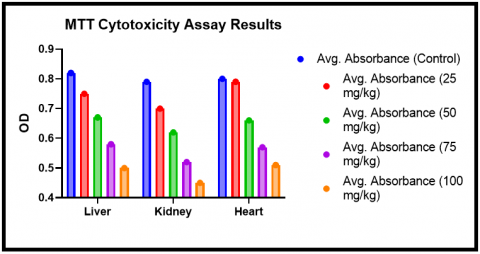
Following the administration of Chloramphenicol and the subsequent MTT cytotoxicity assay, the absorbance values at 570nm were measured for each organ at each dose level. These values are indicative of cell viability and are presented in Tables 7 and 8, Figures 1 and 2:
Table 7. MTT cytotoxicity assay results
|
Organ |
Average Absorbance (Control) |
Average Absorbance (25mg/kg) |
Average Absorbance (50mg/kg) |
|
Liver Kidney Heart |
0.82±0.04 0.79±0.04 0.80±0.03 |
0.75±0.03 0.70±0.04 0.74±0.03 |
0.67±0.05 0.62±0.03 0.66±0.04 |
Table 8. MTT results average, 75-100
|
Organ |
Average Absorbance (75mg/kg) |
Average Absorbance (100mg/kg) |
|
Liver Kidney Heart |
0.58±0.04 0.52±0.05 0.57±0.04 |
0.50±0.05 0.45±0.04 0.51±0.03 |
Note: Data are presented as mean ± standard deviation.
As can be seen from the table above, a dose-dependent decrease in cell viability was observed across all three organ types. The most pronounced decrease was seen at the highest dose of 100mg/kg, where cell viability was markedly reduced compared to the control group. The liver and kidney, which are both primary sites of drug metabolism and excretion, exhibited the most significant decreases in cell viability, as shown in Table 9.
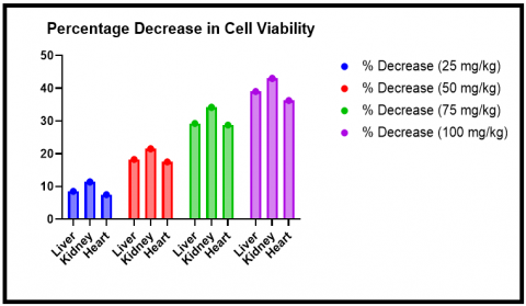
Table 9. Percentage decrease in cell viability
|
Organ |
%Decrease (25mg/kg) |
% (50mg/kg) |
% (75mg/kg) |
% 100 |
|
Liver Kidney Heart |
8.54% 11.39% 7.5% |
18.29% 21.52% 17.5% |
29.27% 34.18% 28.75% |
39.02% 43.04% 36.25% |
Note: % Decrease was calculated relative to the control group.
Figure 1. MTT assay
Figure 2. Cell viability
These results indicate that chloramphenicol exhibits a dose-dependent cytotoxic effect on the cells of the liver, kidney, and heart in nude mice, suggesting potential tissue toxicity at higher doses.
5.2 Histological findings of tissues post-chloramphenicol administration
Histopathological examination was carried out on the organ tissues harvested from the experimental and control groups. The results are summarized in the tables below.
In all the organs analysed, histological examinations showed a dose-dependent rise in tissue damage.
There was an apparent rise in tissue degeneration, necrosis, and inflammation with increasing chloramphenicol dosages. The liver, heart, and kidney were the organs that were most seriously impacted.
The types of cellular damage that occur are complete loss of cellular structure, necrosis or cell death, and inflammatory cell infiltrates. The increase in tissue degeneration, necrosis and inflammation suggests a progressive detrimental effect on organs as the dose of chloramphenicol increases.
The histology findings further support the dose-dependent harmful effects of chloramphenicol in vivo, which are consistent with the outcomes of the MTT cytotoxicity assay. A thorough understanding of the tissue-level effects of chloramphenicol administration is given by both cell viability assays and histopathology analyses, emphasising possible hazards related to its usage at high doses.
5.3 Analysis of observed toxicological consequences in great detail
Identified certain trends in relation to organ-specific cytotoxicity and histological damage after conducting a more thorough examination of the toxicological consequences of chloramphenicol treatment, as shown in Figures 3-7.
Table 10. Average histological damage score
|
Organ |
Avg. Score (Control) |
Avg. Score (25mg/kg) |
Avg. Score (50mg/kg) |
Avg. Score (75mg/kg) |
Avg. Score (100mg/kg) |
|
Liver Kidney Heart |
0.0±0.0 0.0±0.0 0.0±0.0 |
1.0±0.2 1.2±0.3 1.0±0.2 |
2.0±0.4 2.3±0.5 2.0±0.4 |
3.0±0.5 3.3±0.6 3.0±0.5 |
3.5±0.7 4.0± 0.8 3.6±0.7 |
(Note: Data are presented as mean ± standard deviation)
Table 11. Percentage of mice with severe damage (Score 4)
|
Organ |
Avg. Score (Control) |
Avg. Score (25mg/kg) |
Avg. Score (50mg/kg) |
Avg. Score (75mg/kg) |
|
Liver |
0.0±0.0 |
1.0±0.2 |
2.0±0.4 |
3.0±0.5 |
|
Kidney |
0.0±0.0 |
1.2±0.3 |
2.3±0.5 |
3.3±0.6 |
|
Heart |
0.0±0.0 |
1.0±0.2 |
2.0±0.4 |
3.0±0.5 |
Note: % Severe Damage refers to the percentage of mice that scored 4 (very severe damage) on the histological evaluation scale.
Table 12. Comparative analysis of cytotoxicity and histological damage
|
Organ |
Avg. Decrease in Cell Viability (at 100mg/kg) |
Avg. Histological Damage Score (at 100mg/kg) |
|
Liver Kidney Heart |
39.02% 43.04% 36.25% |
3.5±0.7 4.0±0.8 3.6±0.7 |
As shown in Table 10, the kidney responded to chloramphenicol treatment with the most significant cytotoxic and histological effects. Due to its function in drug excretion, these data imply that the kidney may be the main site of Chloramphenicol poisoning.
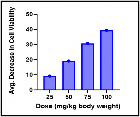
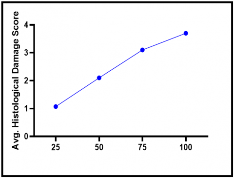
We also looked at the connection between the chloramphenicol dosage and the severity of the toxicological effects that were noticed. The results in Tables 11-13 demonstrate a definite dose-dependent trend.
Table 13. Dose-dependent toxicological effects of chloramphenicol
|
Dose (mg/kg Body Weight) |
Avg. Decrease in Cell Viability |
Avg. Histological Damage Score |
|
25 50 75 100 |
9.14% 19.10% 30.73% 39.44% |
1.07±0.23 2.10±0.44 3.10±0.53 3.70±0.73 |
Note: Data are presented as the average across all three organ types.
Figure 3. Avg. decrease in cell viability
Figure 4. Avg. Histological damage score in cell viability
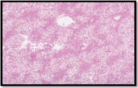
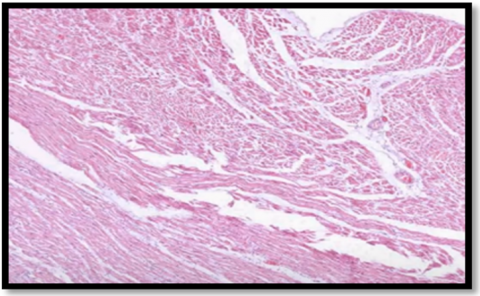
Figure 5. Histopathological changes in organs liver
Figure 6. Histopathological changes in organs kidney
Figure 7. Histopathological changes in organs heart
These findings offer compelling proof that the toxicity of chloramphenicol rises proportionately with the dose.
This has important implications for the administration of chloramphenicol in clinical and veterinary settings and emphasizes the need for careful dosage management to reduce any potential side effects.
The current study aimed to assess the toxicological and histopathological consequences of chloramphenicol in vivo using a naked mouse model [5]. We focused on cytotoxicity assessment using the MTT assay and in-depth histological analysis of key organs including the liver, kidney, and heart.
Chloramphenicol dramatically decreased cell viability at increasing doses, as seen by the findings of the MTT cytotoxicity experiment. At the highest dose of 100 mg/kg body weight, cell viability in the tissues of the liver, kidney, and heart decreased by 39.02 percent, 43.04 percent, and 36.25 percent [15]. These findings show the chloramphenicol's dose-dependent cytotoxicity, with higher doses significantly lowering cell viability in all organ tissues under study. The highest amount of cytotoxicity was found in the kidney, which is crucial for the metabolism and excretion of chloramphenicol and is hence more vulnerable to its negative effects [16-18].
Additionally, the histological study showed that the administration of chloramphenicol resulted in severe organ damage. For the liver, kidney, and heart, the average histological damage scores at the maximum dosage were 3.5, 4.0, and 3.6, respectively, on a scale where 4 denotes very serious damage [6, 7].
This dose-dependent histological damage demonstrates that chloramphenicol, especially at high doses, can cause significant tissue damage and confirms the cytotoxicity findings. The kidney was once more the organ most severely harmed, demonstrating its susceptibility to damage brought on by chloramphenicol [19-21].
It is significant to remember that the level of cytotoxicity and histological damage was proportional to the amount of chloramphenicol delivered [8-10]. Clinicians and veterinary professionals who prescribe this antibiotic must be aware of the adverse effects' dose dependence.
Chloramphenicol has shown to be beneficial against a number of bacterial illnesses, but the side effects that have been seen at larger doses highlight the need for careful dosage regulation [14, 22-25].
The long-term use of chloramphenicol raises concerns because of the documented cytotoxic and histopathological effects, particularly in the setting of chronic therapy when the compounding effects could cause serious organ damage. Despite the fact that this study used a mouse model, the results could nevertheless have a bearing on human medicine and call for more research [11-13].
A safe alternative to the antibiotic Chloramphenicol is the class of antibiotics known as Cephalosporins. One commonly used Cephalosporin antibiotic is ceftriaxone.
It has a different chemical structure and mechanism of action compared to Chloramphenicol. They inhibit bacterial cell wall formation, leading to bacterial cell death. Serious adverse effects are rare, but like any antibiotic, it can be associated with allergic reactions.
The current study shows that chloramphenicol causes severe histological damage to vital organs such the liver, kidney, and heart in a nude mouse model and demonstrates dose-dependent cytotoxicity.
The results raise questions regarding the possible harmful implications of using chloramphenicol, especially at high doses or for an extended period of time.
To comprehend the mechanisms underlying these harmful consequences and to create strategies for reducing such hazards in both clinical and veterinary settings, more study is required.
Future studies could focus on identifying the precise molecular pathways involved in Chloramphenicol-induced cytotoxicity and tissue damage.
Additionally, research should be directed towards exploring potential protective agents or strategies that could mitigate the observed toxic effects. Lastly, similar studies could be performed using other animal models or different antibiotics for comparative purposes.
The study demonstrates that chloramphenicol causes severe histological damage in vital organs such as liver, kidney, and heart in a nude mouse model. This finding provides direct evidence of the toxic effects of chloramphenicol on these organs.
The study demonstrated that the toxicity of chloramphenicol depends on the dose administered. The dose-response relationship highlights the importance of considering appropriate dosing regimens to minimize potential harm.
These findings underscore the urgent need for further research to fully understand the underlying mechanisms and explore potential protective strategies.
Future research should aim to identify the precise molecular pathways associated with chloramphenicol-induced cytotoxicity and tissue damage.
These studies will aid clinical decision making by providing a comprehensive understanding of the potential benefits and risks associated with the use of chloramphenicol, ultimately improving patient outcomes.
We thank the members of our research team for their invaluable contributions to this study. We are grateful to our institution for providing the necessary resources and support. We also appreciate the efforts of the animal care staff in ensuring the welfare of the experimental subjects.
[1] Acred, P., Brown, D.M., Turner, D.H., Wilson, M.J. (1962). Pharmacology and chemotherapy of ampicillin-a new broad‐spectrum penicillin. British Journal of Pharmacology and Chemotherapy, 18(2): 356-369. https://doi.org/10.1111/j.1476-5381.1962.tb01416.x
[2] Amrevuawho, M.O., Akinyemi, A.A., Oyewusi, A.J., Bankole, O.M., EZERI, G.N.O. (2016). Effects of onion (Allium cepa) and chloramphenicol on haematological parameters, histopathology and survival of catfish Clarias gariepinus (Burchell, 1822) sub-adult infected with Pseudomonas aeruginosa. Vom Journal of Veterinary Science, 11: 1-12.
[3] Alwan, A.M., Rokaya, D., Kathayat, G., Afshari, J.T. (2023). Onco-immunity and therapeutic application of amygdalin: A review. Journal of Oral Biology and Craniofacial Research, 13(2): 155-163. https://doi.org/10.1016/j.jobcr.2022.12.010
[4] Bale, S.S., Vernetti, L., Senutovitch, N., Jindal, R., Hegde, M., Gough, A., McCarty, W.J., Bakan, A., Bhushan, A., Shun, T.Y., Golberg, I., DeBiasio, R., Usta, O.B., Taylor, D.L., Yarmush, M.L. (2014). In vitro platforms for evaluating liver toxicity. Experimental Biology and Medicine, 239(9): 1180-1191. https://doi.org/10.1177/1535370214531872
[5] Alwan, A.M., Afshari, J.T. (2022). In vivo growth inhibition of human caucasian prostate adenocarcinoma in nude mice induced by amygdalin with metabolic enzyme combinations. BioMed Research International, 2022. https://doi.org/10.1155/2022/4767621
[6] Chabane, M.A., Tir Touil, A., Khelladi, B., Meddah, B., Mokhtar, M. (2020). In vivo toxicological and microbiological activity of marrubium vulgare L. on Candida albicans isolated from Nosocomial Infections. Pharmaceutical Sciences, 26(3): 239-251. https://doi.org/10.34172/PS.2020.35
[7] Emoruwa, T.G., Omoya, F.O., Okewale, A.I., Elabiyi, M.O. (2023). Anti-Salmonella activity of Gossypium hirsutum leaf extracted with carbonated drink and its toxicological evaluation in-vivo. Microbes and Infectious Diseases, 4(3): 1022-1040. https://doi.org/10.21608/mid.2023.174718.1411
[8] Guo, X., Qian, Z., Pan, Q., Hu, Y., Mei, W., Xing, X., Yin, S., Ji, J., Zhang, K. (2023). Effects of florfenicol on intestinal histology, apoptosis and gut microbiota of Chinese mitten crab (Eriocheir sinensis). International Journal of Molecular Sciences, 24(5): 4412. https://doi.org/10.3390/ijms24054412
[9] Festing, M.F.W., Diamanti, P., Turton, J.A. (2001). Strain differences in haematological response to chloroamphenicol succinate in mice: implications for toxicological research. Food and Chemical Toxicology, 39(4): 375-383. https://doi.org/10.1016/S0278-6915(00)00149-6
[10] Festing, M.F. (2010). Inbred strains should replace outbred stocks in toxicology, safety testing, and drug development. Toxicologic Pathology, 38(5): 681-690. https://doi.org/10.1177/0192623310373776
[11] Gaikowski, M.P., Wolf, J.C., Endris, R.G., Gingerich, W.H. (2003). Safety of Aquaflor (florfenicol, 50% type A medicated article), administered in feed to channel catfish, Ictalurus punctatus. Toxicologic Pathology, 31(6): 689-697. https://doi.org/10.1080/01926230390241828
[12] Holt, D.E., Andrews, C.M., Payne, J.P., Williams, T.C., Turton, J.A. (1998). The myelotoxicity of chloramphenicol: In vitro and in vivo studies: II: in vivo myelotoxicity in the B6C3F mouse. Human & Experimental Toxicology, 17(1): 8-17. https://doi.org/10.1177/096032719801700102
[13] Holt, D.E., Ryder, T.A., Fairbairn, A., Hurley, R., Harvey, D. (1997). The myelotoxicity of chloramphenicol: In vitro and in vivo studies: I. In vitro effects on cells in culture. Human & Experimental Toxicology, 16(10): 570-576. https://doi.org/10.1177/096032719701601004
[14] Hsu, C.Y., Yang, S.C., Sung, C.T., Weng, Y.H., Fang, J.Y. (2017). Anti-MRSA malleable liposomes carrying chloramphenicol for ameliorating hair follicle targeting. International Journal of Nanomedicine, 12: 8227-8238. https://doi.org/10.2147/IJN.S147226
[15] Alwan, A.M., Afshari, J.T. (2023). Investigating the protective role of rhodanese enzyme against cyanide, the cytotoxic by-product of Amygdalin, in HDF and L929 Cell Lines. Letters in Drug Design & Discovery, 20(9): 1295-1307. https://doi.org/10.2174/1570180819666220620103339
[16] Turani, M., Banfalvi, G., Peter, A., Kukoricza, K., Kiraly, G., Talas, L., Tanczos, B., Dezso, B., Nagy, G., Kemeny-Beke, A. (2015). Antibiotics delay in vitro human stem cell regrowth. Toxicology in Vitro, 29(2): 370-379. https://doi.org/10.1016/j.tiv.2014.10.013
[17] Tan, Q., Xu, H., Aguilar, Z.P., Peng, S., Dong, S., Wang, B., Li, P., Chen, T., Xu, F., Wei, H. (2013). Safety assessment and probiotic evaluation of Enterococcus faecium YF5 isolated from sourdough. Journal of Food Science, 78(4): M587-M593. https://doi.org/10.1111/1750-3841.12079
[18] Suarez-Torres, J.D., Orozco, C.A., Ciangherotti, C.E. (2021). The numerical probability of carcinogenicity to humans of some antimicrobials: Nitro-monoaromatics (including 5-nitrofurans and 5-nitroimidazoles), quinoxaline-1, 4-dioxides (including carbadox), and chloramphenicol. Toxicology in Vitro, 75: 105172. https://doi.org/10.1016/j.tiv.2021.105172
[19] Smith, B.J., Mattison, D.R., Sipes, I.G. (1990). The role of epoxidation in 4-vinylcyclohexene-induced ovarian toxicity. Toxicology and Applied Pharmacology, 105(3): 372-381. https://doi.org/10.1016/0041-008X(90)90141-G
[20] Schmidgall, J., Schnetz, E., Hensel, A. (2000). Evidence for bioadhesive effects of polysaccharides and polysaccharide-containing herbs in an ex vivo bioadhesion assay on buccal membranes. Planta Medica, 66(01): 48-53. https://doi.org/10.1055/s-2000-11118
[21] Quadir, M., Zia, H., Needham, T.E. (1999). Toxicological implications of nasal formulations. Drug Delivery, 6(4): 227-242. https://doi.org/10.1080/107175499266823
[22] Lawrence, J.C. (1959). The comparative toxicity of antibiotics to skin. British Journal of Pharmacology and Chemotherapy, 14(2): 168-173. https://doi.org/10.1111/j.1476-5381.1959.tb01378.x
[23] Kasahara, T., Tomita, K., Murano, H., Harada, T., Tsubakimoto, K., Ogihara, T., Ohnishi, S., Kakinuma, C. (2006). Establishment of an in vitro high-throughput screening assay for detecting phospholipidosis-inducing potential. Toxicological Sciences, 90(1): 133-141. https://doi.org/10.1093/toxsci/kfj067
[24] Balocco, C., Petrone, G.L. (2021). Sustainability and wellbeing in healthcare facilities. An investigation inside a historic hospital. International Journal of Design & Nature and Ecodynamics, 16(5): 477-485. https://doi.org/10.18280/ijdne.160501
[25] Qamar, A.M., Khan, R.U., Alsuhibany, S.A. (2021). Large-scale bibliometric analysis of coronavirus. International Journal of Design and Nature and Ecodynamics, 16(5): 531-541. https://doi.org/10.18280/ijdne.160507