Encapsulation of Kecombrang (Etlingera elatior) Flowers Extract Using the Ionic Gelation Method

Sri Wahjuni*![]() | Ni Wayan Bogoriani
| Ni Wayan Bogoriani![]() | Ni Made Puspawati
| Ni Made Puspawati![]() | Ida Bagus Putra Manuaba
| Ida Bagus Putra Manuaba![]() | Ahmad Fudholi
| Ahmad Fudholi![]()
© 2024 The authors. This article is published by IIETA and is licensed under the CC BY 4.0 license (http://creativecommons.org/licenses/by/4.0/).
OPEN ACCESS
The extract flower of Etlingera elatior (kecombrang) is one of herb interested for research. This flower known to have a beneficial antioxidant activity. This antioxidant effect was obtained as a results of high flavonoid content. Photosensitivity and low bioavailability and their fast metabolism were the main limitations for its use, which can be overcome through encapsulation of the extract (flavonoids) into nanoparticle-based chitosan-tripolyphosphate by ionic gelation method. This is an observational study aims to encapsulate kecombrang flowers extract that is already form in nanoextract and assess the characterization. The nanoparticle extract of this flower obtained has a particle size of 312.7 nm with a zeta potential of -16.2 mV and a polydispersity index value of 0.502, amorphous (non-uniform) morphology, has a tendency to be spherical, and has an imperfectly spherical shape, has functional groups –OH, -NH, C=O, and a phosphate group. Overall Kecombrang flowers extract is able to be encapsulated in chitosan-tripolyphosphate.
Etlingera elatior, ionic gelation, nanoparticle
Antioxidants are substances that have the ability to either absorb or neutralize free radicals, thereby it can be applied for preventing a variety of degenerative diseases, including stroke, diabetes mellitus, cardiovascular disease, and cancer, and among others. Synthetic antioxidants are frequently utilized, but since they sometimes may have negative effects on the body, therefore, natural antioxidants derived from natural sources must be employed in their place. One natural source that may be used as a natural antioxidant is the kecombrang flower (Etlingera elatior). Secondary metabolites including phenolics, flavonoids, triterpenes, saponins, tannins, steroids, alkaloids, and glycosides are found in all parts of the kecombrang plant, including the roots, stems, leaves, and flowers. Many researchers are starting to pay attention to kecombrang flowers because of its potential as a traditional medicine through its antioxidant activity. Kecombrang flowers has a diversity of chemical compounds where the methanol extract of kecombrang flowers contains secondary metabolite compounds such as phenolic compounds and flavonoids [1-3]. Kecombrang flowers and stems are also reported to have antidiabetic and antioxidant activity in obese rats due to their secondary metabolite content [4, 5].
Flavonoids are a group of natural products that have a variety of biological activities, and are widely used as therapeutic agents to treat diseases, and play an important role in the pharmaceutical industry [6]. Several studies have shown that flavonoids are known to exhibit strong antioxidant activity [7]. Secondary metabolites, one of which is flavonoids, have low bioavailability under conditions of large particle size, fast metabolism and are unstable to the influence of high light intensity so they are easily oxidized, so one potential strategy that can be applied is to encapsulate them into nanoparticle preparations that are more easily dispersed and stable in methanol [8].
The ionic gelation method is one of the methods applied to encapsulate extracts into nanoparticles using a cross-linking mechanism between chitosan using polyanion salts such as NaTPP. Kecombrang flowers extract can have a negative charge in water because the hydroxyl groups of flavonoid compounds can hold ionic interactions with the positively charged amine groups of chitosan [9], and the addition of NaTPP which functions as a crosslinker agent to stabilize the nanoparticles formed [10].
Based on the background above, this research is conducted in order to gain kecombrang flowers extract nanoparticles by applying the ionic gelation method. In addition, this research also aims to characterize the nanoparticle obtained, in term of particle size, also their phytochemical determination was assessed. Finally, in this study an encapsulation of the nano extract was carried out.
2.1 Material
The main material used in this research was kecombrang flower (Etlingera elatior) obtained from local markets in Denpasar. The chemicals used were aqua DM, glacial acetic acid, chitosan, sodium tripolyphosphate (NaTPP), 10% sodium hydroxide (NaOH), concentrated sulfuric acid (H2SO4), concentrated hydrochloric acid (HCl), and magnesium powder.
2.2 Equipment
The equipments used in this research included a set of maceration containers, filter paper, spatula, magnetic stirrer, centrifugation, rotary evaporator (Buchi), particle size analyzer (PSA) of the Zetasizer Advanced Pro Blue Mlvern brand, scanning electron microscope (SEM) of the JSM 6510LA, infrared spectrophotometer (IR) of the Shimadzu IRPrestige-21, and a set of laboratory glassware.
3.1 Sample extraction
As usual, a standard procedure was carried to determine the water content of the kecombrang flower. For extraction, a number of 800 g of Kecombrang flowers powder with a water content of 7.56% was extracted by maceration using methanol solvent. Maceration was carried out overnight for four times. After macerated along overnight, a brownish yellow filtrate of 8.0 L was obtained. The filtrate was then evaporated using a press dryer produced a crude product ready for continues step.
3.2 Preparation of kecombrang flower nanoparticles
The encapsulation process was carried out initially by preparing 0.5% chitosan solution in 1% glacial acetic acid and 1% NaTPP solution in aquadest. Then 50 mL of 0.5% chitosan solution in a beaker glass was added with 50 mL of 1% NaTPP solution drop by drop, resulting in an observable precipitation indicating that nano chitosan-NaTPP was formed. Then methanol extract of kecombrang flower (2%) was added to the nano chitosan-NaTPP drop by drop, and stirred using a magnetic stirrer at a speed of 3800 rpm for 120 minutes. The formed nanoparticle colloids were separated by centrifugation for 15 minutes at a speed of 3000 rpm. The formed nanoparticle precipitate was dried in the refrigerator until kecombrang flower nanoparticle crystal was obtained. The nanoparticle crystal was then analyzed using FTIR, SEM, and PSA to determine the characteristics of the nano encapsulant.
3.3 Phytochemical test
A total of 3 pairs of test tubes were prepared. A pair of test tubes contained as much as 2.0 mL solution of kecombrang flower extract and 2.0 mL solution of kecombrang nanoparticles in each separate tube. In the first pair of test tubes, 3 drops of concentrated HCl and a little magnesium powder were added in each separate tube, in the second pair, 3 drops of concentrated H2SO4 were added and heated for 15 minutes, and in the third pair, 3 drops of 10% NaOH were added. The occurring color changes were then observed and recorded.
4.1 Plant anatomical determination
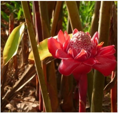
The sample used in this research was kecombrang flowers obtained from Klaci Marga Village, Tabanan, Bali-Indonesia. The plant determination process was carried out at the LIPI-UPT Plant Conversion Center of the Bali "Eka Karya" Botanical Garden. Kecombrang plant as can be seen in Figure 1, is a plant traditionally in Indonesia used as an herb or food sour. The anatomical determination of this plant was presented as follow: Kingdom: Plantae; Subkingdom: Spermatophyta; Division: Magnoliophyta; Classs: Liliopsida; Subclass: Commelinidae; Ordo: Zingiberales; Familiae: Zingiberaceae; Genus: Etlingera; and Species: Etlingera elatior (Jack) R.M.Sm.
Figure 1. Kecombang plan
4.2 Simplicia extraction
A number of 800 g of kecombrang flowers powder was macerated with methanol at a temperature of 70-80℃, in order to maximize the maceration process due to higher temperature of the solvent leads to the plant tissue damage more quickly. Finally, the bioactive compounds contained can be more quickly solved by the solvent [11, 12]. The macerate obtained is then evaporated using a press dryer to remove the maceration solvent. The crude extract obtained was 45.32 g.
4.3 Phytochemical test
The results of phytochemical tests using 10% Sodium Hydroxide (NaOH), Bate Smith-Metcalfe and Wilstater reagents produced a specific color in the kecombrang flowers extract and nanoextract of kecombrang flowers. All of phytochemical tests were presented in Table 1. The kecombrang flowers crude extract obtained was yellow in color reacted with 10% NaOH reagent, red with the reagent Bate Smith-Metcalfe and Wilstater's reagent. In the nanoextract of kecombrang flowers, a yellow-orange color was obtained with 10% NaOH, with Bate Smith-Metcalfe and Wilstater reagents yellow. From these results, it can be seen that the most obvious intensity of color change is produced by the nano extract of kecombrang flowers, so it can be said to have a higher flavonoid content than the extract of kecombrang flowers [10].
Table 1. Phytochemical test results of kecombrang flowers extract and nanoextract
|
Fraction |
Color Test |
Remark |
||
|
NaOH 10% |
Bate smith-Metcalfe |
Wilstater |
||
|
Extract |
Yellow |
Red |
Red |
++ |
|
Extract Nano |
Yellow-orange |
Yellow |
Yellow |
+++ |
4.4 Kecombrang flowers extract nanoparticles
Kecombrang flower nanoparticles were made using the ionic gelation method, namely polyelectrolyte complexation between positively charged chitosan and negatively charged tripolyphosphate. This method was chosen because the process is simpler, avoids the use of high temperatures, and can be controlled easily [13]. The bioactive substances contained in the kecombrang flower extract were cross-encapsulated in chitosan-tripolyphosphate nanoparticles. Chitosan is dissolved in a dilute acid solution to obtain chitosan cations, namely the amine group. The positively charged amine group will cross-link with the negative group of NaTPP polyanion to form a complexation between these different charges which causes the resulting chitosan nanoparticles to be more stable [14].
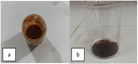
The NaTPP polyanion is formed because it dissociates in water releasing sodium ions and tripolyphosphate ions which then react with NH3+ from chitosan. NaTPP salt is used to function as a cross-linking agent, because this material tends to have non-toxic properties, so this additional material has been approved by the United States Food and Drug Administration [10]. The addition of the NaTPP solution drop by drop accompanied by stirring using a magnetic stirrer aims to prevent solidification so that the resulting nanoparticles do not form large lumps, as well as to obtain a constant stirring speed [13]. The nanoparticles obtained can be seen in Figure 2.
Figure 2. Kecombrang flowers nanoparticle (a), kecombrang flowers (b)
4.5 Characterization results with Particle Size Analyzer (PSA)
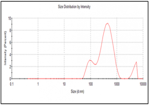
Particle Size Analyzer (PSA) is used to see whether a formula has become a nanoparticle which can be determined from the particle size distribution, zeta potential value and polydispersity index. The test was carried out 3 times, the resulting particle size was 312.7 ± 3.09 nm as presented in Figure 3.
Figure 3. Size of kecombrang flowers nanoextract particles
According to Umair et al. [15] a size of 200 to 300 nm is considered suitable for crossing biological barriers, avoiding the RES (Reticulo Endothelial System), and avoiding glomerular filtration. Sizes up to 400 nm are also considered acceptable (evaluated in the case of liposomes) [16]. A study reported a suitable size for the permeability and retention (EPR) effect which is in the range of 10–500 nm [17].
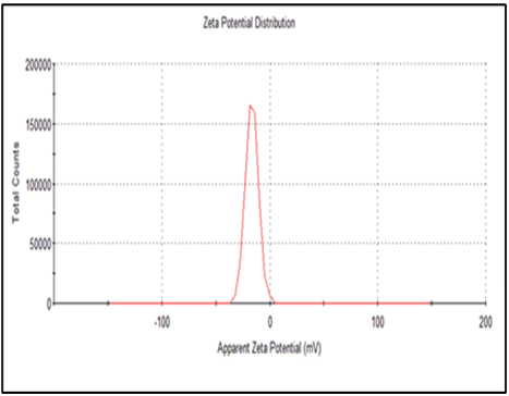
The zeta potential value obtained from kecombrang flowers extract nanoparticles is -16.1 mv which provides short-term stability as can be depicted from Figure 4. As a rule of thumb, zeta potential values of ±30 mV will provide good stability and ±60 mV has excellent stability.
Figure 4. Zeta potential value of kecombrang flowers nanoextract
The average value of the polydispersity index (PI) obtained was 0.502, which indicates that the particle size is well controlled with narrow dispersion. PI is a measure of particle homogeneity, ranging from 0 to 1, where the closer the PI value is to zero, the more homogeneous the system becomes (mono-dispersed) while the higher the PI value produced, the more unstable the formula.
4.6 Characterization results using Scanning Electron Microscopy (SEM)
Scanning Electron Microscopy (SEM) is a material characterization technique that is widely used to view the surface morphology of particles down to 1 nm in size. The SEM test results of agarwood leaf nanoextract can be seen in Figure 5 has a tendency to be spherical, and there is an imperfect round shape. Spherical shaped nanoparticles have greater cellular absorption than rod shaped ones. The less spherical shape of the nanoparticles will facilitate contact between particles, leading to aggregation [17]. The instrument SEM is also equipped with EDX (Energy Dispersive X-Ray).
EDX is a detector found in SEM tools. EDX is used to analyze the chemical composition on the surface of the sample.
The chemical composition based on EDX in Table 2, is known to consist of 43.62% oxygen (O), 24.53% carbon (C), 18.15% sodium (Na) and 6.66% nitrogen (N). as the main component. This indicates the formation of nanoparticles in the presence of chitosan biopolymer, NaTPP, and the active compound contained in the ethylacetate extract. Other elements were found in small amounts such as P, K, Si, Al, while the element hydrogen (H) was not detected on EDX. This is most likely because the hydrogen element is lighter than other elements, so it is above the surface and cannot be detected by instruments [18, 19].
Figure 5. The result SEM particle nano kecombrang flowers with Electron Microscopy results of combrang flower nanoparticles with a magnification of 7500x (a), magnification of 1000x (b)
Table 2. Surface chemical composition of nanoparticles kecombrang flowers extract
|
Atom Number |
Element Symbol |
Element Name |
% Atom Content |
% Total Weight |
|||||
|
8 |
O |
Oxygen |
43.62 |
37.56 |
|||||
|
6 |
C |
Carbon |
24.53 |
15.86 |
|||||
|
11 |
Na |
Sodium |
18.15 |
22.45 |
|||||
|
7 |
N |
Nitrogen |
6.66 |
11.11 |
|||||
|
15 |
P |
Phosphorus |
3.46 |
6.45 |
|||||
|
19 |
K |
Potassium |
1.97 |
4.14 |
|||||
|
14 |
Si |
Silicon |
1.50 |
2.27 |
|||||
|
13 |
Al |
Aluminium |
0.11 |
0.16 |
|||||
4.7 Characterization results with Fourier Transform Infra Red (FTIR)
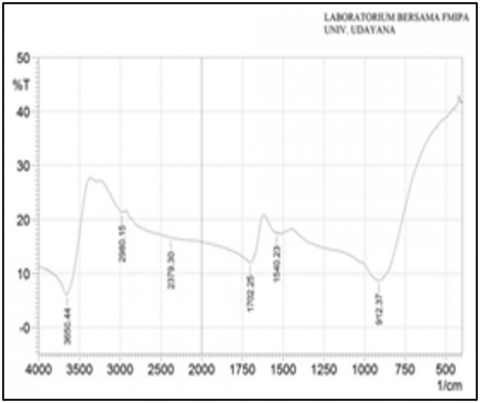
Characterization of kecombrang flowers extract nanoparticles using FTIR serves to determine change in functional groups that indicate chemical interactions. The spectrum result of chitosan,methanol extract and n-heksan extract nanoparticles of kecombrang flowers using FTIR are shown in Figure 6, Figure 7 and Figure 8, indicates that kecoombrang flowers extract nanoparticles have been formed using the ionic gelation method.
Figure 6 shows the stretching vibration of the O-H group in chitosan with a wave number of 3563 cm-1 and the stretching vibration of the N-H group appears at a wave number of 1639 cm-1. In the FTIR results of nanoparticles of kecombrang flower extract, the wave number of N-H has shifted to 1540 cm-1. This wave number shift is thought to be due to the interaction between chitosan and kecombrang flower extract, namely cross-linking between the NH2 group of chitosan and Na-TPP as well as interactions with the –OH group phenolics from flavonoid compounds in kecombrang flower extract [1, 4]. The discovery of a wave number of 912 cm-1 in nanoparticles indicates the presence of a phosphate group which is a typical group of NaTPP [18]. In the spectrum of kecombrang flower extract, the O-H group was detected at a wave number of 3658 cm-1, whereas in the spectrum of kecombrang flower nanoparticles extract, it shifted to 3650 cm-1. This is due to the interaction between the -OH group in chitosan and the phenolic -OH group from the flavonoid compounds in kecombrang flower extract [14]. Absorption at a wave number of 1700 cm-1 with weak intensity indicates the presence of the C=O group [20].
Figure 6. FTIR spectrum of chitosan
Figure 7. FTIR spectrum kecombrang flowers extract
Figure 8. FTIR spectrum of kecombrang flowers nanoextract
Chitosan-tripolyphosphate-Kecombrang flowers extract nanoparticles were successfully made using the ionic gelation method with a particle size of 312.7 ± 3.09 nm. The color change in the results of the phytochemical test showed that the flavonoid content in the nanoextract of the kecombrang flower was higher than that of the kecombrang flower extract. Further research needs to be carried out to increase the stability of kecombrang flowers nanoextract produced with the addition of surfactants.
[1] Bogoriani, N.W., Sri, W., Maria, K.E., (2023). Antioxidant activity of kecombrang flowers (Etlingera elatior) methanol extract and identification of its compound using LCMS/MS. Indonesia Journal of Biomedical Science (IJBS), 17(1): 172-177. https://doi.org/10.15562/ijbs.v17i1.480
[2] Agustiarini, V., Wijaya, D.P. (2022). Uji aktivitas antioksidan ekstrak etanol-air (1:1) bunga rosella (Hibiscus Sabdariffa L.) dengan metode DPPH (1,1-difenil-2-pikrilhidrazil). Jubnal Puneittian Satns, 24(1): 29-32. http://doi.org/10.56064/jps.v24i1.679
[3] Parwata, I.M.O.A., Kusuma, I.N.A., Dewi, I.G.A.K.S. P. (2022). Kadar total flavonoid dan uji aktivitas antioksidan fraksi etil asetat daun gaharu (Gyrinops Versteegii). Jurnal Kimia (Journal of Chemistry), 16(1): 20-25. https://doi.org/10.24843/JCHEM.2022.v16.i01.p03
[4] Sukadana, I.M., Bogoriani, N.W., Ariani,M. (2023). Compounds in the stem of Etlingera elatior can reduce the levels of free fatty acid and Blood Glucose in Obesity Wistar Rats. Research Journal of Pharmacy and Technology, 16(10): 4530-4536. https://doi.org/10.52711/0974-360X.2023.00738
[5] Bogoriani, N.W., Ariati K., and Pratiwi, I G.A.P.E. (2022) Potenccy of balinese kecombrang (Etlingera elatior) extract as antioxidant against the activity of superoxide dismutase (SOD), glutathione (GSH) and fatty liver in obese rats. Biomedical and Pharmacology Journal, 15(1): 337-344. https://doi.org/10.13005/bpj/2372
[6] Obefemi, T.O., Akinmoladun, A.C., Olaleye, M.T., Agboade, S.O., Onasanya, A.A. (2017). Antidiabetic potential of methanolic and flavonoid-rich leaf extracts of Synsepalum dulcificum in type 2 diabetic rats. Journal of Ayurveda and Integrative Medicine, 8(4): 238-246. https://doi.org/10.1016/j.jaim.2017.01.008
[7] Liu, Q., Jing, Y., Han, C., Zhang, H., Tian, Y. (2019). Encapsulation of curcumin in zein/ caseinate/sodium alginate nanoparticles with improved physicochemical and controlled release properties. Food Hydrocolloids. 93(2): 432-442. https://doi.org/10.1016/j.foodhyd.2019.02.003
[8] Detsi, A., Kavetsou, E., Kostopoulou, I., Pitterou, I., Pontillo, A.R.N., Tzani, A., Christodoulou, P., Siliachli, A., Zoumpoulakis, P. (2020). Nano systems for the encapsulation of natural products: the case of chitosan biopolymer as a matrix. Pharmaceutics, 12(669): 1-48. https://doi.org/10.3390/pharmaceutics12070669
[9] Kafshgari, M.H., Khorram, M., Khodadoost, M., and Khavari, S. (2011). Reinforcement of chitosan nanoparticels obtained by an ionic crosslinking process. Iranian Polymer Journal, 20(5): 445-456.
[10] Parwata, I.M.O.A., Putra Manuaba, I.B., Putu Sutirtayasa, I.W. (2018). Gaharu Leaf Water Extract Reduce MDA and 8-OHdG Levels and Increase Activities SOD and Catalase in Wistar Rats Provided Maximum Physical Activity. Bali Medical Journal, 5(3): 79-83.
[11] Vilmakumar, C.S., Hosagaudar, V.B., Suja, S.R., Vilash, V., Krishnakumar, N.M., Latha, P.G. (2014). Comparative preliminary phytochemical analysis of (ethanolic extracts of leaves of Olea dioica Roxb., infected with the rust fungus Zaghouania oleae (E.J. Butler) Cummins and non-infected plants. Journal of Pharmacognosy and Phytochemistry, 3(4): 69-72.
[12] Kurniasari, D., Atun, S. (2017). Pembuatan dan karakterisasi nanopartikel ekstrak etanol temu kunci (Boesnbergia pandurata) pada berbagai variasi komposisi kitosan. Jurnal Sains Dasar, 6(1): 31-35. https://doi.org/10.21831/jsd.v6i1.13610
[13] Putri, A.I., Sundaryono, A., dan Candra, I.N. (2018). Karakterisasi Nanopartikel Kitosan Ekstrak Daun Ubijalar (Ipomoea batatas L.) Menggunakan Metode Gelasi Ionik. Jurnal Pendidikan dan Ilmu Kimia. 2(2): 203-207. https://doi.org/10.33369/atp.v2i2.7561
[14] Suryadi, Y., Susilowati, D.N., Samudra, I.M. (2021). Antifungal activity of chitosan-tripolyphosphate formula to Colletotrichum spp. Infection on Chilli. Jurnal Riset Sains dan Teknologi. 5(2): 2549-9750. https://doi.org/10.30595/jrst.v5i2.10225
[15] Umair, M., Javed, I., Rehman, M. (2016). Nanotoxicity of inert materials: the case of gold, silver and iron. Journal of Pharmacy & Pharmaceutical Sciences, 19(2): 161-180. https://doi.org/10.18433/J31021
[16] Alasvand, N., Urbanska, A.M., Rahmati, M., Saeidifar, M., Gungor-Ozkerim, P.S., Sefat, F., Rajadas, J., Mozafari, M. (2017). Therapeutic nanoparticles for targeted delivery of anticancer drugs. Multifunctional Systems for Combined Delivery, Biosensing and Diagnostics, 245-259. https://doi.org/10.1016/B978-0-323-52725-5.00013-7
[17] Wu, J., Wang, Y., Yang, H., Liu, X., Lu, Z. (2017). Preparation and biological activity studies of resveratrol loaded ionically cross-linked chitosan-TPP nanoparticles. Carbohydrate Polymers, 175: 170-177. https://doi.org/10.1016/j.carbpol.2017.07.058
[18] Oksal, E., Pangestika, I., Muhammad, T.S.T., Mohamad, H., Amir, H., Kassim, M.N.I., Andriani, Y. (2020). In vitro and in vivo studies of nanoparticles of chitosan-Pandanus tectorius fruit extract as new alternative treatment for hypercholesterolemia via Scavenger Receptor Class B type 1 pathway. Saudi Pharmaceutical Journal, 28(10): 1263-1275. https://doi.org/10.1016/j.jsps.2020.08.017
[19] Hakim, A.R. (2018). Kajian produksi nanopartikel dari arang akasia dengantumbukan bola baja diameter 5/16,1/4,3/16,5/32 inchi. Publikasi Ilmiah Universitas Muhammadiyah Surakarta, 3(2): 1-13. http://eprints.ums.ac.id/id/eprint/66778.
[20] Sastrohamidjojo, H. (2007). Spektroskopi. Liberty. Yogyakarta.