Zulkaidhah Zulkaidhah*![]() | Wardah Wardah
| Wardah Wardah![]() | Rukmi Rukmi
| Rukmi Rukmi![]() | Abdul Hapid
| Abdul Hapid![]() | Dewi Wahyuni
| Dewi Wahyuni![]() | Hamka Hamka
| Hamka Hamka![]()
© 2024 The authors. This article is published by IIETA and is licensed under the CC BY 4.0 license (http://creativecommons.org/licenses/by/4.0/).
OPEN ACCESS
The Nutmeg tree (Myristica fragrans Houtt.) serves as a Multi-Purpose Tree Species (MPTS) in high demand due to its multitude of applications. Despite its potential, Indonesia's nutmeg productivity lags behind the global average, with a yield of only 98.9 kg per hectare. Among the various impediments to productivity, the limited expertise of local farmers in nutmeg cultivation and the prevalence of leaf diseases in nutmeg seedlings in nurseries are paramount. Leaf diseases can be lethal to the seedlings and significantly impact their quality, which, in turn, affects the growth and productivity of the mature plants. This study was conducted to assess the prevalence of leaf diseases in nutmeg nurseries and identify the causal pathogens. The average percentage of damage across all disease types was found to be as follows: leaf spot (3.95%), leaf blight (4.42%), leaf rust (7.27%), and powdery mildew (1.025%). Pathogenic fungi were identified as the causative agents, with Nigrospora sp. causing leaf spot, Rhizoctonia sp. causing leaf blight, Oidium tingitanium causing powdery mildew, and Pestalotia sp. causing leaf rust. The overall average intensity of pathogen attack was 6.52%, classified in the mild damage category. Our findings suggest that fungal pathogens predominantly cause leaf diseases in nutmeg seedlings. Therefore, effective microclimate management strategies should be adopted to mitigate the impacts of these diseases in nutmeg nurseries.
nutmeg seedlings, leaf disease, percentage of attack, attack intensity
Nutmeg (Myristica fragrans Houtt.) is an evergreen, medium-stemmed plant, capable of reaching up to 18 meters in height [1]. This perennial species, with a lifespan exceeding a century, has been identified as a multipurpose tree species. It is increasingly sought after for its essential oil, spice, and wood [2, 3]. While Java Island remains the primary cultivation hub for nutmeg in Indonesia, a steady initiation of production has been observed on Sulawesi Island [4].
Among the various nutmeg types found in Indonesia, Myristica fragrans Houtt. stands out as the dominant species, both in terms of quality and productivity [5]. The plant reaches its peak production within a span of 60 - 70 years. Despite Indonesia's status as one of the world's leading producers and exporters of nutmeg, its productivity lags considerably behind the global average, yielding only 98.9 kg per hectare compared to the worldwide average of 451 kg per hectare [6]. Disease infestation in nutmeg plants has been identified as a significant contributor to this sub-optimal productivity. Common diseases include white root fungus (caused by Rigidoporus lignosus) and fruit rot (caused by Stigmina myristicae) [3, 7, 8].
Disease manifestations are not confined to field cultivation but can also inflict damage in nurseries, significantly impacting the quality of seedlings and subsequent plant growth [5, 9]. Leaf diseases, including sooty dew, powdery mildew, leaf blight, leaf rust, and anthracnose, are commonly observed in seedlings [10]. Reports from West Java indicate that necrotic spot, light leaf spot, and sooty mildew are the most prevalent foliar diseases in nutmeg seedlings [11].
The types of diseases that generally attack the leaves are diseases that are easily transmitted in a relatively fast time so that almost 75% of the nutmeg seedlings are affected, ranging from mild to severe symptoms and many even cause the death of the seedlings. This condition can cause problems because it can cause quite large economic losses. Low productivity due to disease infections is currently an obstacle to nutmeg production [9, 11].
To effectively mitigate disease attacks on nutmeg seedlings in nurseries, initial efforts should focus on identifying the pathogens responsible and assessing the extent of the damage [10, 12]. In light of this, the present study aims to ascertain the prevalence of leaf diseases in nutmeg nurseries and to identify the causative pathogens.
2.1 Research sites
The research was carried out in the Nutmeg Plant Nursery, Tropical Plants Field Laboratory, Palu, Central Sulawesi, Indonesia. Disease identification activities were carried out at the Laboratory of Plant Pest and Disease, Faculty of Agriculture, Universitas Tadulako, Indonesia.
2.2 Identification of the causes of nutmeg disease
Each nutmeg nursery block was divided into 3 equal-sized sections to serve as disease observation sites. In each section, 3 symptomatic seedlings were purposely taken (2 from the edge and 1 from the center). Observations were made by looking at the signs and symptoms that appeared on the leaves. The collection of symptomatic material (leaves) is needed as a source of inoculum for pathogen identification.
Observations in the laboratory were made by observing the leaves of symptomatic plants on Potato Dextrose Agar (PDA) media. Isolation begins with planting diseased leaf tissue. Tissues/leaf parts were taken at the border of healthy and diseased and cut into small pieces. then put in 3% sodium hypochlorite solution for 2 minutes and cleaned with sterile water. After drying, it was transferred into a petri dish containing PDA. Pathogens that appear are observed and purified by transferring to another petri dish containing PDA, until a pure culture is obtained. Pure cultures obtained were identified macroscopically and microscopically.
Pathogen identification was carried out by matching and comparing the characteristics seen in the growth of pathogens with the characteristics in existing references [13-15]. Pathogens obtained from pure culture were described macroscopically and microscopically. Macroscopically observed were the growth rate of colonies in petri dishes and the color of the colonies. Microscopic observation by looking at the shape of the pathogen which includes hyphae shape, hyphae color, spore shape and spore box shape as an object of identification [16].
2.3 Data analysis
Data analysis was carried out by calculating the percentage of incidence of each type of disease on nutmeg plant seedlings. The equation is as follows [17]:
Disease Incidence $=\frac{\text { The number of diseased plants }}{\text { The total number observed }} \times 100 \%$
Meanwhile, the severity caused by the leaf disease attacks on nutmeg plant seedlings is determined based on the intensity of the disease. The intensity is calculated using the following formula:
Disease Intensity $==\frac{\mathrm{X} 1 \mathrm{Y} 1+\mathrm{X} 2 \mathrm{Y} 2+\mathrm{X} 3 \mathrm{Y} 3+\mathrm{X} 4 \mathrm{Y} 4}{\mathrm{XY} 4} \times 100 \%$
X=The total number of plants observed.
X1-X4=The total number of diseased plants – mild damage (score 1) to dead plants (score 4).
Y1-Y4=Score of plants – mild damage (score 1) to dead plants (score 4).
The category of the disease severity is determined based on the disease intensity. It is presented in Table 1 below.
Table 1. The category of severity based on the disease intensity on stands
|
Intensity (%) |
Condition of Stands |
|
0 – 1 |
Healthy |
|
> 1 – 25 |
Mildly damaged |
|
> 25 – 50 |
Moderately damaged |
|
> 50 – 75 |
Heavily damaged |
|
> 75 – 100 |
Extremely damaged or dead |
The results of the calculation of attack intensity were tested using analysis of variance with 3 replications. If the results show a real effect, it will be continued with the honest real difference test (BNJ). Statistical analysis was carried out using Statistical Package for Social Sciences (SPSS) for windows 16.0 [18-20].
3.1 Types of plant leaf disease and pathogenic fungi
The identification of plant leaf diseases on nutmeg seedlings in the nursery indicated four types of diseases were found in the four observation plots. They include leaf spots, blight, powdery mildew, and rust. The types of plant leaf fungal diseases and related information are presented in Table 2.
Table 2. Types of plant leaf disease and pathogenic fungi
|
No. |
Observation Plots |
Disease |
Pathogenic Fungi |
Incidence (%) |
|
1 |
Plot 1 |
· Leaf Spot |
Nigrospora sp. |
3.3 |
|
· Blight |
Rhizoctonia sp. |
5.1 |
||
|
· Powdery mildew |
Oidium tingitanium |
1.5 |
||
|
2 |
Plot 2 |
· Rust |
Pestalotia sp. |
14.0 |
|
· Blight |
Rhizoctonia sp. |
1.0 |
||
|
· Leaf Spot |
Nigrospora sp. |
3.8 |
||
|
3 |
Plot 3 |
· Leaf Spot |
Nigrospora sp. |
8.7 |
|
· Powdery mildew |
Oidium tingitanium |
2.6 |
||
|
4 |
Plot 4 |
· Blight |
Rhizoctonia sp. |
11.6 |
|
· Rust |
Pestalotia sp. |
15.1 |
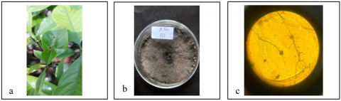
The observations of plant leaf diseases on nutmeg seedlings in the nursery reveal that the leaf spot caused by Nigrospora sp. is identified from the symptoms of black-edged lesions and brown spots with yellow halos. The sites are irregular on both upper and lower leaf surfaces (Figure 1). The results of macroscopic observations indicate that initially, the colony is white, and it turns grey to dominant black as the infection progresses. Meanwhile, the rough and thick colonies generally have a diffuse distribution pattern without concentric rings. The microscopic observations of Nigrospora sp. indicate that the hyphae are septate and brown, and conidiophores are short, simple and brown. At the same time, conidia are black, globose, 7.56µm in length and 5.31µm in width. According to the study by Barnett and Hunter [15], conidiophores are simple and short, and conidia are globose and black. The symptoms and growth of Nigrospora sp. are illustrated in Figure 1.
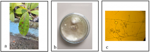
Rust disease on nutmeg seedlings caused by Pestalotia sp. is indicated by irregular yellow-brown spots. Subsequently, the spots enlarge and merge into blotches that cover the leaf surface, partially or entirely (Figure 2). In severe symptoms, the leaves experience chlorosis, namely premature yellowing of leaves in which the plant eventually dies [21-23]. The isolate of Pestalotia sp. is identified from its morphological characteristic: white cottony mycelium that turns light grey when mature. Similarly, study [24] also reported that the mycelium of Pestalotia sp. is white, like a cotton ball. The microscopic observation shows that the conidia are oval to elliptical with one slightly tapered end, and the other has three to five hair-like structures or whip feathers. According to the study by Barnett and Hunter [15], conidiophores consist of several dark-colored cells and a pointed tip of hyaline, while the tip of the cell contains two or more protrusions. At the tip of the conidiophores, there are antenna-like structures of 25-40 m in length and 2-3 in number [25]. The symptoms and growth of Pestalotia sp. are presented in Figure 2.
Figure 1. Leaf spot on nutmeg leaf (a) A symptom identified on diseased plant, (b) Isolate of Nigrospora sp. on PDA media, and (c) Microscopic observation of Nigrospora sp.
Figure 2. Rust on nutmeg leaf (a) A symptom identified on the diseased plant, (b) Isolate of Pestalotia sp. on PDA media, and (c) Microscopic observation of Pestalotia sp.
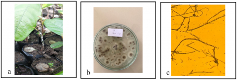
Figure 3. Powdery mildew on nutmeg leaf (a) A symptom identified on diseased plant, (b) Isolate of Oidium tingitanium on PDA media, and (c) Microscopic observation of Oidium tingitanium
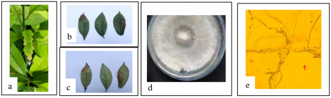
Figure 4. Blight on nutmeg leaf (a) A symptom identified on diseased plant, (b) A symptom on the upper surface of leaf, (c) A symptom on the lower surface of leaf, (d) Isolate of Rhizoctonia sp. on PDA media, and (e) Microscopic observation of Rhizoctonia sp.
Powdery mildew on nutmeg seedlings is caused by Oidium tingitanium. The symptoms include powdery splotches on the leaves and stems of infected plants. The critical stage in the infection process of powdery mildew is the formation or germination. The presence of white powdery mass on the leaves, shoots, and stems of seedlings indicates the germinating conidia of Oidium tingitanium penetrates the plants, causing the fungal pathogen is known as powdery mildew (Figure 3). Halfeld-Vieira and Nechet’s [26] research shows that, powdery mildew that attacks A. mangium is only found in nurseries with a shaded environment. The powdery mildew colonies are white to brown, irregular pattern, epiphytic mycelium, amphigenous (growing along all sides), hyphae clearly insulated, filamentous, irregularly branched, straight to wavy. Kasiamdari et al. [27] reported that the hyphae of Oidium tingitanium has small, single and nipple-shaped appressoria with short linear chains of conidia.
Blight is caused by Rhizoctonia sp. It causes necrotic symptoms in the form of spots on the leaves that spread rapidly and enlarge gradually, causing the leaves to wither and completely fall off [28]. The early symptoms include wet spots on one or both sides of the leaf a few cm from the leaf tips (Figure 4(a)). The leaf edges commonly curl upward and dry up (Figures 4(b) and 4(c)). The lesions develop as water-soaked stripes on leaf blades. The macroscopic observations on PDA media indicate that the colonies of thread-like hyphae begin to appear on the first day. These hyphae are cloudy white and brownish, and arranged into a network called a mycelia. On the third day, white and small irregular masses appear on the surface of the mycelia (Figure 4(d)). On the fifth day, the masses turn dark brown called sclerotia. Microscopically, the fungus appears as branching, perpendicular hyphae, septate or insulated, without any conidia or spores (Figure 4(e)). Fungal cultures grow rapidly, the colonies have spread to fill the media in just three days.
3.2 Types of plant leaf disease and pathogenic fungi
The incidence of plant leaf disease in all observation plots in the nursery and the category of severity are presented in Table 3.
The percentage of damage shows the ratio between the number of infected seedlings and the total number of seedlings. The calculation results show that the average percentage of damage to nutmeg seedlings for all types of diseases tends to be low, which is below 25% [29]. The statistical test results also showed that the intensity of pathogen attack on the research block was not significantly different. The percentage distribution of damage for leaf spot was 3.95%, leaf blight was 4.42%, leaf rust was 7.27% and powdery mildew was 1.025%. The average percentage of leaf rust was higher than the other diseases, while powdery mildew was the disease with the lowest percentage of attack. Although all of these leaf diseases are relatively low, they need to be a concern because these four types of diseases have a very easy way of spreading, namely through wind, water, insects and even humans. The low average percentage of attack on all types of diseases is due to environmental conditions that are less favorable for pathogen development. Relative humidity in the nursery ranged from 68%-72% with a minimum average temperature of 29℃ and a maximum average temperature of 32℃. Anggraeni and Wibowo [30] stated that the development of disease pathogens from the fungi group will experience conidium germination and infection in the temperature range of 20℃-25℃, so temperatures above 30℃ can inhibit the development of fungi.
Table 3. The incidence and severity of leaf diseases on nutmeg seedlings
|
Plot |
Incidence (%) |
Disease Intensity (%) |
Severity |
|||
|
Leaf Spot |
Blight |
Rust |
Powdery Mildew |
|||
|
1 |
3.3 |
5.1 |
0 |
1.5 |
3.8 |
Mild |
|
2 |
3.8 |
1.0 |
14. 0 |
0 |
9.3 |
Mild |
|
3 |
8.7 |
0 |
0 |
2.6 |
3.7 |
Mild |
|
4 |
0 |
11.6 |
15.1 |
0 |
9.3 |
Mild |
|
Total |
15.8 |
17.7 |
29.1 |
4.1 |
26.1 |
|
|
Average |
3.95 |
4.42 |
7.27 |
1.025 |
6.52 |
Mild |
The average intensity of attack by pathogens causing leaf diseases on nutmeg seedlings as a whole was 6.52% with a light damage category. The severity of pathogen attack on plants is largely determined by environmental factors. The nutmeg seedling block in the green house causes the microenvironment to be more controlled. Apart from the temperature and humidity that do not favor the development of pathogens, land sanitation is also a contributing factor to the low disease attack. Routine cleaning of the nursery blocks can inhibit the development of disease inoculum. The disease is strongly influenced by plant resistance factors, the number of pathogens, the virulence of pathogens, the suitability of environmental conditions for pathogen development and the duration of suitable conditions for pathogens pathogens [29, 30].
The presence of pathogen attacks that cause damage to all nutmeg seedling blocks even though they are still classified as lightly damaged is evidence that nutmeg seedlings in the nursery are starting to be attacked by disease. If left unchecked, it is likely that the disease will continue to develop and harm the seedlings in the nursery, because seedlings have a high susceptibility to disease attacks and their adaptability is still very low [31]. In addition, if prevention is not done early on, the spread of the disease can occur more quickly. Damage to the leaves causes the photosynthesis process to be disrupted and at the nursery level, this damage can cause significant losses because it can result in leaf fall which ultimately results in the death of the seedlings [32].
Observation of disease development in nutmeg nurseries, especially diseases caused by fungi, can be carried out in two seasonal periods, namely the dry season and the rainy season, so that it can be used as a comparison for observing the level of leaf disease attack. In addition, observations of damage to leaves caused by diseases can be made on more samples to find out more about the types of diseases that attack and the types of pathogens that cause them [33].
Four types of leaf diseases were found in four observation blocks of nutmeg seedlings: leaf spot (Nigrospora sp.), leaf blight (Rhizoctonia sp.), powdery mildew (Oidium tingitanium) and leaf rust (Pestalotia sp.). The average percentage of damage to nutmeg seedlings for all diseases was 3.95% for leaf spot, 4.42% for leaf blight, 7.27% for leaf rust and 1.025% for powdery mildew. The average intensity of attack by pathogens causing leaf diseases on nutmeg seedlings as a whole was 6.52% with a mild damage category.
We thank the Dean of the Faculty of Forestry, Tadulako University for the research funding support that has been given to us through the flagship research scheme through contract number 742.j/UN28.2/PL/2022. Researcher also expressed his gratitude to the manager of the Tropical Plant Cultivation Field Laboratory for the permission given to conduct research.
[1] Thangaselvabai, T., Sudha, K.R., Selvakumar, T., Balakumbahan, R. (2011). Nutmeg (Myristica fragrans Houtt)-The twin spice-A review. Agricultural Reviews, 32(4): 283-293.
[2] Naeem, N., Rehman, R., Mushtaq, A., Ghania, J.B. (2016). Nutmeg: A review on uses and biological properties. International Journal of Chemical and Biochemical Sciences, 9: 107-110. https://www.researchgate.net/publication/336825717_Nutmeg_A_review_on_uses_and_biological_properties.
[3] Pramudita, L., Widajati, E., Suwarno, F.C., Surahman, M. (2017). Karakteristik morfologi benih sebagai parameter untuk penentuan pohon induk sumber benih Pala (Myristica fragrans Houtt). Indonesian Journal of Agronomy, 45(1): 64-70. https://doi.org/10.24831/jai.v45i1.13755
[4] Rahardiyan, D., Poluakan, M., Moko, E.M. (2020). Physico-chemical properties of nutmeg (Myristica fragrans houtt) of North Sulawesi nutmeg. Fullerene Journal of Chemistry, 5(1): 23-31. https://doi.org/10.37033/fjc.v5i1.146
[5] Samantha, R. (2020). Preservation of nutmeg tree (Myristica fragrans Houtt) used as Bogor’s local culinary ingredient for the strengtheness of urban landscape identity. IOP Conference Series: Earth and Environmental Science, 12013.
[6] Oktavia, D., Pratiwi, S.D., Munawaroh, S., Hikmat, A., Hilwan, I. (2022). The potential of medicinal plants from heath forest: Local knowledge from Kelubi Village, Belitung Island, Indonesia. Biodiversitas Journal of Biological Diversity, 23(7): 3553-3560. https://doi.org/10.13057/biodiv/d230731
[7] Subhan, M., Basri, H. (2019). Klasifikasi mutu buah pala (Myristica Fragrans Houtt) berbasis pengolahan citra menggunakan metode deep learning arsitektur faster R-CNN. INTEK: Jurnal Penelitian, 6(2): 106. https://doi.org/10.31963/intek.v6i2.1566
[8] Arrizqiyani, T., Sonjaya, N., Asty, A. (2017). Optimalisasi potensi tanaman pala sebagai antibakteri Escherichia coli menggunakan metode ekstraksi. In Prosiding Seminar Nasional Publikasi Hasil-Hasil Penelitian dan Pengabdian Masyarakat, pp. 375-382.
[9] Morales-Cedeño, L.R., Orozco-Mosqueda, Ma. del C., Loeza-Lara, P.D., Parra-Cota, F.I., de los Santos-Villalobos, S., Santoyo, G. (2021). Plant growth-promoting bacterial endophytes as biocontrol agents of pre- and post-harvest diseases: Fundamentals, methods of application and future perspectives. Microbiological Research, 242: 126612. https://doi.org/10.1016/j.micres.2020.126612
[10] Haase, D.L., Davis, A.S. (2017). Developing and supporting quality nursery facilities and staff are necessary to meet global forest and landscape restoration needs. REFORESTA0, 4: 69-93. https://doi.org/10.21750/REFOR.4.06.45
[11] Susanna, S., Sinaga, M.S., Wiyono, S., Triwidodo, H. (2020). Diagnosis of dieback disease of the nutmeg tree in Aceh Selatan, Indonesia. Walailak Journal of Science and Technology, 17(8): 801-810. https://doi.org/10.48048/wjst.2020.4379
[12] Abid, R., Butt, S. (2015). Repellent activity of cardamom, ginger and nutmeg against certain insect pests. International Journal of Zoology, 5(6): 1-6.
[13] Geetharamani, G., Pandian, A. (2019). Identification of plant leaf diseases using a nine-layer deep convolutional neural network. Computers & Electrical Engineering, 76: 323-338. https://doi.org/10.1016/j.compeleceng.2019.04.011
[14] Liu, B., Zhang, Y., He, D., Li, Y. (2017). Identification of apple leaf diseases based on deep convolutional neural networks. Symmetry, 10(1): 11. https://doi.org/10.3390/sym10010011
[15] Barnett, H.L., Hunter, B.B. (1998). Illustrated Genera of Imperfect Fungi. US Department of Agriculture, Agricultural Research Service, Washington State University, APS Press. St. Paul, Minnesota USA.
[16] Munshi, M., Sohrab, M.H., Begum, M.N., Rony, S.R., Karim, M.A., Afroz, F., Hasan, M.N. (2021). Evaluation of bioactivity and phytochemical screening of endophytic fungi isolated from Ceriops decandra (Griff.) W. Theob, a mangrove plant in Bangladesh. Clinical Phytoscience, 7(1): 1-10. https://doi.org/10.1186/s40816-021-00315-y
[17] Schuh, W. (1990). Influence of tillage systems on disease intensity and spatial pattern of Septoria leaf blotch. Phytopathology, 80(12): 1337-1340.
[18] Zulkaidhah, Z., Malik, A., Hapid, A., Hamka, H., Ariyanti, A., Rahman, N. (2021). The diversity of termite species on natural forest and agroforestry land in Sulawesi tropical forests in Indonesia. Annals of Silvicultural Research, 46(2): 141-147. https://doi.org/10.12899/asr-2228
[19] Zulkaidhah, Z., Rahma, M., Wardah, W., Wahyun, D., Hapid, A. (2022). Respon pertumbuhan semai tanjung (Mimusops elengi Linn.) terhadap intensitas cahaya Di Arboretum Fakultas Kehutanan Universtas Tadulako. Jurnal Penelitian Hutan Tanaman, 19(2): 137-148.
[20] Zulkaidhah, Z., Wardah, W., Saleh, S., et al. (2022). Soil macrofauna diversity and litter decomposition rate in the buffer zone of lore lindu biosphere reserve Indonesia. International Journal of Design & Nature and Ecodynamics, 17(5): 753-760. https://doi.org/10.18280/ijdne.170513
[21] Hidayati, N., Nurrohmah, S.H., Ardhany, F. (2020). Isolasi, identifikasi dan karakterisasi penyebab penyakit karat daun pada semai pinus di perum perhutani bkph purworejo, KPH Kedu Selatan. Jurnal Pemuliaan Tanaman Hutan, 14(1): 21-32.
[22] Suharti, T., Kurniaty, R. (2013). Inventarisasi penyakit daun pada bibit di stasiun penelitian Nagrak. Jurnal Perbenihan Tanaman Hutan, 1(1): 43-49.
[23] Bambang, Y., Diba, F., Anwari, M.S. (2019). Identifikasi serangga dan penyakit di areal persemaian Pt. Sari Bumi Kusuma Di kecamatan bukit raya kabupaten katingankalimantan tegah. Jurnal Hutan Lestari, 7(3). https://doi.org /10.26418/jhl.v7i3.37624
[24] Madhi, Q.H. (2016). Isolating and diagnose of the fungus Pestalotia spp that causes spotted leaves for four plants collected from some nurseries of the province of Maysan/Iraq. European Academic Research, 4(3): 2760-2772.
[25] Rahman, S., Adhikary, S.K., Sultana, S., Yesmin, S., Jahan, N. (2013). In vitro evaluation of some selected fungicides against Pestalotia palmarum (Cooke.) Causal agent of grey leaf spot of coconut. Journal of Plant Pathology & Microbiology, 4(9): 100197. https://doi.org/10.4172/2157-7471.1000197
[26] Halfeld-Vieira, B.D., Nechet, K.D. (2009). First report of powdery mildew of Acacia mangium in Brazil. Summa Phytopathologica, 35(3): 237. https://doi.org/10.1590/S0100-54052009000300016
[27] Kasiamdari, R.S., Riefani, M.K., Daryono, B.S. (2016). The occurrence and identification of powdery mildew on melon in Java, Indonesia. In AIP Conference Proceedings, 1744(1): 020050. https://doi.org/10.1063/1.4953524
[28] Herliyana, E.N., Sakbani, L., Herdiyeni, Y., Munif, A. (2020). Identifikasi cendawan patogen penyebab penyakit pada daun jabon merah (Anthocephalus macrophyllus (Roxb.) Havil). Journal of Tropical Silviculture, 11(3): 154-162. https://doi.org/10.29244/j-siltrop.11.3.154-162
[29] Saragi, S.M., Firdara, E.K., Putir, P.E. (2019). Identifikasi, frekwensi dan intensitas serangan hama penyakit pada shorea balangeran (Korth.) burck pada persemaian BPDASHL Kahayan, Tumbang Nusa, kalimantan tengah (Identification, frequency and intensity of pets attacks on Shorea balangeran (Korth.) Bur. Jurnal Hutan Tropika, 14(1): 51-59. https://doi.org/10.36873/jht.v14i1.332
[30] Anggraeni, I., Wibowo, A. (2006). Serangan Penyakit embun tepung dan karat daun pada Acacia Auriculiformis a. Cunn. Ex Benth. di kediri, Jawa Timur. Jurnal Penelitian Hutan Dan Konservasi Alam, 3(1): 45-53. https://doi.org/10.20886/jphka.2006.3.1.45-53
[31] Harrison, M.E., Ottay, J.B., D’Arcy, L.J., et al. (2020). Tropical forest and peatland conservation in Indonesia: Challenges and directions. People and Nature, 2(1): 4-28. https://doi.org/10.1002/pan3.10060
[32] Zulkaidhah, Z., Kusumawati, A., Yusran, Y., Rahmawati, D.W., Wardah, W., Wulandari, R. (2023). Inventarisasi penyakit daun pada tegakan Gmelina (Gmelina arborea Roxb) Di Kabupaten Sigi Sulawesi Tengah. Agrifor: Jurnal Ilmu Pertanian dan Kehutanan, 22(1): 123-132.
[33] Pataky, J.K., Williams, M.M., Headrick, J.M., Nankam, C., du Toit, L.J., Michener, P.M. (2011). Observations from a quarter century of evaluating reactions of sweet corn hybrids in disease nurseries. Plant Disease, 95(12): 1492-1506.