Immunological Response in Asthmatic Patients: The Role of Iron Oxide Nanoparticles Against Klebsiella pneumoniae

Fatima Rammadan Abdul![]() | Mohammed T. Abdul Hussein
| Mohammed T. Abdul Hussein![]() | Firas N. Jaafar*
| Firas N. Jaafar*![]()
© 2023 IIETA. This article is published by IIETA and is licensed under the CC BY 4.0 license (http://creativecommons.org/licenses/by/4.0/).
OPEN ACCESS
Asthma, a prevalent respiratory disorder, is distinguished by airway obstruction and respiratory distress stemming from acute and chronic pulmonary inflammation. In this study, 150 sputum and nasopharyngeal swabs were collected from asthma patients, comprising 87 males and 63 females, specifically to isolate Klebsiella pneumoniae (K. pneumoniae). Additionally, blood samples were obtained from 35 patients at the Allergic Specialized Center, Baghdad, and 25 healthy individuals served as controls. All diagnoses were established by specialist respiratory physicians, and serums were preserved for later serological testing. A sandwich enzyme-linked immunosorbent assay was employed to ascertain the levels of total IgE, IL-4, and IL-22.The pathophysiology of bronchial asthma is intertwined with T-cell activation and fluctuations in cytokine levels. It was hypothesized that serum levels of total Immunoglobulin E (IgE) would be elevated in patients. Indeed, significant differences (p=0.001) were observed between patients' total IgE serum levels (304.33 ± 21.3 IU/mL) and controls (36.54 ± 0.69 IU/mL). Conversely, IL-4 and IL-22 levels in asthma patients (66.77 ± 4.1 pg/mL and 41.83 ± 0.37 pg/mL respectively) differed significantly from healthy controls for IL-4 (22.75 ± 0.68 pg/mL, p<0.01) but not for IL-22 (40.89 ± 1.35 pg/mL).Klebsiella pneumoniae was isolated from 12 sputum and nasopharyngeal swab samples from asthmatic patients. Post-incubation, 83.3% of the isolates were found to be biofilm-forming, while 16.7% did not form biofilms. Iron oxide nanoparticles, with an average diameter of 85.9 nm, were utilized to inhibit K. pneumoniae biofilm formation. At a concentration of 5 mg/mL, these nanoparticles exhibited a significant augmentation of about 58% (p≤0.05) in inhibiting biofilm formation. However, at concentrations of 0.5mg/mL and 50 mg/mL, the augmentation was 42.9% (P>0.05) and 37.1% (p>0.05) respectively, which were not statistically significant.This study underscores the potential of iron oxide nanoparticles in impeding biofilm formation by K. pneumoniae and the pivotal role of cytokine levels in the pathophysiology of asthma.
asthma, K. pneumoniae, immunoglobulin E, interleukin, iron oxide nanoparticle
Asthma, a pervasive, chronic inflammatory disease of the pulmonary airways, poses a significant global health challenge. It impacts individuals of all ages, with a predilection for onset in early childhood. Allergic asthma, in particular, is characterized by airway hyper-reactivity, elevated levels of allergen-specific IgE in the circulation, and/or positive skin prick tests to various allergens. The introduction of an allergen triggers a cascade of type 2 immune responses, resulting in an increase in IgE, type 2 cytokines, and eosinophils.
The etiology of asthma has long been hypothesized to stem from deregulated T-helper2 (TH2) immune responses to environmental allergens [1]. Asthma is a heterogeneous disease with multiple inflammatory pathways, as confirmed by clinical studies. Despite the established associations between prominent or deregulated TH2 responses and allergic disorders and asthma, our understanding of the mechanisms underlying this disease's origin is constantly evolving as we delve deeper into the complexity of effector T-cell subsets [2].
IL-4, IL-5 and IL-13 are cytokines produced by TH2 cells and together they support the initiation and persistence of allergic inflammation. Important pro-inflammatory activities of interleukin-4 in asthma, such as the production of the IgE isotype switch [3]. Expression of the VCAM-1 protein, which facilitates eosinophil transmigration through endothelium. IgE, also known as immunoglobulin E, has long been recognized as the primary mediator of type 1 hypersensitivity reactions. The results demonstrate a strong connection between particular Ig E or total IgE antibodies and asthma diseases, which has been established in studies to be a relationship between allergens and allergic disease [4]. According to research on IL-22 role in asthma sufferers, IL-22 prevents human bronchial epithelial cells from expressing pro-inflammatory chemokine and adhesion molecules when IFN is present [5]. Also, they have demonstrated an inverse relationship between the levels of pro-inflammatory chemokine and the levels of IL-22 in asthma patients, pointing to IL-22 potential protective effects in these individuals [6].
Respiratory bacterial infection has been closely associated with asthma development. While many studies have primarily focused on viruses, bacterial colonization has also been shown to exacerbate the disease. Various bacterial species have been implicated in the pathogenesis of asthma.
K. pneumoniae, a non-fermenting, Gram-negative bacillus, is considered a significant pathogen in nosocomial infections due to its anti-phagocytosis capsule [7]. Although Klebsiella species are typically part of the natural flora in the human mouth, nose, and gastrointestinal system, they can also act as opportunistic pathogens [8]. Possible infections include diarrhea, septicemia, pneumonia, urinary tract infections, and soft tissue infections [9]. The pathogenicity of K. pneumoniae is enhanced by several factors, including enzyme production and biofilm formation. K. pneumoniae is encapsulated and primarily resides in the lower gastrointestinal tract of mammalian hosts, where its population can significantly increase following antibiotic therapy that reduces the diversity of commensal microbiota [10].
The primary aim of this study is to quantify the levels of total IgE, IL-4, and IL-22 in asthma patients and to evaluate their correlation with clinical outcomes. We aim to assess the potential utility of these cytokines as independent prognostic markers and the propensity of K. pneumoniae, isolated from asthmatic patients, to form biofilms. We also intend to examine how the concentration of nanoparticles influences the destruction of bacterial biofilms by the effect of Iron Oxide Nanoparticles (IONPs). A relationship between the time required for biofilm suppression and the concentration of the nanoparticles is thus anticipated.
2.1 Study group
A total of one hundred-fifty sputum and nasopharyngeal swabs from patients with asthma (males 87 and 63 females) to isolate K. pneumoniae. Blood samples were collected from Thirty-five patients, the sera were separated and stored until use for serological test. Patients with asthma diseases are revise in the Allergic Specialized Center\Baghdad and during the period four month. specialized physicians diagnosed these cases. The individual control were same age and gender with patients, samples collected from twenty-five with no signs of asthma diseases or any other disease (15 men, 10women).
2.2 Determination of total IgE in patients and controls serum
Total serum IgE was determined by an immune-enzymetric assay using the total IgE ELISA kit (Euroimmum/German). Reference range of total IgE levels according to age, from newbornto over 16 years old (1.2~100) IU/mL.
2.3 Measurement of IL-4 and IL-22 levels in patients' and control sera
The presence of cytokines has been found in patient sera with asthma patients and healthy control. IL-4 and IL-22, two cytokines, have been identified by the (My biosource/USA) and (Elabscience/China) respectively, Detection Range (15.6~1,000) pg/ml.
2.4 Identification and sources bacteria
Sputum samples and nasopharyngeal swabs collected from asthmatic patients resulted in the isolation of twelve K. pneumoniae isolates. Initial diagnostic depending on Gram reaction and morphological characteristic of the colonies based on bacterial growth on Macconkey, Eosin Methylene Blue and Blood agar, as well as the numbers of biochemical test and the final diagnosis of the isolated bacteria is done by VITEK2 compact system (Bio Merieux France) [11].
2.5 Biofilm formation
The method mentioned in study [12] was used to investigate the capability of the isolates to procedure a biofilm, which is the micro -titration plate’s method. The following values in Table 1 served to distinguish between the isolates under study on the basis of their ability on biofilm formation.In the following is a briefly of the procedure.
Wells –flat bottom polystyrene were filled with 200µL of the diluted culture and 200µL BHI +1% glucose.After being incubated for 24hat 37℃, plates washing by PBS to remove unbounded bacteria.Adherent bacteria forming biofilm in plate were fixed with sodium acetate, stained by crystal violet and incubate at room temperature for 20 min., then washed three times with PBS.All wells were filled by 200µL ethanol (95%) to release the dye from the cell.Finally, optical density by using micro ELISA auto reader at wavelength 550 nm.
Table 1. Determination of the capacity to biofilm development according to the absorbance
|
OD |
Adhesion Ability |
Biofilm Formation |
|
<0.120 |
No adhesion |
unproductive |
|
0.120-0.240 |
weak |
weak |
|
>0.240 |
strong |
strong |
The degree of biofilm creation was estimated in relation to the following equation.
$Biofilm \ Capacity = Average \ Absorption \ of \ the \ Test \ Sample- Average \ Absorption \ of \ the \ Control$
2.6 Iron oxide nanoparticles preparation
Iron oxide nanoparticles were synthesized using [13] technique. The production of IONPs was suggested by the black precipitate, which 3.1736 g FeCl2•4H2O (0.016 mol) and 7.5684 g FeCl3•6H2O (0.028 mol) and 320 mL of deionized water, Fe2+/Fe3+=1/1.75. For one hour, the mixture was stirred at 80℃ under a nitrogen atmosphere. Then ammonia solution (25%) 40 ml was added to the mixture quickly, agitated under nitrogen atmosphere for additional hour and cooled to room temperature.The deionized water was used to wash the black precipitate five times and collected via magnet. Finally, it was dried in an oven at 70℃. Purity was assessed using Scanning Probe Microscope (SPM) study at the University of Baghdad's Faculty of Science. Nanoparticles were studied using Fourier Transform Infrared Spectroscopy (FTIR) at the department of chemistry, Mustansiryiah University. Iraq's Baghdad. With a 2400S spectrophotometer, totally samples were examined over a range of 600-4000 cm.
2.7 Anti-biofilm activity of IONPs nanoparticles
In this work, the adherence of bacteria to polystyrene well plates without IONPs and bacteria with IONPs was compared. Each well received 20 l of the K. pneumoniae culture suspension.The iron-oxide nanoparticles were then added to 180 μl at three different concentrations (0.5, 5 and 50 mg/ml). After that, biofilms were allowable to raise for 20 hours. Next, wells were washed to eliminateboundlessmicrobesat that time, Each well added 200 L of a 0.1% safranin staining solution.For 10 minutes, plates were incubated. The wells were cleaned with sterile water &given time to fully dry. Using a BioTic ELISA reader (ELx800), optical density (absorbance at 490 nm) was measured to gauge the development of the biofilm [14]. Researches wereachieved in triplicate bydistinctly cultured bacteria.
2.8 Bio-statistical analysis
Statistical Analysis System- SAS (2012) software was utilized to examine how the influences of study differed.
3.1 Population of study
According to this study, men suffer from asthma more often than females. Male patients made up 87 (58%), while female patients made up 63 (42%), showed in Figure 1. This result was associated with several studies conducted by each [15-17] showing the incidence of asthma was greater in males than in females.The late onset of asthma among female leads to a change in the ratio between the two genders. The latter constitutes a large part of the cases of asthma in adults [18].
Figure 1. Proportions between males and females in asthmatic patients
It is commonly known that women are more than males to infect with asthma frequently, because of different hormones, this is the case [19]. Hence, it has been postulated that hormonal changes during the menstrual cycle may be extremely important in the pathophysiology of asthma, leading to cyclic aggravation of the condition. Women suffer from an increase in asthma symptoms during premenstrual or menstrual phases. This phenomenon is called premenstrual asthma (PMA) [20].
3.2 Immunological study
The activation of cells and levels of some cytokines contribute to the occurrence of asthma. In addition, the role of cytokines and circulating lymphocytes in the occurrence of acute asthma attacks is still unclear.
3.2.1 Serum Level of total IgE in asthma patients
The current study showed that the total IgE serum levels were (304.33 ± 21.3) in patients and (36.54 ± 0.69) IU/mL controls. Statistical analysis showed that there is a significant difference between the two groups (p=0.001).
Table 2. Distribution of total IgE levels in study groups
|
Parameters |
Mean ± SE |
Probability |
|
|
Patients group (n=35) |
Healthy group (n=25) |
||
|
Total IgE level (IU/ml) |
304.33±21.3 |
36.54±0.69 |
|
ANOVA test was used to compare between data.
** Significant at<0.01
SE: Standard Error, P: Probability
When asthmatic patients were compared to controls In this research, significant correlation (P=0.001) between serum total IgE levels, As shown in the Table 2 and Figure 2. This result is compatible with a previous study [21], who found a strong association between asthma and the level of total IgE in the blood in children.Likewise, a study attained [22] who demonstrated that patients with uncontrolled asthma had considerably higher serum total IgE values.
Figure 2. Case-control differences according to total IgE
Maintaining symptom severity in asthma diseases. IgE has an important role in the pathophysiology of allergic asthma, which leads to the liberation of different groups of mediators as a result of its binding to receptors on the surfaces of many immune cells, mast cells, or basophils, which leads to enhancing airway hyper- responsiveness, mucus secretion, and increased vascular permeability [23].
3.2.2 Levels of IL-4 in the sera
The results of the present study in relative to the IL- 4 asthma patients were (66.77 ± 4.1) pg/ml. While healthy control individuals were (22.75 ± 0.68) pg/ml. Significant variations existed between the studied groups, as the demonstrates Table 3 and Figure 3.
Table 3. Distribution of IL-4 levels in the sera of the study group
|
Parameters |
Mean ± SE |
Probability |
|
|
Patients group (n=35) |
Healthy group (n=25) |
||
|
IL-4 pg/ml (Mean±SE) |
66.77±4.1 |
22.75±0.68 |
|
ANOVA test was used to compare between data.
** Significant at<0.01
SE: Standard Error, P: Probability
The IL-4 outcome is consistent with study [24]. But the findings of study [25] pointed to children's consistent low levels of IL-4. An important cytokine in the emergence of allergic inflammation is interleukin (IL-4).It is associated with the isotope switch's induction and B cells' excretion of IgE. As a result, the formation of allergic diseases is influenced by the level of IL-4, even though IL-4 causes CD4 T cells to differentiate into Th2 cells, which produce IL-4, IL-5, IL-9 and IL-13 via the activation of IgE production. Th2 cytokines are essential for the rise of asthma disorders and the treatment infections and activate eosinophil, basophils and mast cells. The basis for comprehending the mechanisms behind immune responses were provided by the hypothesis of a Th1/Th2 balance [26].
Figure 3. Case-control differences according to IL-4
3.2.3 Interleukin-22 level assessment
The findings of this investigation showed that asthma patients' serum levels of IL-22 had increased.Interleukin-22 level in patients were (41.83 ± 0.37) pg/ml and (40.89 ± 1.35) pg/ml in the healthy. According to the Table 4 and Figure 4 statistical analysis, no significant between patients' and the healthy.
Table 4. Serum IL-22 levels in study groups
|
Parameters |
Mean ± SE |
Probability |
|
|
Patients group (n=35) |
Healthy group (n=25) |
||
|
IL-22 pg/ml |
41.83±0.37 |
40.89±1.35 |
0.44 NS |
ANOVA test was used to compare between data.
NS: Non-significant
SE: Standard Error, P: Probability
Figure 4. Case-control differences according to IL-22
According to this study findings (P value=0.44), no significant differences between asthmatic patients and the normal group.
Numerous studies exhibited the different result, such as study [27] who exhibited that in a mouse asthma model, IL-22 is essential for the development of antigen sensitization. Another study [28] there is a positive relationship between the severity of the disease and the increase in the level of IL-22 in the serum of asthmatic patients. These results vary as a result of the difference in raceof population.
3.2.4 Study of sensitivity and specificity of immunology
In relation to the ROC consequences, positive true results were perceived in total IgE (AUC=1.00, P=0.003), IL-4 (AUC=1.00, P=0.0032). While, IL-22 (AUC=0.613, P=0.346) referred to in the Table 5 and Figure 5.
Table 5. Area under curve (AUC) of studied parameters
|
Consequence Variable(s) |
AUC |
Cut off Value |
P Value |
|
total IgE |
1.00 |
82.90 IU/ml |
|
|
IL-4 |
1.00 |
36.03 pg/ml |
|
|
IL-22 |
0.613 |
39.54 pg/ml |
0.346 NS |
Figure 5. ROC curve of (A) IgE; (B) IL-4; (C) IL-22
ROC curve, in our opinion, ought to be the special standard for identifying the characteristics that are sensitive and specific sufficient to provision an asthma diagnosis. Whereas, further research is needed to determine its use in prognosis, risk assessment, and therapeutic intervention evaluation. Significant statistical variances between patients and healthy are shown using ROC curves. When comparing various biomarkers, the Area Under the Curve (AUC) gives an effective statistic. Though, an AUC value near to 1 indicates an excellent marker, a curve located close to the diameter is of no diagnostic utility. Value of AUC close 1.00 is almost often attended via acceptable sensitivity and specificity values for the biomarker [29]. To interpret the use of ROC curves in understanding biomarkers to clinical practice, when biomarkers are studied for the disease being considered, high sensitivity means that asthma will be identified in most cases, high specificity indicates that few, if any, healthy subjects will test positive [30].
3.3 Isolation and Identification of K. pneumoniae
From a total of 150 sputum samples and nasopharyngeal swabs of patientscharacteristics predisposing to asthma infection, twelvespecimenshave a positive result on Macconkey agar and blood agar, 8(66.7%) were sputum, and 4(33.3%) were from nasopharyngeal,as shown in the Figure 6. Comparison of patients sampleswith K. pneumoniae. infections revealed isolation percentage of bacteria in sputum is more than in nasopharyngeal.
Figure 6. Percentage of K. pneumoniae isolates depending on the type of sample
Figure 7. K. pneumonaie on (A) Macconkey and (B) Eosin methylene blue agar
K. pneumoniae can be easily isolated from clinical specimens using general or specific media at 37℃ in an aerobic environment. Grow on blood and macconkey agar.Larg, mucoid, pink colour lactose fermenting colonies on macconkey agar.Biochemical testing and microscopical identification are also employed to identify and distinguish it from other gram-negative bacilli. This study showed that motility, oxidase, indole and methyl red were negative. While, catalase, urease, vogas- proskaur and citrate utilization were positive, ferment glucose. The confirmed identification by VITEK2 compact system which indicated that (12) isolates were belongs to K. pneumoniaeas shown in Table 6 and Figure 7.
Table 6. Microscopic and biochemical characterization of K. pneumoniae
|
Characteristic |
||
|
Biochemical Tests |
Indole |
- |
|
Methyl red |
- |
|
|
Vogas- Proskaur |
+ |
|
|
Citrate utilization |
+ |
|
|
Catalase |
+ |
|
|
Coagulase |
- |
|
|
Oxidase |
- |
|
|
H2S |
|
|
|
Gas production |
+ |
|
|
Glucose fermentation |
+ |
|
|
Urease |
+weak |
|
|
Motility |
- |
|
3.4 Biofilm production assay
The Table 7 and Figure 8 show the results of the biofilm formation test according to the method plates micro-titration, K. pneumoniae isolates after the incubation period showed that 83.3% of the isolates showed a positive result for biofilm formation.The percentage of strong isolates was (50%), while of the weak biofilm formation was (33.3%), although the percentage of negative isolates was (16.7%).
Table 7. Biofilm formation detection test for K. pneumoniae isolates
|
Isolate No. |
Optical Density Rate |
Optical Density Rate - Density Rate Control |
|
Control (negative) |
0.127 |
0.127 |
Figure 8. Capacity of K. pneumoniae to biofilm formation using MTP method
Figure 8 appears a comparison between the percentages of the strong, weak and negative positive result of this method. I used these as a standard technique for the speed of cell adhesion and biofilm formation in various types of microorganisms. Gram stain or positive gram stain or yeast, as the dye used is associated with negatively charged molecules on the surface of the cell bacteria from polysaccharides, which gives a full measure of biofilm formation. A study [31] showed that the MTP method is the most sensitive and ease of recognition of biofilm creation and that detection of mucus production depends on several factors such as the method of investigation used, the type of medium used and incubation conditions.
3.5 Iron oxide nanoparticles' description
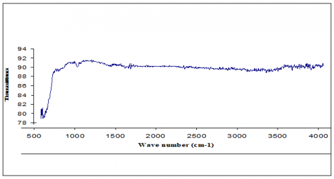
Fourier Transform–Infrared spectra (FTIR), Figure 9 showed that iron oxide nanoparticles were successfully formed. It is clear that certain bands have characteristic appearances, for example the incidence of the Fe-O bond (850 cm-1). It appears that the vibrations of iron oxide's Fe-O bonds are what caused the low wave numbers between 850 and 400 cm-1 to absorb frequencies [32, 33].
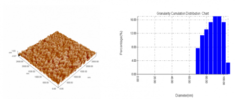
SPM measurements of particle sizes revealed that the nanoparticles are almost spherical and mono- dispersed. The histogram and particle size distribution shown in Figure 10(B). Having a range of (70–105) nm and average sizes of 85.9 nm according to Figure 10(A).
Figure 9. Iron oxide nanoparticle Fourier Transform –Infrared Spectra
(A) (B)
Figure 10. (A) Scanning Probe MicroscopeIONPs; (B) SPM particle size distribution histogram of IONPs
3.6 Influence of IONPs on K. pneumoniaebiofilm development
The results showed in Tables 8 and 9 concentration of 50, 5, 0.5 mg/ml of iron oxide nanoparticle on K. pneumoniaebiofilm, inductionin the quantity of biofilm biomass in the 20hr. dealing time compared with control.
Iron oxide nanoparticles achieved had an average diameter 85.9 nm, the particles were used to inhibit K pneumoniae biofilm formation. It showed the highest augmentation in concentration 5 mg/ml IONP about 58% significant (p≤0.05), while at concentration 0.5mg/ml exhibited 42.9% augmentation (P>0.05). Percentage augmentation of K. pneumoniaein concentration of 50 mg/ml gave 37.1 % non- significant (p˃0.05) compared with the control. Thus, IONPs utilized in bacteria biofilm formation at low concentration for 20 hours produced consequences that were consistent with study [34]. Someone has indicated that the size of the IONPs affects the increasing biofilm mass regulating biofilms with iron [35]. K. pneumoniae had the lowest enhancement rate (37.1%) at a concentration of 50 mg/ml. Therefore, decreased biofilm development was caused by increased IONPs concentration [36]. Thus,we have seendiverse results reliant on the concentrations and the mechanisms to inter the IONPs.
Table 8. Result of concentration of IONPs on K. pneumoniae biofilm development
|
Concentration mg/ml |
% Amplification |
|
50 |
37.1% |
|
5 |
58.9% |
|
0.5 |
42.9% |
|
Amplification was determined by the formula: $\%$ Amplification $=\frac{\text { Test O.D - Control O.D }}{\text { Control O.D }} * 100$ |
|
Table 9. K. pneumoniae biofilm on polystyrene surface with IONPs
|
Concentration mg/ml |
OD at 490nm (Mean+SD) |
P-Value |
T-Test |
Standard Error |
|
50 |
0.201±0.05 |
0.06⃰⃰ |
2.56 |
0.028 |
|
5 |
0.180±0.013 |
0.005⃰⃰⃰ ⃰ ⃰ |
5.38 |
0.007 |
|
0.5 |
0.171±0.029 |
0.064⃰⃰ |
2.53 |
0.016 |
|
Control |
0.125±0.012 |
|
|
|
⃰ ⃰ significant
⃰ ⃰ ⃰ very significant
control/ bacteria without nanoparticles
In difference with study [37], it demonstrated a 16-hour rise in P. aeruginosa biofilm biomass when 0.2 mg was present [38]. The effectiveness of IONPs to eliminate bacteria biofilm relies on the incubation period and nanoparticle concentration since findings were comparable to recent research using iron oxide nanoparticles on bacteria biofilms within 20 hours. Therefore, decrease the time with increasing nanoparticle concentration to inactivate bacterial biofilm and vice versa. Other unknown factors could also be associated with particle shape and surface charge, as well as surface feature playing an important role in intercellular bonding.
There are extremely significant differences between the asthma group and control in total IgE and IL-4, but no significant in IL-22. The result suggest that IgE and IL-4 are important parts of the markers for asthma disease. The varying levels of certain cytokines play an important role in arousing asthma, inflammatory response could be a common pathway adjusting asthma. Decrease biofilm formation were seen for K. pneumoniae with increase concentration iron oxide nanoparticles and other unknown factors could also be associated with particle shape and surface charge, as well as surface feature playing an important role in intercellular bonding.
Expanding the immunological study by using a larger number of interleukins and chemokines, in order to know their direct effect on asthma. More attention should be given to the bacterial role in asthma patients, because they have different and varied virulence factors such as biofilms, as well as their possession of external antigens that may trigger allergic diseases, including asthma.
We thank the support of the College of Science / Mustansiriyah University for the research project and for facilitating all project requirements.
[1] Weidner, J., Bartel, S., Kılıç, A., et al. (2021). Spotlight on microRNAs in allergy and asthma.Allergy, 76(6): 1661-1678. https://doi.org/10.1111/all.14646
[2] Breiteneder, H., Peng, Y.Q., Agache, I., et al. (2020). Biomarkers for diagnosis and prediction of therapy responses in allergic diseases and asthma. Allergy, 75(12): 3039-3068. https://doi.org/10.1111/all.14582
[3] Godbout, M. (2016). Influence de l’interleukine-4 sur le recrutement des neutrophiles équins dans un modèle inflammatoire sous-cutané. http://hdl.handle.net/1866/13382
[4] Ptaschinski, C., Lukacs, N.W. (2018). Acute and chronic inflammation induces disease pathogenesis. Molecular Pathology, 25-43. https://doi.org/10.1016/B978-0-12-802761-5.00002-X
[5] Wu, C.T., Lee, Y.T., Ku, M.S., Lue, K.H. (2020). Role of biomarkers and effect of FIP-fve in acute and chronic animal asthma models.Journal of Microbiology, Immunology and Infection, 53(6): 996-1007. https://doi.org/10.1016/j.jmii.2020.07.006
[6] Ulu, A., Sveiven, S., Bilg, A., et al. (2022). IL-22 regulates inflammatory responses to agricultural dust-induced airway inflammation.Toxicology and Applied Pharmacology, 446: 116044. https://doi.org/10.1016/j.taap.2022.116044
[7] Esaiassen, E. (2018). Antibiotics and probiotics to neonates-Adverse effects, impact on gut microbiota and antibiotic resistome, and Bifidobacterium pathogenicity. https://hdl.handle.net/10037/15334
[8] Sanders, D.J., Inniss, S., Sebepos-Rogers, G., Rahman, F.Z., Smith, A.M. (2021). The role of the microbiome in gastrointestinal inflammation. Bioscience Reports, 41(6). https://doi.org/10.1042/BSR20203850
[9] Khan, H.A., Ahmad, A., Mehboob, R. (2015). Nosocomial infections and their control strategies.Asian Pacific Journal of Tropical Biomedicine, 5(7): 509-514. https://doi.org/10.1016/j.apjtb.2015.05.001
[10] Wang, G., Zhao, G., Chao, X., Xie, L., Wang, H. (2020). The characteristic of virulence, biofilm and antibiotic resistance of Klebsiella pneumoniae.International Journal of Environmental Research and Public Health, 17(17): 6278. https://doi.org/10.3390/ijerph17176278
[11] Baron, E.J., Fingold, S.M. (1999). Diagnostic Mirobiology. 9th ed. Baily and Scotts, The C.V. Mosby company.
[12] Bose, S., Khodke, M., Basak, S., Mallick, S.K. (2009). Detection of biofilm producing staphylococci: Need of the hour. Journal of Clinical and Diagnostic Research, 3(6): 1915-1920.
[13] Kedar, E., Palgi, O., Golod, G., Babai, I., Barenholz, Y. (1997). Delivery of cytokines by liposomes. III. Liposome-encapsulated GM-CSF and TNF-alpha show improved pharmacokinetics and biological activity and reduced toxicity in mice. Journal of Immunotherapy, 20(3): 180-193. http://www.editorialmanager.com/jit/
[14] O'Toole, G.A., Kolter, R. (1998). Flagellar and twitching motility are necessary for Pseudomonas aeruginosa biofilm development. Molecular Microbiology, 30(2): 295-304. https://doi.org/10.1046/j.1365-2958.1998.01062.x
[15] Hameed, S., Khan, F.I., Hameed, B. (2019). Understanding security requirements and challenges in Internet of Things (IoT): A review. Journal of Computer Networks and Communications, 1-14. https://doi.org/10.1155/2019/9629381
[16] Wright, P.J., Neat, F.C., Gibb, F.M., Gibb, I.M., Thordarson, H. (2006). Evidence for metapopulation structuring in cod from the west of Scotland and North Sea. Journal of Fish Biology, 69: 181-199. https://doi.org/10.1111/j.1095-8649.2006.01262
[17] Zein, J.G., Erzurum, S.C. (2015). Asthma is different in women. Current Allergy and Asthma Reports, 15: 1-10. https://doi.org/10.1007/s11882-015-0528-y
[18] Sansone, F., Attanasi, M., Di Pillo, S., Chiarelli, F. (2020). Asthma and obesity in children. Biomedicines, 8(7): 231. https://doi.org/10.3390/biomedicines8070231
[19] Goldsby, T.J., Closs, D.J. (2000). Using activity‐based costing to reengineer the reverse logistics channel. International Journal of Physical Distribution & Logistics Management. https://doi.org/10.1108/09600030010372621
[20] Pignataro, F.S., Bonini, M., Forgione, A., Melandri, S., Usmani, O.S. (2017). Asthma and gender: The female lung. Pharmacological Research, 119: 384-390. https://doi.org/10.1016/j.phrs.2017.02.017
[21] Strømgaard, S., Thomsen, S.F., Fenger, M., Backer, V. (2011). Predictors of serum total IgE in a random sample of 7–17 year old children. International Scholarly Research Notices, 169859. https://doi.org/10.5402/2011/169859
[22] Amal, S., Shalaby, S.M., Abdel-Nour, H.M., Sarhan, W.M., Gehad, M.H., Yousif, Y.M. (2022). Impact of cytokines genes polymorphisms and their serum levels on childhood asthma in Egyptian population. Cytokine, 157: 155933. https://doi.org/10.1016/j.cyto.2022.155933
[23] Regateiro, F.S., Botelho Alves, P., Moura, A.L., Azevedo, J.P., Regateiro, F.S. (2021). The diverse roles of t cell subsets in asthma. European Annals of Allergy and Clinical Immunology, 53(05): 201. http://hdl.handle.net/10400.1/17166
[24] Al-Yasiri, M.Y.K. (2014). Study some Immunological and Haematological changes upon workers of Vegetable Oil factory in Baghdad suffering from hypersensitivity Type-1. Collage of Sciences for women. University of Baghdad. Iraq, 1-101.
[25] García, E., Duarte, S., Calderón, C., et al. (2011). Expression of IL-10, IL-4 and IFN-γ in active skin lesions of children with papular urticaria. Biomédica, 31(4): 525-531.
[26] Zenclussen, A.C. (2013). Adaptive immune responses during pregnancy. American Journal of Reproductive Immunology, 69(4): 291-303. https://doi.org/10.1111/aji.12097
[27] Tamasauskiene, L., Gintauskiene, V.M., Bastyte, D., Sitkauskiene, B. (2021). Role of IL-22 in persistent allergic airway diseases caused by house dust mite: A pilot study. BMC Pulmonary Medicine, 21: 1-8. https://doi.org/10.1186/s12890-021-01410-z
[28] Daliri, E.B.M., Ofosu, F.K., Chelliah, R., Lee, B.H., Oh, D.H. (2020). Health impact and therapeutic manipulation of the gut microbiome.High-Throughput, 9(3): 17. https://doi.org/10.3390/ht9030017
[29] Metz, C.E. (1978). Basic principles of ROC analysis. InSeminars in Nuclear Medicine, 8(4): 283-298. https://doi.org/10.1016/S0001-2998(78)80014-2
[30] Bara, A., Manduca, A., Bernabeu, A., et al. (2018). Sex-dependent effects of in utero cannabinoid exposure on cortical function. Elife, 7: e36234. https://doi.org/10.7554/eLife.36234
[31] Abdul, F.R. (2018). Evaluation of some virulence factors, hemagglutination and agglutination of antigens of Acinetobacter baumannii isolated from clinical samples. Journal of Global Pharm. Technology, 10(03): 200-208. https://doi.org/10.1590/s0074-02762004000800010
[32] Hamidi, S., Safi, B., Waheed, A.W. (2022). Study of the method of electro-crystallization and the study of effective factors in the production of nanostructures in this method. Influence: International Journal of Science Review, 4(1): 239-248. https://doi.org/10.54783/influencejournal.v4i1.22
[33] Abdul, F.R., Subhi, H.T., Taher, N.A., Raheem, I.A. (2019). Activity of iron oxide nanoparticles on bacterial biofilm formation. Journal of pharmaceutical Sciences and Research, 11(3): 1126-1130.
[34] Borcherding, J., Baltrusaitis, J., Chen, H., et al. (2014). Iron oxide nanoparticles induce Pseudomonas aeruginosa growth, induce biofilm formation, and inhibit antimicrobial peptide function. Environmental Science: Nano, 1(2): 123-132. https://doi.org/10.1039/C3EN00029J
[35] Wu, Y., Outten, F.W. (2009). IscR controls iron-dependent biofilm formation in Escherichia coli by regulating type I fimbria expression. Journal of Bacteriology, 191(4): 1248-1257. https://doi.org/10.1128/jb.01086-08
[36] Agarwala, M., Choudhury, B., Yadav, R.N.S. (2014). Comparative study of antibiofilm activity of copper oxide and iron oxide nanoparticles against multidrug resistant biofilm forming uropathogens. Indian Journal of Microbiology, 54: 365-368. https://doi.org/10.1007/s12088-014-0462-z
[37] González, A.G., Mombo, S., Leflaive, J., Lamy, A., Pokrovsky, O.S., Rols, J.L. (2015). Silver nanoparticles impact phototrophic biofilm communities to a considerably higher degree than ionic silver. Environmental Science and Pollution Research, 22: 8412-8424. https://doi.org/10.1007/s11356-014-3978-1
[38] Taghizadeh, S.M., Ebrahiminezhad, A., Raee, M.J., Ramezani, H., Berenjian, A., Ghasemi, Y. (2022). A study of l-lysine-stabilized iron oxide nanoparticles (IONPs) on microalgae biofilm formation of chlorella vulgaris. Molecular Biotechnology, 64(6): 702-710. https://doi.org/10.1007/s12033-022-00454-8