Huda Ali Mahdi*![]() | Mohamed Ibrahim Shujaa
| Mohamed Ibrahim Shujaa![]() | Entidhar Mhawes Zghair
| Entidhar Mhawes Zghair![]()
© 2023 IIETA. This article is published by IIETA and is licensed under the CC BY 4.0 license (http://creativecommons.org/licenses/by/4.0/).
OPEN ACCESS
Brain tumors, characterized by the uncontrolled and rapid proliferation of cells, can result in fatal outcomes if not identified and treated promptly. Consequently, the development of a reliable and automated diagnostic system is of paramount importance. In this study, a Fuzzy Convolutional Neural Network (F-CNN) is employed for the efficient diagnosis of brain tumors (Glioma, Meningioma, Pituitary, and non-tumors), leveraging the computational capabilities of Google Colaboratory. The methodology comprises four stages: pre-processing, training, testing, and evaluation. The pre-processing stage entails rescaling the image, resizing, random flipping, and random rotation. The training phase involves the construction of an intelligent model, encompassing four blocks: convolution, ReLU, batch normalization, and max pooling. This is followed by flattening, a fuzzy inferences layer, and a dense layer with dropout. The model was trained using a Kaggle dataset comprising 7022 brain tumor MRI images and validated with a test set of 470 MRI images sourced from the Neurological Wholesale Hospital in Baghdad. The proposed F-CNN model achieved a high accuracy rate of 99.31% while maintaining low computational complexity and time. This work illustrates the potential of Deep Learning approaches, such as F-CNNs, in enhancing the precision and efficiency of medical imaging diagnostics.
Fuzzy Convolutional Neural Network, Deep Learning, MRI images, fuzzy logic, medical image processing, brain tumor
Modern clinical settings heavily rely on medical imaging to facilitate accurate diagnosis and treatment of a myriad of health conditions [1]. These imaging techniques, which include X-rays, ultrasound (US), Computer Tomography (CT), and Magnetic Resonance Imaging (MRI), play a pivotal role in disease detection, clinical monitoring, and treatment planning [2]. These diagnostic tools allow both qualitative and quantitative assessments of symptoms at the lesion site. They find utility in the evaluation of various anatomical structures, such as the heart, brain, liver, lungs, chest, and kidneys. The extraction of crucial information through medical image analysis notably enhances the precision of clinical diagnoses [3].
Brain tumors, growths that form within the brain or tissues under the skull, can have benign or malignant characteristics. These tumors grow erratically, exerting pressure on the brain [4], potentially leading to several neurological complications. The World Health Organization (WHO) reports that in 2020, cancer was the second leading cause of death worldwide, accounting for approximately 10 million fatalities [5]. It is estimated that in 2019, nearly 0.7 million Americans were afflicted with brain tumors, with 0.86 million cases diagnosed. Out of these, 26,170 were identified as malignant, and 60,800 as benign. Notably, only 35% of malignant patients in the US survived [6]. Therefore, the importance of accurate brain tumor MR images for diagnosis and treatment decisions cannot be overstated [7]. However, accurate identification and diagnosis of brain tumors from MR images with comparable structures or characteristics largely depend on the expertise and availability of radiologists. The use of automated classification, which diagnoses brain tumor MR images with minimal influence from human experts, could potentially address this issue [8].
Convolutional neural networks (CNNs), a Deep Learning technique, have been employed to tackle complex classification problems [9]. By employing diverse strategies, CNNs facilitate disease prediction, classification, and decision-making during disease diagnosis [10]. Fuzzy logic, a tool that simulates human thought and perception, is often integrated with convolutional neural networks to create Fuzzy Convolutional Neural Networks. These networks enhance function approximation and data classification, making them more precise and reliable [11]. The combination of fuzzy logic systems and convolutional neural networks has shown promise in improving the accuracy of brain disease prediction and diagnosis.
This study proposes an automatic diagnosis system that prioritizes high accuracy with reduced time complexity. The application of a Fuzzy Convolutional Neural Network in a medical image diagnosis system is explored for the diagnosis of certain types of brain tumors (glioma, meningioma, pituitary, and non-tumor). The primary contributions of this work include the collection of a dataset of actual patient images from the Neurological Hospital in Baghdad for evaluating the performance of the FCNN algorithm, and the implementation of the Batch Normalization technique during model training. This technique offers several advantages, including accelerated training process, improved learning rate, and simplified initialization of layer weights.
Gliomas, meningiomas, and pituitary tumors are the three most typical types of brain tumors:
It takes a lot of time and effort to manually identify and classify brain tumors in vast databases of medical photos for routine clinical work. In order to assist radiologists in the diagnosis of human disease in various body regions, many solutions have been created today that use ML and relate to software that integrates AI with computer vision to evaluate radiological and pathological pictures. The development of CAD systems has been significantly expedited by Machine Learning [18]. Classifying objects of interest, such lesions, into distinct classes based on input attributes is one of the most recent uses of Machine Learning in CAD [19].
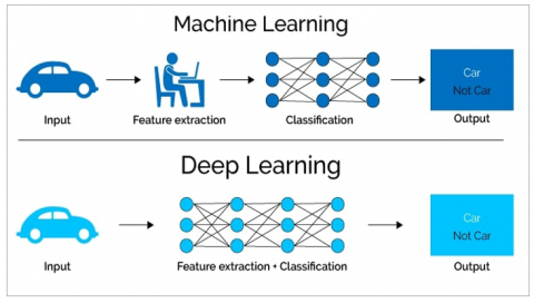
Figure 1. Machine Learning vs. Deep Learning [20]
Machine Learning (ML) serves as the foundational phase that concentrates on uncovering and examining patterns from a dataset. This process enables machines to incrementally improve their performance by identifying or learning informative features that accurately depict regularities or patterns in the data. However, the application of Machine Learning techniques can pose challenges to individuals lacking domain expertise. Traditionally, meaningful or task-relevant features have been primarily generated by human specialists, drawing upon their understanding of the target domain [21].
Deep Learning (DL), an evolution of this concept, encompasses multi-layered neural networks that empower machines to learn independently and make autonomous decisions. Machine Learning evaluates a given situation using distinct features that underscore patterns. This facilitates the "learning" process and allows the application of this acquired knowledge to comparable situations in the future. Predictive technologies, bolstered by Machine Learning, can augment patient care by supporting clinical decision-making [22]. Figure 1 illustrates the distinction between ML and DL.
This section presents a curated compendium of prior studies focused on the diagnosis of tumors. Over the years, numerous researchers have devoted efforts to brain tumor identification, yielding promising results:
Seetha and Raja [23] introduced a method for the classification of convolutional neural networks (CNNs) aimed at detecting brain tumors. The proposed architecture, deeper in nature, utilizes smaller kernels, and the neurons carry comparatively lower weight. As per experimental results, the proposed CNN model demonstrated an accuracy of 97.5%.
Hossain et al. [24] suggested the deployment of the Fuzzy C-Means clustering technique for brain tumor extraction from 2D Magnetic Resonance Brain Images (MRI). This approach was succeeded by traditional classifications and convolutional neural networks. Although the suggested use of CNN achieved an accuracy rate of 97.87%, it was observed to necessitate a longer execution time and larger data storage.
Toğaçar et al. [25] proposed a novel CNN model named Brain MRNet, which incorporates a residual network designed using hypercolumn technology and attention modules. The initial step in Brain MRNet is image preprocessing, followed by the application of image augmentation techniques. After attention modules have selected essential parts of the image, it is received by the convolution layer. The Brain MRNet model was utilized for brain tumor identification using readily available MR images, achieving a classification accuracy of 96.05%.
Van Hai and Amaechi [26] proposed a model that combines convolutional neural networks and fuzzy rules, intended for the classification and detection of medical imaging, including the differentiation between normal and malignant brain cells. The experimental results of the proposed model demonstrated an accuracy of 97.6%.
Lamrani et al. [27] examined a proposed CNN architecture designed for the classification of MRI brain images into tumor-present and tumor-absent categories. These medical images underwent preprocessing and resizing prior to CNN processing. Based on training and testing outcomes, the pre-trained architectural model achieved classification accuracy and precision rates of 96%.
Brain tumor must be diagnosed as soon as possible because there is a lot of overlap between types of brain tumor.so need to:
A reliable automatic classification system is necessary to reduce the death rate of people. Introduce automatic system for detection and diagnosis brain tumors to have good results in a high accuracy within little computing time and not sensitive for noisy data.
Applying computer-aided methods to models that can distinguish between abnormal and normal brain pictures MR imaging will produce better results than manual, conventional diagnostic methods and the objective of this work is:
This work proposed the automatic model to diagnosis some types of the brain tumor (glioma, meningioma, pituitary and non-tumor) used Fuzzy Convolutional Neural Network. That can be achieved by using Google Colab [28] or Colaboratory [29]. The study is composed of several parts, beginning with the importation of the dataset from website, and a number of images in real patients from the Neurological Wholesale Hospital in Baghdad. The pre-processing of the dataset.
The dataset was subsequently split into training and testing sets. On the dataset, the models were trained. The accuracy of the model was calculated for training and testing. Figure 2 shows the proposed system's architectural layout.
Figure 2. System architecture
4.1 Dataset descriptions
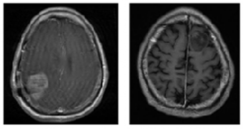
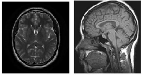
The website dataset used under the name of “Brain Tumor MRI Dataset” is (156 MB) datase comprises 7022 images of human brain MRIs that have been divided into 4 categories, as illustrated in Figure 3: glioma, meningioma, non-tumor, and pituitary. Thereafter, The MRI image dataset has been divided into two sets: a training set and a testing set. The training set contains 5,712 images, which corresponds to 81.4% of the total dataset. The remaining 18.6% of the images, which amounts to 1,311, are included in the testing set. Including subfolders for each class (glioma, meningioma, pituitary, and non-tumor), the dataset has been divided into the Training and Testing folders. There are 1,321 glioma photos,1339 meningioma pictures, 1,595 non-tumor photos and 1,457 images for pituitary in the training folder. The testing folder contains 300 images for glioma, 306 images for meningioma, 405 images for non-tumor and 300 images for pituitary as showed in Table 1.
Also, the proposed model is tested on Neurological Wholesale Hospital in Baghdad database which consists of 470 MRI image to obtain real diagnoses for real patient. It was also tested with taken website dataset.
(a) Glioma tumor
(b) Meningioma tumor
(c) Pituitary tumor
(d) Non-tumor
Figure 3. Brain tumor MRI dataset
Table 1. Dataset details
|
Dataset of Website |
Glioma |
Meningioma |
Pituitary |
Non-Tumor |
|
Training set |
1321 |
1339 |
1457 |
1595 |
|
Testing set |
300 |
306 |
300 |
405 |
4.2 Data process
Medical images, especially images of brain tumors, were processed to help and facilitate their diagnosis and extract features through the use of four pre-processing steps, which are resizing, rescaling, random Flip and random Rotation with fixed size of all image is 128×128.
On these images, we planned to use Deep Learning (DL) Diagnosis techniques. As a result, we created a scale between 0 and 1 that is consistent with the approaches.
4.3 Fuzzy Convolutional Neural Network architecture
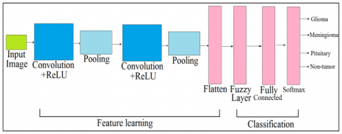
Medical images have great resolution, which makes image processing difficult. FCNN makes decisions throughout disease diagnosis by using a variety of methods and makes it simple to anticipate and categorize the condition [30]. The complete system for categorization is created by combining the data gathered by CNN and fuzzy. CNNs are used to investigate patterns in images. Convoluting an image and searching for patterns are used to accomplish this. Fuzzy layer aids in decision-making for tumor diagnosis and increases precision. This characteristic makes CNNs highly good at identifying objects in pictures [31]. The suggested approach based of MRI images to diagnosis brain tumors using FCNNs. The four fundamental layers that make up the architecture of an FCNN are the convolutional layer, the pooling layer, the fuzzy layer and the fully connected layer. Figure 4 presents the proposed a FCNN model.
Figure 4. Proposed a FCNN model
4.4 Evaluation metrics
One metric is accuracy. The percentage of accurate predictions for a model is how accuracy is defined. A comparison between the output images and the ground truth images that are included in the dataset is done as evaluation metrics for the FCNN model's output analysis. The expert's expertise was used to generate the ground truth photographs. An image that has been correctly classified as a tumor is true negative (TN), whereas an image that has been correctly classified as a non-tumor is true positive (TP) [32]. False positive (FP) denotes photos of tumors that were wrongly identified, whereas false negative (FN) denotes images of non-tumors that were incorrectly diagnosed [33].
The comparison with the real-world photographs is used to determine these factors. A metrics for assessing performance are mentioned below, with numbers ranging from 0 to 100. These evaluation criteria are computed using Eq. (1) [34].
$accuracy$$=\frac{T P+T N}{T P+T N+F P+F N}$ (1)
4.5 Model architecture
The model contains four convolutional layers, with the first layer having 64 filters using the ReLU function. 64 filters in second convolutional layer use the ReLU function, 64 filters in third convolutional layer use the ReLU function, and 64 filters in fourth convolutional layer use the ReLU function. After activation layers (ReLU function), batch normalization layers are utilized. After each batch normalization layers, max-pooling layers in 2×2 dimensions are employed. These layers are followed by a dropout layer (was set to 0.2). There are three dense layers employed, the first and the second layers each having 512 output perceptron’s that use ReLU, with layer fuzzy inference block is added to aid in the decision-making process of diagnosis, Third dense layer with four perceptron. Reduced learning rate to 0.001. The training was stopped after 500 epochs. Table 2 displays the specific elements and parameters of a (FCNN) model. Different hyper-parameters are used in the FCNN model. The hyper-parameter settings for the FCNN model are displayed in Table 3. Classification consists in labeling the constituent elements of an image according to a predefined rule. It involves the use of an algorithm that assigns labels to groups.
Table 2. The proposed FCNN parameters
|
Layers (Type) |
Output of Shape |
Parameter |
|
resizing_1 (Resizing) |
(None,128,128,3) |
0 |
|
rescaling_1 (Rescaling) |
(None,128,128,3) |
0 |
|
random_flip_1 (Random Flip) |
(None,128,128,3) |
0 |
|
random_rotation_1(Random Rotation) |
(None,128,128,3) |
0 |
|
conv2d_4 (Conv2D) |
(None,124,124,64) |
4864 |
|
activation_7 (Activation) |
(None,124,124,64) |
0 |
|
batch_normalization_4 (Batch Normalization) |
(None,124,124,64) |
256 |
|
max_pooling2d_4 (MaxPooling2D) |
(None,62,62,64) |
0 |
|
dropout_6(Dropout) |
(None,62,62,64) |
0 |
|
conv2d_5 (Conv2D) |
(None,60,60,64) |
36928 |
|
activation_8 (Activation) |
(None,60,60,64) |
0 |
|
batch_normalization_5 (Batch normalization) |
(None,60,60,64) |
256 |
|
max_pooling2d_5 (Max Pooling 2D) |
(None,30,30,64) |
0 |
|
dropout_7 (Dropout) |
(None,30,30,64) |
0 |
|
conv2d_6 (Conv2D) |
(None,28,28,64) |
36928 |
|
activation_9 (Activation) |
(None,28,28,64) |
0 |
|
batch_normalization_6 (Batch Normalization) |
(None,28,28,64) |
256 |
|
max_pooling2d_6 (MaxPooling2D) |
(None,14,14,64) |
0 |
|
dropout_8 (Dropout) |
(None,14,14,64) |
0 |
|
conv2d_7 (Conv2D) |
(None,12,12,64) |
36928 |
|
activation_10 (Activation) |
(None,12,12,64) |
0 |
|
batch_normalization_7 (Batch Normalization) |
(None,12,12,64) |
256 |
|
max_pooling2d_7 (Max Pooling2D) |
(None,6,6,64) |
0 |
|
dropout_9 (Dropout) |
(None,6,6,64) |
0 |
|
flatten_1 (Flatten) |
(None,2304) |
0 |
|
dense_3 (Dense) |
(None,512) |
1180160 |
|
activation_11 (Activation) |
(None,512) |
0 |
|
dropout_10 (Dropout) |
(None,512) |
0 |
|
dense_4 (Dense) |
(None,512) |
262656 |
|
fuzzy_inference_block_1 (fuzzy_inference_block) |
(None,512) |
0 |
|
activation_12 (Activation) |
(None,512) |
0 |
|
dropout_11 (Dropout) |
(None,512) |
0 |
|
dense_5 (Dense) |
(None,4) |
2052 |
|
activation_13 (Activation) |
(None, 4) |
0 |
|
The Total params |
1, 561, 540 |
|
|
Trainable params |
1,561, 028 |
|
|
No-trainable params |
512 |
|
Table 3. The developed FCNN model hyper-parameters
|
Dropout Rate |
0.2 |
|
Filter Size |
(5,5)(3,3)(3,3) (64,64,64,64) |
|
Learning Rate |
0.001 |
|
Loss Function |
categorical_crossentropy |
|
Num. of convolution layer |
4 |
|
Num. of epoch |
500 |
|
Num. of fully connected layer |
3(512,512,4) |
|
Num. of Max poling layer |
4(2,2) |
|
Optimizer |
Adam |
The investigated model has been utilized the Google Collaboratory platform based on Python and Keras library on TensorFlow was used. Used of Fuzzy Convolutional Neural Network model to solve the diagnosis task of brain tumor images.
In epoch (1), the elapsed time is 32 second, the parameter of training accuracy is 59.77%, the parameter of loss is 1.6547, the parameter of validation accuracy is 33.03%, and the parameter of validation loss is 2.5674. In epoch (65), the elapsed time is 20 second, the parameter of training accuracy is 93.93%, the parameter of loss is 0.1846, the parameter of validation accuracy is 95.73%, and the parameter of validation loss is 0.1857. In epoch (183), the elapsed time is 19 second, the parameter of training accuracy is 97.15%, the parameter of loss is 0.0912, the parameter of validation accuracy is 98.02%, and the parameter of validation loss is 0.0909.
Table 4. The results of training accuracy and validation accuracy of the FCNN model
|
Epoch |
Time Elapsed |
Loss |
Training Accuracy |
Validation Loss |
Validation Accuracy |
|
1 |
32s |
1.6547 |
0.5977 |
2.5674 |
0.3303 |
|
2 |
20s |
0.8532 |
0.6950 |
2.9656 |
0.3021 |
|
3 |
21s |
0.7228 |
0.7397 |
1.0347 |
0.7292 |
|
9 |
20s |
0.4180 |
0.8479 |
0.6805 |
0.8146 |
|
19 |
21s |
0.3256 |
0.8855 |
0.3308 |
0.8909 |
|
29 |
20s |
0.2748 |
0.9063 |
0.2349 |
0.9306 |
|
40 |
20s |
0.2243 |
0.9251 |
0.2675 |
0.9130 |
|
52 |
20s |
0.1934 |
0.9379 |
0.2871 |
0.9222 |
|
65 |
20s |
0.1846 |
0.9393 |
0.1857 |
0.9573 |
|
86 |
20s |
0.1436 |
0.9533 |
0.0986 |
0.9680 |
|
105 |
21s |
0.1394 |
0.9620 |
0.0921 |
0.9657 |
|
145 |
19s |
0.1164 |
0.9629 |
0.0785 |
0.9725 |
|
168 |
19s |
0.1179 |
0.9648 |
0.0648 |
0.9779 |
|
183 |
19s |
0.0912 |
0.9715 |
0.0909 |
0.9802 |
|
228 |
20s |
0.0772 |
0.9764 |
0.0773 |
0.9840 |
|
260 |
20s |
0.0821 |
0.9750 |
0.0630 |
0.9832 |
|
305 |
20s |
0.0781 |
0.9767 |
0.0359 |
0.9878 |
|
363 |
20s |
0.0582 |
0.9825 |
0.0562 |
0.9863 |
|
412 |
20s |
0.0571 |
0.9820 |
0.0474 |
0.9886 |
|
429 |
20s |
0.0599 |
0.9853 |
0.0374 |
0.9924 |
|
478 |
20s |
0.0473 |
0.9855 |
0.0435 |
0.9931 |
|
500 |
21s |
0.0486 |
0.9860 |
0.0559 |
0.9886 |
Table 5. Comparison of the proposed work with other work
|
REF/Year |
Type of Daises |
Methods |
Accuracy (%) |
|
[23]/2018 |
Brain Tumor |
CNN |
97.5% |
|
[24]/2019 |
Brain Tumor |
FuzzyC-Means+CNN |
97.87% |
|
[25]/2020 |
Brain Tumor |
CNN (Brain MRNet) and hyper column technique+ image augmentation |
96.05% |
|
[26]/2021 |
Brain Tumor |
CNN with fuzzy rules |
97.6% |
|
[27]/2022 |
Brain Tumor |
CNN |
96% |
|
Proposed work |
Brain Tumor |
Fuzzy Convolutional Neural Network |
99.31% |
In epoch (478), the elapsed time is 20 second, the parameter of training accuracy is 98.55%, the parameter of loss is 0.0473, the parameter of validation accuracy is 99.31%, and the parameter of validation loss is 0.0435. Table 4 shows the time elapsed, the loss and training accuracy. Additionally, it displays the suggested model's validation accuracy and validation loss.
There is no need for separate feature extraction stages in the proposed FCNN-based diagnostic. As a result, after train the model to detect brain cancers, complexities and computation time are low its 20 second as well accuracy is high. The diagnosis results of model achieve the 99.31% accuracy for testing and 98.60% accuracy for training. Table 5 presents the compare the proposed work with other researchers.
Cancer constitutes one of the most significant health challenges currently threatening human life, emerging as the second leading cause of mortality subsequent to heart disease. The primary aim of the present research was to design an automatic brain tumor diagnosis system that combines rapidity, accuracy, high performance, low complexity, and time efficiency. The proposed architecture, a Fuzzy Convolutional Neural Network (FCNN), was constructed with the goal of achieving superior accuracy in the classification of glioma, meningioma, pituitary, and non-tumor brain diseases, outperforming other algorithms in terms of speed.
To train and test the model efficiently, an appropriate brain tumor dataset was utilized. Google Colab, built on Keras and TensorFlow, was employed for implementation, utilizing the Brain Tumor MRI Dataset sourced from an online platform and from the Neurological Wholesale Hospital in Baghdad. The evaluation metrics employed included training accuracy, loss, validation accuracy, and validation loss. The model exhibited a validation accuracy of 99.31%, training accuracy of 98.60%, and an execution time per epoch of 20-21 seconds.
Training loss was observed to be exceptionally minimal, while training accuracy was high. The commendable performance of the FCNN suggests its potential utility for medical professionals in the diagnosis of brain tumors in both pediatric and adult populations. By processing a large volume of MRI images swiftly, the system is capable of delivering highly accurate diagnostic results quickly.
Future work aspires to extend this research to 3D brain imaging. The self-contained framework evolved from the model is also planned to be applied to various open-source datasets, including those related to breast cancer, skin cancer, and Alzheimer's disease.
[1] Hussain, S., Mubeen, I., Ullah, N., Shah, S.S.U.D., Khan, B.A., Zahoor, M., Ullah, R., Khan, F.A., Sultan, M.A. (2022). Modern diagnostic imaging technique applications and risk factors in the medical field: A review. BioMed Research International, 2022: 5164970. https://doi.org/10.1155/2022/5164970
[2] Shetty, A., Chandra, S. (2022). Engineered hybrid nanoparticles for multimodal medical imaging and diagnosis. In: Chaughule, R.S., Patkar, D.P., Ramanujan, R.V. (eds) Nanomaterials for Cancer Detection Using Imaging Techniques and Their Clinical Applications. Springer, Cham. https://doi.org/10.1007/978-3-031-09636-5_12
[3] Du, G., Cao, X., Liang, J., Chen, X., Zhan, Y. (2020). Medical image segmentation based on u-net: A review. Journal of Imaging Science & Technology, 64(2): 1-12.
[4] Aastha, Mishra, S., Mohanty, S. (2022). Integration of Machine Learning and IoT for assisting medical experts in brain tumor diagnosis. In: Pattnaik, P.K., Vaidya, A., Mohanty, S., Mohanty, S., Hol, A. (eds) Smart Healthcare Analytics: State of the Art. Intelligent Systems Reference Library, vol 213. Springer, Singapore. https://doi.org/10.1007/978-981-16-5304-9_10
[5] Cancer. https://www.who.int/news-room/fact-sheets/detail/cancer, accessed on Nov. 11, 2022.
[6] Ostrom, Q.T., Cioffi, G., Gittleman, H., Patil, N., Waite, K., Kruchko, C., Barnholtz-Sloan, J.S. (2019). CBTRUS statistical report: primary brain and other central nervous system tumors diagnosed in the United States in 2012-2016. Neuro-oncology, 21(Supplement_5): v1-v100. https://doi.org/10.1093/neuonc/noz150
[7] Zhou, L., Zhang, Z., Chen, Y.C., Zhao, Z.Y., Yin, X.D., Jiang, H.B. (2019). A deep learning-based radiomics model for differentiating benign and malignant renal tumors. Translational Oncology, 12(2): 292-300. https://doi.org/10.1016/j.tranon.2018.10.012
[8] Choudhury, C.L., Mahanty, C., Kumar, R., Mishra, B.K. (2020). Brain tumor detection and classification using convolutional neural network and deep neural network. In 2020 International Conference on Computer Science, Engineering and Applications (ICCSEA), Gunupur, India, pp. 1-4. https://doi.org/10.1109/ICCSEA49143.2020.9132874
[9] Liu, X., Zhang, R., Meng, Z., Hong, R., Liu, G. (2019). On fusing the latent deep CNN feature for image classification. World Wide Web, 22: 423-436. https://doi.org/10.1007/s11280-018-0600-3
[10] Kugunavar, S., Prabhakar, C.J. (2021). Convolutional neural networks for the diagnosis and prognosis of the coronavirus disease pandemic. Visual Computing for Industry, Biomedicine, and Art, 4(1): 12. https://doi.org/10.1186/s42492-021-00078-w
[11] Deepika, J., Rajan, C., Senthil, T. (2021). Security and privacy of cloud-and IoT-based medical image diagnosis using Fuzzy Convolutional Neural Network. Computational Intelligence and Neuroscience, 2021: 1-17. https://doi.org/10.1155/2021/6615411
[12] Sarvamangala, D.R., Kulkarni, R.V. (2022). Convolutional neural networks in medical image understanding: A survey. Evolutionary Intelligence, 15(1): 1-22. https://doi.org/10.1007/s12065-020-00540-3
[13] Liu, L., Zhang, H., Wu, J., Yu, Z., Chen, X., Rekik, I., Wang, Q., Lu, J., Shen, D. (2019). Overall survival time prediction for high-grade glioma patients based on large-scale brain functional networks. Brain Imaging and Behavior, 13: 1333-1351. https://doi.org/10.1007/s11682-018-9949-2
[14] Chieffo, D.P.R., Lino, F., Ferrarese, D., Belella, D., Della Pepa, G.M., Doglietto, F. (2023). Brain tumor at diagnosis: from cognition and behavior to quality of life. Diagnostics, 13(3): 541. https://doi.org/10.3390/diagnostics13030541
[15] Choi, K., Kim, D.Y., Kim, H.J., Hwang, G., Kim, M.K., Kim, H.G., Paik, S. (2022). Imaging features and pathological correlation in mixed microcystic and angiomatous meningioma: A case report. Journal of the Korean Society of Radiology, 83(4): 951. https://doi.org/10.3348/jksr.2021.0119
[16] Alsumali, A., Cote, D.J., Regestein, Q.R., Crocker, E., Alzarea, A., Zaidi, H.A., Bi, W.L., Dawood, H.Y., Broekman, M.L., van Zandvoort, M.J.E., Mekary, R.A., Smith, T.R. (2017). The impact of transsphenoidal surgery on neurocognitive function: A systematic review. Journal of Clinical Neuroscience, 42: 1-6. https://doi.org/10.1016/j.jocn.2017.01.015
[17] Papadomanolakis, T.N., Sergaki, E.S., Polydorou, A.A., Krasoudakis, A.G., Makris-Tsalikis, G.N., Polydorou, A.A., Afentakis, N.M., Athanasiou, S.A., Vardiambasis, I.O., Zervakis, M.E. (2023). Tumor diagnosis against other brain diseases using T2 MRI brain images and CNN binary classifier and DWT. Brain Sciences, 13(2): 348. https://doi.org/10.3390/brainsci13020348
[18] Buchlak, Q.D., Esmaili, N., Leveque, J.C., Bennett, C., Farrokhi, F., Piccardi, M. (2021). Machine learning applications to neuroimaging for glioma detection and classification: An artificial intelligence augmented systematic review. Journal of Clinical Neuroscience, 89: 177-198. https://doi.org/10.1016/j.jocn.2021.04.043
[19] Ahmadi, M., Dashti Ahangar, F., Astaraki, N., Abbasi, M., Babaei, B. (2021). FWNNet: Presentation of a new classifier of brain tumor diagnosis based on fuzzy logic and the wavelet-based neural network using machine-learning methods. Computational Intelligence and Neuroscience, 2021: 8542637. https://doi.org/10.1155/2021/8542637
[20] Deep Learning vs. Machine Learning – What’s The Difference? https://levity.ai/blog/difference-machine-learning-deep-learning, accessed on Mar. 21, 2023.
[21] Schulz, M.A., Yeo, B.T., Vogelstein, J.T., Mourao-Miranada, J., Kather, J.N., Kording, K., Richards, B., Bzdok, D. (2020). Different scaling of linear models and Deep Learning in UKBiobank brain images versus machine-learning datasets. Nature Communications, 11(1): 4238. https://doi.org/10.1038/s41467-020-18037-z
[22] Greenhill, A.T., Edmunds, B.R. (2020). A primer of artificial intelligence in medicine. Techniques and Innovations in Gastrointestinal Endoscopy, 22(2): 85-89. https://doi.org/10.1016/j.tgie.2019.150642
[23] Seetha, J., Raja, S.S. (2018). Brain tumor classification using convolutional neural networks. Biomedical & Pharmacology Journal, 11(3): 1457. http://dx.doi.org/10.13005/bpj/1511
[24] Hossain, T., Shishir, F.S., Ashraf, M., Al Nasim, M.A., Shah, F.M. (2019). Brain tumor detection using convolutional neural network. In 2019 1st International Conference on Advances in Science, Engineering and Robotics Technology (ICASERT), Dhaka, Bangladesh, pp. 1-6. https://doi.org/10.1109/ICASERT.2019.8934561
[25] Toğaçar, M., Ergen, B., Cömert, Z. (2020). BrainMRNet: Brain tumor detection using magnetic resonance images with a novel convolutional neural network model. Medical Hypotheses, 134: 109531. https://doi.org/10.1016/j.mehy.2019.109531
[26] Van Hai, P., Amaechi, S.E. (2021). Convolutional neural network integrated with fuzzy rules for decision making in brain tumor diagnosis. International Journal of Cognitive Informatics and Natural Intelligence (IJCINI), 15(4): 1-23. https://doi.org/10.4018/IJCINI.20211001.oa47
[27] Lamrani, D., Cherradi, B., El Gannour, O., Bouqentar, M.A., Bahatti, L. (2022). Brain tumor detection using mri images and convolutional neural network. International Journal of Advanced Computer Science and Applications, 13(7): 452-460. https://doi.org/10.14569/IJACSA.2022.0130755
[28] Welcome To Colaboratory - Colaboratory. https://colab.research.google.com/, accessed on Oct. 31, 2022.
[29] Musallam, A.S., Sherif, A.S., Hussein, M.K. (2022). A new convolutional neural network architecture for automatic detection of brain tumors in Magnetic Resonance Imaging images. Access, 10: 2775-2782. https://doi.org/10.1109/ACCESS.2022.3140289
[30] Alhussein, M., Muhammad, G., Hossain, M.S., Amin, S.U. (2018). Cognitive IoT-cloud integration for smart healthcare: Case study for epileptic seizure detection and monitoring. Mobile Networks and Applications, 23: 1624-1635. https://doi.org/10.1007/s11036-018-1113-0
[31] Kumar, P., Srivastava, S., Mishra, R.K., Sai, Y.P. (2022). End-to-end improved convolutional neural network model for breast cancer detection using mammographic data. The Journal of Defense Modeling and Simulation, 19(3): 375-384. https://doi.org/10.1177/1548512920973268
[32] Mishra, N.K., Singh, P., Joshi, S.D. (2021). Automated detection of COVID-19 from CT scan using convolutional neural network. Biocybernetics and Biomedical Engineering, 41(2): 572-588. https://doi.org/10.1016/j.bbe.2021.04.006
[33] Choi, B.K., Madusanka, N., Choi, H.K., So, J.H., Kim, C.H., Park, H.G., Bhattacharjee, S., Prakash, D. (2020). Convolutional neural network-based MR image analysis for Alzheimer’s disease classification. Current Medical Imaging, 16(1): 27-35. https://doi.org/10.2174/1573405615666191021123854
[34] Serte, S., Demirel, H. (2021). Deep learning for diagnosis of COVID-19 using 3D CT scans. Computers in Biology and Medicine, 132: 104306. https://doi.org/10.1016/j.compbiomed.2021.104306