Maniraj Subramanian Pitchiah* | Thamizhamuthu Rajamanickam
© 2022 IIETA. This article is published by IIETA and is licensed under the CC BY 4.0 license (http://creativecommons.org/licenses/by/4.0/).
OPEN ACCESS
Skin cancer is the most prevalent and deadliest kind of cancer. Melanoma is the most dangerous type of skin cancer, but it can be detected earlier and successfully treated. The dermoscopic image classification using Machine Learning (ML) approaches in identifying melanoma is increased over the last two decades. The proposed classification system involves three stages. Initially, the pre-processing employs the median filter and thresholding approach aids to remove the hairs and unwanted noise. Then, the shape components, Asymmetry, Border Irregularity, Colour and Dermoscopic structure (ABCD) rule, and Grey Level Co-occurrence Matrix (GLCM) features are utilized to extract the skin lesion region. After that, the K-Nearest Neighbor (KNN), Random Forest (RF), and Support Vector Machine (SVM) classifiers are employed to perform melanoma skin classification from the skin lesion. The dermoscopic skin images used in this study are obtained from the PH2 database. Finally, the SVM classifier outperformed the other classifiers in identifying melanoma skin cancer, providing 94.81% efficiency.
dermoscopic images, machine learning, support vector machine, K-Nearest Neighbor, ph2 database, grey level co-occurrence matrix
Cancer, also known as malignant tumours, is the uncontrolled growth of cells in a specific region in the human body. Any malignancy that develops from skin cells is referred to as skin cancer. The skin is the thickest and most substantial organ in the human body, making up about one-twelfth of the total mass of the human body. It is one of the biggest organs since, in adults, it takes up around two square meters of space. In addition, it accounts for between 18% and 40% of the body's overall water content. The functions of skin include (i) protection against damage caused by both mechanical and chemical agents is one of the skin's primary tasks, (ii) the experiences of touch, discomfort, and temperature, (iii) the maintenance of an appropriate internal temperature, and (iv) the establishment of a barrier against the evaporation of bodily water.
The most prevalent form of skin cancer, known as basal cell carcinoma, accounts for around 70 percent of all occurrences. Squamous Cell Carcinoma (SCC) is characterized by the invasion of epidermal cells into the dermis, which causes the basal layer and its basement membrane to become disorganized. SCC does not spread. However, it does cause areas of minor scaling within an otherwise erythematous and well-defined lesion. Last but not least, malignant melanoma (MM) is a rare form of the neoplasm known as melanoma that develops at the junction of the dermis and the epidermis as a tumour of the melanocytes. Melanocytes are cells that generally occupy positions in the basal layer and are responsible for producing the pigment known as melanin. The presence of many moles is an essential constitutional risk factor for MM. It also includes freckling and a family history of MM.
Skin cancer mortality rate has risen dramatically in recent years. The survival rate has also improved as detection and treatment methodologies have advanced. According to the American Cancer Society, skin cancer will be diagnosed in 60190 men and 40160 women in 2020, with 6850 fatalities. It's common in men, but more prevalent in women under 50 [1]. Also, the incidence of skin cancer in whites (2.6%) is 20 times more than in blacks (0.1%).
Even though there are more than a hundred distinct types of cancer, the earlier any form of cancer is diagnosed, the higher the chance may be cured by therapy. The signs of skin cancer often don't appear until the illness has progressed to a more advanced stage. This makes skin cancer a very deadly disease. Any therapy to remove the malignant tissue at this time has a significant mortality and morbidity rate due to the advanced stage of the disease. Patients with a high risk of acquiring skin cancer are examined reqularly to identify pre-cancerous cells in the hope that they may be removed before they progress into a cancerous tumour. On the other hand, these pre-cancerous cells are not visible to the naked eye, and their distribution is, by nature, unpredictable. Because of this, the traditional practice of randomly obtaining biopsies to discover any malignant cells that may be present is a procedure that is both incorrect and time-consuming.
Skin cancer classification based on visual information necessitates using highly competent dermatologists and is also a time-consuming technique. An automated Machine Learning (ML) classification system is being developed to examine skin cancer more accurately and detect it at the earliest. A reliable image processing method has been developed in this work for skin cancer diagnosis using dermoscopic image classification. It uses three different features, such as shape components, ABCD rule, and GLCM features, and three classifiers, such as KNN, RF, and SVM, are employed. The highest classification accuracy achieved by the proposed system is 94.81% on the PH2 database while using the SVM classifier.
This paper is organized as follows: Section 2 focuses on the literature survey. It reviews various techniques covering skin cancer diagnosis using dermoscopic images. Section 3, explains the approach for the computer-based melanoma classification system utilizing the ML methodology. Section 4 presents the findings and explanation of the proposed approach described in section 3, and a comparative analysis to other classification systems. The overview of the work is included in the final section with future scope.
SVM and Neural Network (NN) classifiers providing the automatic lesion detection system are discussed by Farooq et al. [2]. The dermoscopic images are preprocessed to detect the skin lesions automatically. The skin lesion class labels comprise two components: the hidden layers in which the features are extracted at the end of processing. Then, the Fully Connected (FC) layers [3] of the NN model are used for the actual classification task. The NN ensemble model is described in the study of Anas et al. [4] for dermoscopy image classification. Initially, a self-generating neural network is used to segment the skin lesion. Color, shape, and texture are extracted as feature descriptors. For classification, a network ensemble model that combines back propagation and a fuzzy-based network is employed [5].
Menzies scale method with ABCD rule-based checklist approach for pattern analysis and early cancer diagnosis is explained in the study of Kasmi and Mokrani [6]. The preprocessing includes the Gabor filters to remove the skin hairs and the geodesic active contour approach to find the lesion boundaries. Then, the ABCD rule is employed to extract the features from the image, and the total dermoscopy score is calculated for classifying malignant and benign skin lesions. SVM classifier with ABCD rule-based feature diagnosis of melanoma skin cancer is explained in the study of Firmansyah et al. [7]. The preprocessing includes the median filters to remove the skin hairs and the geodesic active contour approach to find the lesion boundaries. Then, the GLCM algorithm [8] is employed to extract the features from the image, and the total dermoscopy score is calculated and the SVM classification is presented. This involves incorporating dermoscopic image processing methods with the Stolz algorithm [9]. The detection is carried out on an image captured using a mobile camera device and the testing is done on the same device. The ABCD is used to detect and separate benign tumours from melanoma, potentially allowing for early identification of melanoma [10]. The pre-processing provides for the automated recognition of hair based on lesion borders, whereas Gabor filters exploit the geodesic of active contours.
The Artificial NN (ANN) based [11] back-propagation neural network technique employed to classify skin cancer using a weighted least squares architecture with texture and colour feature extraction is discussed by Thurnhofer-Hemsi and Dominguez [12]. The edge-preserving decomposition information of the original image contributes to the normalized symmetrical GLCM technique, and the histogram of oriented gradients approach is used in the neural network-based extreme learning machine model. A new approach to the problem involves drawing a bounding box around the impacted areas and using regression techniques to reduce the search space [13]. The three phases of the system are to (i) enhance the data, ii) boundary extraction, iii) Deep Convolutional Neural Network (DCNN) feature extraction, and selection using exclusive with regression to generate a bounding box around the afflicted portions of the skin lesion. It aids in the reduction of search space, classification accuracy, and feature extraction processing time.
Multiclass skin cancer classification using a two-stream DCNN information fusion framework is presented in the study of Attique Khan et al. [14]. It has two steps. The first step is a fusion-based contrast enhancement approach that feeds improved images to the DenseNet201 architecture that is already trained. A skewness-controlled moth flame optimization approach is then used to optimize the extracted feature characteristics. The suggested feature selection approach extracts and down-sample deep features from the fine-tuned MobileNetV2 pretrained network in the second stream. Finally, the multi-max coefficient correlation approach with the most discriminant characteristics from both networks is fused. Classifying lesion images is done with a multiclass extreme learning machine classifier.
Convolution Neural Network (CNN) model with Transfer Learning (TL) approach and skin cancer classifier is explained in the study of Balaji et al. [15]. For the classification, this paper employed a GoogLeNet, Inception v3 model that was pre-trained using the ImageNet with huge images. It also included comparative studies of edge detection, segmentation, and image pre-processing techniques. Using TL, the CNN model utilizes fine-tuned end-to-end learning to classify the skin lesions [16]. DCNN and TL based approach for skin cancer classification is discussed in the study of Dutta et al. [17]. The suggested framework was trained and tested using the normalized and data-augmented images in the preprocessing stage. Then, the CNN model classifies the images using the weights of the predicted label in the convolutional and pooling layers. Finally, the FC layer with the TL method classifies the skin cancer images utilizing the global average pooling approach.
The automated diagnostic supporting network system is intended to reduce time and resources in diagnosing skin cancer are explained in the study of Masood et al. [18]. In ANN learning algorithms for skin cancer diagnosis, the most critical processes are feature extraction utilizing the segmentation process with pattern recognition and skin cancer detection. SVM is used for classification, and the Levenberg-Marquardt algorithm helps distinguish between malignant and benign melanoma lesions.
Dermoscopy, a non-invasive skin imaging technology, is a visualization approach for identifying characteristics utilizing the pigmented melanocytic neoplasm [19]. The colour characteristic information is used to distinguish between the malignant and benign varieties of melanoma illnesses, which are the most lethal types of skin cancer. The ABCD rule and the current colour, architecture, symmetry, and homogeneity algorithm are used to directly or indirectly include colour as a diagnostic characteristic in the dermoscopy system. The determination of the malignancy score is used in this algorithm-based method. Contourlet transform is employed for melanoma classification [20]. It uses Contourlet sub-band energy features for the classification by Bayesian classifier.
A self-supervised clustering approach for unlabeled skin lesion classification is discussed in the study of Wang et al. [21]. Combining two and three-dimensional wavelet features might aid in the earlier detection of melanoma, the fatal kind of skin cancer. The differential box-counting approach with a fractal feature-based probabilistic classifier is explained in the study of Jacob and Rosita [22]. The fractal properties are obtained using a differential box-counting method. The fractal images utilized as features for the given image are broken down using the energy features generated from the empirical wavelet transform approach.
After a scan of the relevant literature, it is discovered that only a limited quantity of research material about combining several features and classifiers is involved in effective skin image feature extraction and categorization. Nevertheless, it has been shown that the image processing algorithms described in this section are capable of performing well in the area of skin cancer image classification. Because there is a shortage of study data accessible into a probable relationship between the normal and abnormal types of dermoscopic images and their combination of features, texture analysis has been used in diagnosing skin cancer. The following section discusses an efficient skin cancer classification method using three distinct texture features with three different classifiers.
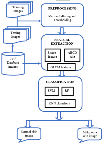
The image processing and pattern recognition algorithms used to classify skin cancer using dermoscopic images are discussed in this section. The research approach is based on the hierarchical model of computer vision, which includes preprocessing, feature extraction stage, and classification using the dermoscopic images from the PH2 database. The proposed architecture of our research work is depicted in Figure 1.
Figure 1. Melanoma skin cancer classification system - block diagram
After sending an image to the system, it is preprocessed using the median filtering approach. The watershed algorithm will be used to segment the pre-processed image. Then, the segmented images are subjected to feature extraction. Finally, classification techniques are employed to discover the best outcomes; three different forms of classification are employed. Features such as shape, GLCM approach and ABCD rules are extracted. Then, they are classified using KNN, RF and SVM classifiers to diagnose melanoma in dermoscopic images. The SVM classifier approach is examined to provide high performance among all the classifiers.
3.1 Image preprocessing
In the preprocessing phase, the median filter is used for the entire image to improve reliability and remove dermoscopic hairs. The median filter is widely employed in medical image analysis because it is thought to be the best and most successful way to reduce noise from skin cancer images. A median filter is used to maintain the amplitude and location of edges. The median filters smooth the images by applying median values. In contrast to the mean channel, the median channel is frequently used to reduce image disorder. Median filter (MF) is employed in this study due to its advantages over other filters. It is an order statistic filter which operates on the data inside a window of size (2m+1, 2m+1) [23]. It is defined in Eq. (1).
$M F_{i j}=\operatorname{median}\left\{I_{i+k, j+l}: k, l=-m, . . m\right\}$
for $i, j=(m+1) \ldots(n-m)$ (1)
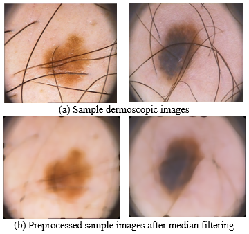
Figure 2 exhibits preprocessed PH2 dermoscopic images with window widths of 21x21 pixels.
Figure 2. Preprocessed sample PH2 dermoscopic image
The median is the average of all the standards of the pixels in the immediate vicinity. Median filtering is particularly effective at ignoring various types of disturbance. This stage results in an output providing the upper quartile frequency image. The median vector distribution results from the median filtering and thresholding of skin lesions images. Noise and hairs are removed from dermoscopic images using a median filter.
3.2 Feature extraction
The feature extraction stage of the recognition and classification approach is as follows: From the segmented image, features are extracted using shape, GLCM, and ABCD rule techniques.
3.2.1 Shape features
Shape-based feature extraction is essentially the comparison of forms represented by their attributes. Shapes may be described using specific superficial geometric characteristics. Because basic geometric characteristics can only distinguish forms with considerable variances, they are often employed to remove false positives or supplemented with additional shape descriptors to distinguish shapes. The image shape is the pixel position listed with the assistance of an arbitrary beginning pixel in shape features derived from digital images. These coordinates may be expressed in Eq. (2) as a vector as follows [24]:
$\vec{s}(r)=u_r \hat{u}+v_r \hat{v}$ (2)
where, $r \in[0, N-1]$ represents the $r^{\text {th }}$ pixel with the contour, and $N-1$ is the total number of pixels. It means that the discrete shape function is time periodic; $\vec{s}(r)=\vec{s}(r+N)$. With the help of each pixel and the equal weight of contour contributing, the shape's centroid and the mass of the shape's centre are determined in Eq. (3) [24]:
$\vec{c}_o=\frac{1}{N} \sum_{r=0}^{N-1} \vec{s}(r)$ (3)
The distance from the centroid (c) images to each point, as well as the image contour are determined. The algorithm for extracting shape features is as follows:
Input: A binary image resulting from the segmentation step of the morphological process. Shape attributes vector is obtained as an output.
Step 1: Determine the size of the image's area.
Step 2: Determine the extent of the lesion.
Step 3: Determine the area distinguished between the full image and the lesion region.
Step 4: Using the equation, calculate the position (2).
Step 5: In the binary image, find the exterior borders of substances, as well as the boundaries of space within these substances.
Step 6: Determine the edge's pixels.
Step 7: Determine the Lesion's border.
Step 8: Determine the extent of the lesion.
Step 9: Using the equation, calculate the centroid position of the lesion region (3).
3.2.2 GLCM features
The GLCM approach is a technique for obtaining second-order statistical texture information. A GLCM is a matrix with the same number of rows and columns as grey pixel levels that analyses the dimensional relationship of pixels using an arithmetical technique. It calculates the combination of pixels in an image with definite principles and appearing in a specific dimensional association. The energy and entropy statistics are defined in Eqns. (4) and (5) respectively [25, 26];
Energy $=\frac{1}{R C} \sum_{i=1}^R \sum_{j=1}^C|\operatorname{skin}(i, j)|$ (4)
Entropy $=-\sum_i P i * \log (p i)$ (5)
where, R and C are considered as the height and width of the skin lesion images at the location i and j.
3.2.3 ABCD rule based features
Skin lesion recognition based on the ABCD rule extracts the four characteristics - Asymmetry (A), Border (B), Color (C), and D (Diameter). These features are retrieved in the following sequence on the preprocessed image. In computerized skin cancer analysis, feature extraction is done utilizing the ABCD characteristics.
1) Asymmetry-Melanoma lesions tend to be asymmetric. The asymmetry index is used to influence the entity's level of symmetry. This is accomplished by splitting the image into parallel or upright halves.
2) Border-The border of melanoma is uneven, ragged, and indistinct. The boundary irregularity is determined using the compactness index.
3) Color-Melanoma is not always the same colour as a normal mole. The colour consistency is determined by the normalized Euclidean distance between each pixel.
4) Diameter-The melanoma lesion is measured to be larger than 6 mm in diameter.
In the next section, all the extracted features in this stage are combined to form the feature space to analyze them independently using KNN, RF, and SVM classifiers.
3.3 Classification
Classification is a critical and significant phase based on the feature extraction, and the skin image lesions should be categorized as melanoma or normal skin images. The classification of skin cancer images utilizes the KNN, RF, and SVM classifiers to diagnose melanoma skin cancer in dermoscopic images.
3.3.1 KNN classifier
The KNN classifier is used as one of the melanoma classification approaches. The Euclidean distance is used to examine the KNN method for classification, which is non-parameterized. The form of nearest neighbor pixel values is the technique of the KNN approach. The recognition of training samples and test sample images are used to complete the skin cancer classification. The samples are classified based on their proximity to the training case. The KNN classifier improves on the approach by selecting the k closest points and reporting the majority sign to diagnose melanoma. It chooses k values that are specific to the categorization. The k output value performs through cross-validation from different subsets. It is better at limiting the noisy impacts of levels in pixels rate within the set of training data.
3.3.2 RF classifier
RF is a supervised learning strategy that overcomes the variance problem to categorize and predicts data. However, it is usually used to tackle categorization problems. A forest is made up of trees, and having more trees means the forest is healthier. Similarly, the RF technique builds decision trees from data samples, extracts forecasts from each data set, and then votes on the best alternative. It's an ensemble method that avoids overfitting by averaging the outcomes, making it superior to a single decision tree algorithm. The working of the RF algorithm is explained using the following steps:
Step 1: Start by selecting random samples from a dataset.
Step 2: For each sample, this algorithm will create a decision tree. The prediction result from each decision tree will be obtained.
Step 3: Voting will be done for each predicted outcome in this step.
Step 4: Finally, chooses the prediction result with the most votes as the final prediction result.
3.3.3 SVM classifier
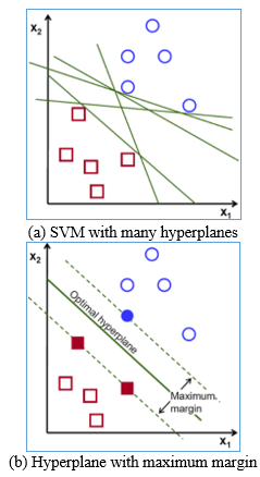
The SVM classifier creates a hyperplane that divides the feature space with the most significant possible margin. The classification step in this study was able to separate the input image into two unique classes: normal and malignant skin images. Features retrieved from the skin lesion site must be used as inputs for the classification step. A classifier with sufficient characteristics must be capable of classifying the dermoscopic lesion features into the desired categories. In many pattern recognition systems, an SVM classifier is utilized. The feature space is formed by combining all the collected features from the feature extraction stage. The ABCD, GLCM, and shape feature spaces are used to classify the provided skin images. The SVM classifier system is shown in Figure 3 with sample parameter classification utilizing many hyperplanes. Therefore, the maximum margin classifier that optimally separates the two classes is the one that greatly saves processing time.
Figure 3. SVM classification
A trained SVM model must predict whether the provided image is a normal image or melanoma skin cancer using the extracted temporal data and their classifications. Separating the multiple classes is done using the weight vector 'w' and a bias 'b' yields a hyperplane. The hyperplane function is written in Eq. (6) [26]:
$w^T x+b=0$ (6)
The data points are classified as normal or malignant depending on the value of the feature space and hyperplane function, it is represented in Eq. (7) as:
Normal if $w^T x+b<0$ and Cancerous if $w^T x+b>0$ (7)
The largest margin classification parameter is used to choose the hyperplane. The points on these two hyper-planes are referred to as support vectors, and the maximum margin classifier will always be parallel and equidistant. Because there is no over-fitting, data in high-dimensional space may be evaluated more quickly with SVM.
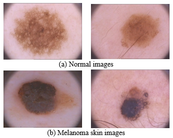
The dermoscopic image classification system results, with a brief discussion are provided in this section. This study employed 200 images from the PH2 database (80 normal, 80 benign and 40 melanoma skin scans) [27, 28]. The dermoscopic images are obtained from the Portugal Hospital, Dermatology Service of Hospital Pedro Hispano. A 20x magnified 8-bit RGB images are acquired using the Tuebinger Mole Analyzer system. The dermoscopic images are colour images with a resolution of 750x520 pixels. Figure 4 shows a selection of images from the PH2 database. The images of skin cancer show that a visual inspection alone is insufficient to detect cancer, necessitating a technology with a more discriminating capacity.
Figure 4. PH2 database sample images
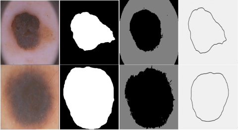
Figure 5 shows the skin lesion image extraction by novel shape features approach. This shows a clear bounded lesion image extraction fed to the classification stage. The shape, color, contour, and texture parameters are employed to extract the features from the dermoscopic images with a clear boundary.
The proposed system with SVM, KNN, and RF classifier approaches is compared using accuracy, sensitivity, and specificity performance measures [29, 30].
Accuracy: The total number of skin images accurately identified according to their categories is referred to as accuracy, and is represented as:
Accuracy $=\frac{T P+T N}{T P+F N+T N+F P}$ (8)
Sensitivity: The capacity of various classifiers in recognizing +ve situations is defined.
Sensitivity $=\frac{T P}{T P+F N}$ (9)
Specificity: The capacity of the classifiers in recognizing -ve situations is defined.
Specificity $=\frac{T N}{T N+F P}$ (10)
where, TP (True Positive) – classification of positive cases as +ve class, TN (True Negative) - negative cases as –ve class, FN (False Negative)- positive cases as –ve class and FP (False Positive) - negative cases as +ve class. Table 1 shows the classification accuracy evaluation of the SVM classifier using the different features.
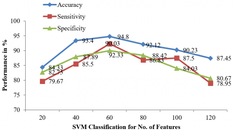
From Table 1, the accuracy is examined for ten runs of the proposed melanoma classification system using ABCD, GLCM, and shape features are evaluated as 94.81%. The SVM classifier provides efficient classification. Figure 6 shows the SVM classifier performance evaluation curve of accuracy, specificity, and sensitivity measure.
Figure 5. Skin lesion image extraction by novel shape features approach
Figure 6. SVM classifier with efficient feature performance curve
Table 1. Accuracy evaluation of the SVM classifier using the special features
|
#Run for SVM classifier using various features |
Accuracy (%) |
||
|
ABCD features |
GLCM features |
Shape features |
|
|
1 |
95 |
95 |
95 |
|
2 |
96 |
94 |
94 |
|
3 |
97 |
93 |
94 |
|
4 |
96 |
95 |
96 |
|
5 |
95 |
95 |
95 |
|
6 |
96 |
96 |
93 |
|
7 |
95 |
95 |
91 |
|
8 |
97 |
94 |
95 |
|
9 |
97 |
96 |
93 |
|
10 |
95 |
96 |
94 |
|
Average |
96 |
94.45 |
94 |
|
Accuracy= 94.81 |
|||
The performance of the melanoma skin image classification system using KNN, RF and SVM classifiers employing the ABCD, GLCM and shape features is shown in Table 2.
Table 2. Accuracy evaluation of the different classifier using the special features
|
# Run using the Features (ABCD rule, GLCM, Shape) |
Accuracy performance (%) |
||
|
KNN |
RF |
SVM |
|
|
1 |
84.36 |
81.64 |
88.93 |
|
2 |
90.98 |
86.45 |
96.57 |
|
3 |
87.69 |
84.33 |
98.95 |
|
4 |
91.72 |
87.5 |
93.89 |
|
5 |
92.98 |
90.94 |
89.86 |
|
6 |
88.45 |
85.37 |
95.79 |
|
7 |
93.79 |
94.55 |
95.72 |
|
8 |
89.94 |
91.88 |
99.99 |
|
9 |
91.92 |
79.99 |
88.92 |
|
10 |
98.86 |
85.5 |
98.95 |
|
Average |
91.169 |
87.615 |
94.817 |
It is evident from Table 2 that the accuracy performance of 86.61% for KNN, and 91.2% for the RF classifier. Both classifiers have lesser performance than the SVM classifier, which has 94.81% accuracy. The accuracy, sensitivity, and specificity measures obtained by SVM, KNN, and RF classifiers are shown in Table 3.
Table 3. Classification system performance for PH2 database images for various classifiers
|
Classifiers |
Accuracy (%) |
Sensitivity (%) |
Specificity (%) |
|
SVM |
94.81 |
92 |
89.88 |
|
RF |
86.61 |
85 |
82.5 |
|
KNN |
91.2 |
90 |
87 |
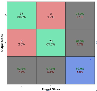
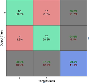
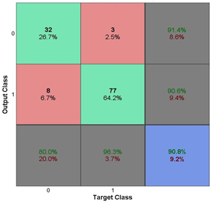
It can be seen from Table 3 that the melanoma classification system provides more promising results for the SVM classifier than other classifiers. The confusion matrices for the two-class prediction using KNN, RF, and SVM classifiers are shown in Figure 7.
The performance evaluation using the features extracted from the ABCD, GLCM, and shape algorithm is inferred from confusion matrices in Figure 7. The confusion matrices depict the number of TP, TN, FP, and FN measures using the Eqns. (8) to (10) for SVM, RF, and KNN classifiers. It can be seen from Figure 8 that the maximum accuracy performance is achieved for the SVM classifier of the system.
(a) SVM
(b) RF
(c) KNN
Figure 7. Confusion matrices of classification system
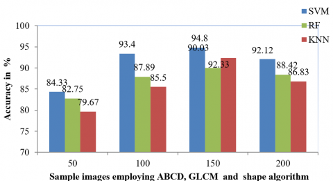
Figure 8. Accuracy analysis of SVM, KNN and RF classifier for melanoma classification
As redundant features are added to the feature subset, performance degrades after achieving maximum accuracy. For performance evaluation of KNN, RF, and SVM classification of melanoma skin cancer, the system takes the fusion of all characteristics. Table 4 shows the comparative analysis of accuracy performance with other existing systems using the PH2 database.
Table 4. Comparative analysis with other existing works using PH2 database images
|
S.No |
Authors |
Techniques and Classifiers using PH2 database |
Performance |
|
1 |
Xie et al. [3] |
Self-generating neural network for color, texture and border features, NN classifier. |
Accuracy – 91.11% |
|
2 |
Anas et al. [4] |
K-means clustering approach, LBP texture feature analysis, SVM classifier. |
Accuracy – 83.33% |
|
3 |
Gurung and Gao [13] |
Data augmentation, Boundary extraction and DCNN feature extraction and CNN classifier |
Accuracy-89.8% |
|
4 |
Balaji et al. [15] |
Fuzzy k-means approach, Firefly optimization and NN classifier. |
Accuracy-91.3% |
|
5 |
Proposed system (2021) |
ABCD features, GLCM and shape features, SVM classifier |
Accuracy- 94.81% |
From Table 4, the proposed methodology with an SVM classifier provides the highest performance than the existing works using PH2 database images. This classification performance is also increased due to more hybrid features. By comparing the results with the existing algorithm, it is observed that the accuracy of SVM classification accuracy is 94.81%.
In this study, three significant modules allow for the precise categorization of dermoscopic images leading to a correct skin cancer diagnosis. Dermoscopic image processing is the initial phase, accomplished using a median filtering method. Hair and sounds are removed with median filtration before feature extraction with a bigger window. The skin cancer diagnostic method is shown utilizing their separate feature descriptors; ABCD, GLCM, and the shape feature vector, respectively from the PH2 database images analyzing the performance of skin cancer diagnosis. Finally, the images are classified using KNN, SVM, and RF classifiers. The SVM, RF, and kNN classifiers are compared, and the SVM outperforms the others. SVM has several advantages, including being accurate and robust even when the training sample is affected. Because the optimality issue is convex, it yields a unique solution. Results prove that the SVM classification approach provides 94.81% classification accuracy, 92% sensitivity, and 89.88% specificity.
[1] Tumpa, P.P., Kabir, M.A. (2021). An artificial neural network based detection and classification of melanoma skin cancer using hybrid texture features. Sensors International, 2: 1-15. https://doi.org/10.1016/j.sintl.2021.100128
[2] Farooq, M.A, Azhar, M.A.M., Raza, R.H. (2016). Automatic lesion detection system (ALDS) for skin cancer classification using SVM and neural classifiers. IEEE 16th International Conference on Bioinformatics and Bioengineering, pp. 301-308. https://doi.org/10.1109/BIBE.2016.53
[3] Xie, F., Fan, H., Li, Y., Jiang, Z., Meng, R., Bovik, A. (2016). Melanoma classification on dermoscopy images using a neural network ensemble model. IEEE Transactions on Medical Imaging, 36(3): 849-858. https://doi.org/10.1109/TMI.2016.2633551
[4] Anas, M., Gupta, K., Ahmad, S. (2017). Skin cancer classification using K-means clustering. International Journal of Technical Research and Applications, 5(1): 62-65. https://doi.org/10.5958/0976-5506.2019.01780.7
[5] Maron, R.C. (2021). A benchmark for neural network robustness in skin cancer classification. European Journal of Cancer, 155: 191-199. https://doi.org/10.1016/j.ejca.2021.06.047
[6] Kasmi, R., Mokrani, K. (2016). Classification of malignant melanoma and benign skin lesions: Implementation of automatic ABCD rule. Implementation of Automatic ABCD Rule. IET Image Processing, 10(6): 448-455. https://doi.org/10.1049/iet-ipr.2015.0385
[7] Firmansyah, H.R., Kusumaningtyas, E.M., Hardiansyah, F.F. (2017). Detection melanoma cancer using ABCD rule based on mobile device. International Electronics Symposium on Knowledge Creation and Intelligent Computing, 127-131. https://doi.org/10.1109/KCIC.2017.8228575.
[8] Maurya, R., Singh, S.K., Maurya, A.K., Kumar, A. (2014). GLCM and multi class support vector machine based automated skin cancer classification. International Conference on Computing for Sustainable Global Development, pp. 444-447. https://doi.org/10.1109/IndiaCom.2014.6828177
[9] Murugan, A., Nair, S.A.H., Kumar, K.S. (2019). Detection of skin cancer using SVM, random forest and kNN classifier. Journal of Medical Systems, 43(8): 1-9. https://doi.org/10.35940/ijeat.B5117.129219
[10] Manne, R., Kantheti, S., Kantheti, S. (2020). Classification of skin cancer using deep learning ConvolutionalNeural networks-opportunities and vulnerabilities-A systematic Review. International Journal for Modern Trends in Science and Technology, 2455-3778. https://doi.org/10.46501/IJMTST061118
[11] Kanimozhi, T., Murthi, A. (2016). Computer aided melanoma skin cancer detection using artificial neural network classifier. Journal of Selected Areas in Microelectronics, 8(2): 35-42. https://doi.org/10.1016/j.sintl.2021.100128
[12] Thurnhofer-Hemsi, K., Dominguez, E. (2021). A convolutional neural network framework for accurate skin cancer detection. Neural Processing Letters, 53(5): 3073-3093. https://doi.org/10.1007/s11063-020-10364-y
[13] Gurung, S., Gao, Y.R. (2020). Classification of melanoma (Skin cancer) using convolutional neural network. International Journal of Advanced Computer Science and Applications, 1-8. https://doi.org/10.1109/CITISIA50690.2020.9371829
[14] Attique Khan, M., Sharif, M., Akram, T., Kadry, S., Hsu, C.H. (2021). A two-stream deep neural network based intelligent system for complex skin cancer types classification. International Journal of Intelligent Systems. https://doi.org/10.1002/int.22691
[15] Balaji, M., Saravanan, S., Chandrasekar, M., Rajkumar, G., Kamalraj, S. (2021). Analysis of basic neural network types for automated skin cancer classification using Firefly optimization method. Journal of Ambient Intelligence and Humanized Computing, 12(7): 7181-7194. https://doi.org/10.1007/s12652-020-02394-0
[16] Brinker, T.J., Hekler, A., Utikal, J.S., Grabe, N., Schadendorf, D., Klode, J., Berking, C., Steeb, T., Enk, A.H., Kalle, C.V. (2018). Skin cancer classification using convolutional neural networks: Systematic review. Journal of Medical Internet Research, 20(10): 11936. https://doi.org/10.2196/11936
[17] Dutta, A., Hasan, M.K., Ahmad, M. (2021). Skin lesion classification using convolutional neural network for melanoma recognition. Proceedings of International Joint Conference on Advances in Computational Intelligence, pp. 55-66. https://doi.org/10.1101/2020.11.24.20238246
[18] Masood, A., Al-Jumaily, A.A., Adnan, T. (2014). Development of automated diagnostic system for skin cancer: Performance analysis of neural network learning algorithms for classification. International Conference on Artificial Neural Networks, pp. 837-844. https://doi.org/10.1007/978-3-319-11179-7_105
[19] Celebi, M.E., Zornberg, A. (2014). Automated quantification of clinically significant colors in dermoscopy images and its application to skin lesion classification. IEEE Systems Journal, 8(3): 980-984. https://doi.org/10.1109/JSYST.2014.2313671
[20] Sonia, R. (2016). Melanoma image classification system by NSCT features and Bayes classification. International Journal of Advances in Signal and Image Sciences, 2(2): 27-33. https://doi.org/10.29284/ijasis.2.2.2016.27-33.
[21] Wang, D., Pang, N., Wang, Y., Zhao, H. (2021). Unlabeled skin lesion classification by self-supervised topology clustering network. Biomedical Signal Processing and Control, 66: 102428-102436. https://doi.org/10.1016/j.bspc.2021.102428
[22] Jacob, S., Rosita, J.D. (2021). Fractal model for skin cancer diagnosis using probabilistic classifiers. International Journal of Advances in Signal and Image Sciences, 7(1): 21-29. https://doi.org/10.29284/ijasis.7.1.2021.21-29
[23] Maniraj, S.P., Sardarmaran, P. (2021). Classification of dermoscopic images using soft computing techniques. Neural Computing and Applications, 33(19): 13015-13026. https://doi.org/10.1007/s00521-021-05998-5
[24] Hussaindeen, A., Iqbal, S., Ambegoda, T.D. (2022). Multi-label prototype based interpretable machine learning for melanoma detection. International Journal of Advances in Signal and Image Sciences, 8(1): 40-53. https://doi.org/10.29284/ijasis.8.1.2022.40-53
[25] Vidya Lakshmi, V., Leena Jasmine, J.S. (2021). A hybrid artificial intelligence model for skin cancer diagnosi. Computer Systems Science and Engineering, 37(2): 233-245. https://doi.org/10.32604/csse.2021.015700
[26] Cheong, K.H., Tang, K.J., Zhao, X., Koh, J.E., Faust, O., Gururajan, R., Ciaccio, E.J., Rajinikanth, V., Acharya, U.R. (2021). An automated skin melanoma detection system with melanoma-index based on entropy features. Biocybernetics and Biomedical Engineering, 41(3): 997-1012. https://doi.org/10.1016/j.bbe.2021.05.010
[27] Mendonça, T., Ferreira, P.M, Marques, J.S., Marcal, A.R., Rozeira, J. (2013). PH2-A dermoscopic image database for research and benchmarking. International Conference on Engineering in Medicine and Biology Society, pp. 5437-5440. https://doi.org/10.1109/EMBC.2013.6610779
[28] PH2 Database Link: https://www.fc.up.pt/addi/ph2%20database.html, accessed on 12 June 2022.
[29] Balamurugan, E., Jackson, A. (2021). Genetic algorithm with bagging for DNA classification. International Journal of Advances in Signal and Image Sciences, 7(2): 31-39. https://doi.org/10.29284/ijasis.7.2.2021.31-39
[30] Arshaghi, A., Ashourian, M., Ghabeli, L. (2020). Detection of skin cancer image by feature selection methods using new buzzard optimization (BUZO) algorithm. Traitement du Signal, 37(2): 181-194. https://doi.org/10.18280/ts.370204