Tamara A. Dawood*![]() | Ashwaq T. Hashim
| Ashwaq T. Hashim![]() | Ahmed R. Nasser
| Ahmed R. Nasser![]()
© 2023 IIETA. This article is published by IIETA and is licensed under the CC BY 4.0 license (http://creativecommons.org/licenses/by/4.0/).
OPEN ACCESS
Skull stripping is regarded as an important pre-processing step by many neuroimaging processing applications. An appropriate skull stripping is crucial because of the complex anatomical makeup of the brain and variations in brain MRI intensity. The removal of the skull region for clinical analysis in brain segmentation tasks is essentially the process of "skull stripping," and its accuracy and effectiveness are very important for diagnostic purposes. It is thought to be a difficult task because it calls for more precise and thorough methods for separating the different regions of the brain and the skull. Consequently, a technique is suggested for skull stripping by improving the contrast of the brain image using Adaptive gamma correction (AGC), which sets its settings dynamically based on the properties of the input image. In addition, the largest connected components, morphological image processing technique, and image multiplications are used in the proposed skull stripping method. The Br35H::Brain Tumor Detection 2020 dataset and Brain MRI Images for Brain Tumor Detection dataset have been used for the experimentation. The results of the experiments show that the proposed image enhancement and skull removal techniques work effectively with an accuracy rate of 96%.
gamma correction, medical image, morphological operation, MRI brain, skull stripping
Medical imaging is crucial to the healthcare system since it aids in diagnosis and decision-making [1, 2]. It enables health doctors to view the body without intrusive treatments, which can significantly enhance patient outcomes. Medical imaging procedures include X-rays, CT scans, MRIs, and ultrasounds, to name a few. These methods provide precise images of inside organs, bones, and other structures using a variety of energies, including electromagnetic radiation and sound waves. Then, these images are employed to aid in disease diagnosis and monitoring, therapy planning and monitoring, and treatment efficacy assessment. Cancer, heart disease, and other dangerous illnesses can all be detected and diagnosed with the help of medical imaging. In general, medical imaging is a vital tool in contemporary medicine that helps patients get better results and saves lives. The brain is one of the most intricately designed organs in the human body and has a staggering number of cells. When cells divide uncontrollably and irregularly, the risk of developing brain tumors rises. The group of cells will harm the healthy cell and impair the behavior of the brain's normal cells, affecting their regular functionality [3, 4]. Many cancer research institutions throughout the world have identified brain tumors as a complex health issue [3].
Due to its benefits of safety and relatively high spatial resolution, the MRI is commonly employed in neonatal diagnostics and research on the development of the brain [5]. The diagnosis of anomalies in the brain, such as brain tumors, is greatly aided by magnetic MRI. The processing quality is harmed by artifacts and unwanted tissues, which could result in unclear diagnoses. Therefore, one of the crucial stages in processing brain imaging is skull stripping, in which the brain tissue is entirely divided from the skull [6]. It is done to reduce unnecessary information in the MR images and to remove non-brain backgrounds. Due to the complicated structure of the brain and the presence of intensity inhomogeneity artifacts in MRI, the task of skull striping for brain imaging is not simple. Cerebrospinal fluid (CSF) space and the skull are black on T1-weighted images. Because of this, the margins between the brain and the skull are well-defined. However, even sharp edges may become distorted during MRI acquisition due to limited resolution or the existence of other anatomical partial structures in the brain (connections between the brain and optic nerves or brainstem). Since about ten years ago, the stripping of the skull has been a crucial pre-processing step in brain imaging. Due to low contrast, hazy brain boundaries, and pixel similarity, automatically extracting the skull from a brain MRI is a difficult task. Using MRI databases with pathological issues makes overall brain extraction more difficult and problematic [7].
There are many algorithms for extracting the brain. However, the outcomes are not sufficient. As a result, a reliable and fully automated brain extraction technique successfully separates the brain part from a brain MRI dataset. is needed. Because of the restrictions of material diversity and imaging modality's spatial resolution, MRI brain volume exhibits a variety of imaging errors, including blurring, noise, partial volume effect, inhomogeneity, and others. It is more challenging to retrieve the brain because of this imaging artifact [8].
The quality of MRI images may suffer due to the limits of image-capturing technologies or the presence of an unfavorable environment. Therefore, there is a need to improve the contrast of the magnetic imaging images before starting the process of skull stripping. In this research, a precise and reliable skull-stripping algorithm is presented. It is possible to use the Gamma Correction technique, but each image needs a different Gamma value according to the specifications of the image itself. As a result, a pre-processed and enhanced MRI image is done using the Adaptive gamma correction (AGC) technique, where the parameters are dynamically set based on the image data. Then the skull is removed from the image using the largest linked components, morphological image processing, and image multiplications. The proposed method is tested using two publicly available datasets: Brain MRI Images for Brain Tumor Detection and 3060 JPG-formatted brain MRI images from the Br35H::Brain Tumor Detection 2020 dataset, which were offered on Kaggle. The method proposed in this study successfully extracts the brain and eliminates non-brain tissues with 96 % accuracy.
The rest of the paper is known as follows: Section 2 discusses similar researches, Section 3 discusses the proposed method of skull stripping, Section 4 presents the results of the experiment, and Section 5 concludes the paper.
Because the skull is a redundant part, it must be removed from the image as an important pre-processing step before the process of brain tumors segmentation and classification to obtain more efficient and accurate results. Numerous techniques are applied in skull stripping studies. In 2015, Roy and Maji [9] developed a method to remove the skull from T1-weighted brain MR images. This technique is entirely intensity-based and incorporates adaptive threshold computation followed by morphological operations. The drawback of this strategy is that it doesn't work on all images with varying contrast levels. In 2016, Benson et al. [10] presents a practical approach based on mathematical morphology for the removal of the skull from brain MRI images. As a Contrast enhancement methodology, histogram equalization was applied. Histogram equalization may not be effective with all MR picture sequences, which is the main issue with this approach [11, 12]. afterward, the skull-stripping attempt will fail. In 2018, Alwawi et al. [13] devised an approach based on global Otsu thresholding and adaptive iterative thresholding. The analysis and deletion of related components come after the global thresholding. To create the brain mask, morphological operations are then used. by choosing the number of pixels required to remove the skull and keep only the brain mask, remove the non-brain component. Although the method convincingly works in the case of 2 dimension brain scans, its only drawback is that it cannot be used in the case of 3 dimension images. In 2018 Laha et al. [14] devised a skull-stripping approach based on morphological procedures, histogram thresholding, and denoising of MRI images. Due to the contrast differences, this procedure does not operate with all MRI pictures, which causes significant holes in the brain mask for some images that cannot be filled in by morphological operations and are thus removed from the original brain image. In 2019, Gao et al. [5] suggested a three-step method for skull stripping that makes use of morphological processing, anisotropic diffusion filtering, and edge detection, and the findings demonstrate that it can properly segment the brain. Without removing important image components, such as edges or other details, anisotropic diffusion minimizes image noise. Finding the appropriate morphology for brain tissue separation can be challenging at times since mathematical morphological methods are sometimes sensitive to little fluctuations in data. In 2019, Akshath and Sheshadri [15] proposed a technique for skull stripping called Threshold and Morphological Operations based Segmentation (TMOS) (Magnetic Resonance Images). It consists of two phases: in the first, the brain region is localized using a Region of Interest (RoI), and in the second, morphological techniques are used to precisely extract the brain region by removing the skull. The disadvantage of this approach is that the RoI is determined using the elliptical mask. This works well with brain MRI images that have ellipse shape and fail with other shapes of brains. In 2019, Naganandhini and Shanmugavadivu [16] suggested an automatic seeded point selection region expanding algorithm and clustering technique to handle MRI image segmentation issues more precisely. Skull stripping separates the brain from the skull and additional meningeal tissues that are visible in the brain picture. A histogram-based threshold-oriented technique was used to perform the skull stripping. The disadvantage of this skull stripping method includes its sensitivity to small data variances and the challenge of determining the ideal morphological size for differentiating brain tissues from other tissues [17]. In 2020, Ullah et al. [7] developed a strategy utilizing histogram equalization techniques for improving the contrast of brain MRI. While skull stripping from an MR brain image is done using morphological image processing techniques. The fundamental drawback of this method is the propensity of the AHE approach to overamplify noise in relatively homogeneous portions of an image. which may lead to failing the stripping of the skull [17]. In 2020, Kalavathi and Prasath [18] Created a system that uses the skull-stripping method and U-Net architecture to segment the brain and automatically detect brain tumors. The threshold value of the probability map's combined grey and white matter has been used to perform skull stripping, but if the estimation and initialization are done incorrectly, poor results are obtained [17]. In 2020, Hussain and Khunteta [19] suggested a technique to isolate the tumor region from MRI images. followed by GLCM methods to extract the features. Using morphological operation, erosion, and dilation, the skull is stripped as a preprocessing step. The employed skull stripping technique is that it can be sensitive to small data fluctuations, and it can be challenging to determine the ideal morphological size for separating brain tissues from other tissues [17]. In 2021, Biratu et al. [20] To identify the abnormality region on brain pictures, they modified the well-known and established region-growing segmentation technique. as a preprocessing step, the skull was removed from each input brain image using a thresholding technique. The drawback of the used skull stripping strategy is that it doesn't work on all brain MRI images with varying contrast levels, so it needs a contrast enhancement before. In 2022, Salman et al. [21] presented an automated brain tumor system that uses hybrid image processing techniques to isolate adjacent organs and other brain tissue from the afflicted location in order to improve localization. The method of removing the skull is initially presented as a pre-processing step to separate the selected brain areas from the undesignated ones. In the suggested method for skull stripping, correct gamma with gamma value 2.5, morphological and mathematical processes are generally used. The drawback of the used skull stripping strategy is that it doesn't work on all images because each image needs a different gamma value depending on its characteristics.
In accordance with the above we proposed our method for skull stripping using adaptive gamma correction to improve the contrast of the dimmed and bright images by selecting gamma values based on the characteristics of each image, otsu thresholding used to binarize the image, extracting the components that are connected and selecting the biggest one which is the brain, applying morphological processes and using region filling to fill the holes to get the brain mask, and finally extract the brain by multiplying the brain mask by the original image. The skull stripping is suggested to automatically remove the unusable regions, such as the meninges, the skull bone, and subcutaneous fat, by using contrast enhancement by adaptive gamma transform, thresholding, arithmetic operation, and morphological operations.
The skull stripping is suggested to automatically remove the unusable regions, such as the meninges, the skull bone, and subcutaneous fat, by using contrast enhancement by adaptive gamma transform [8], thresholding, arithmetic operation, and morphological operations. In order to remove the designated brain regions, the skull stripping operation is a crucial pre-processing step that separates unspecified brain regions. To identify a brain tumor, the resultant images of the affected brain region are evaluated. The proposed skull stripping procedure involves enhancing the image contrast using AGC, applying Otsu’s global thresholding, extracting the components that are connected and finding the biggest one, binaries the largest connected component, applying morphological operations to create the brain mask, and then extracting the brain using mathematical operations. Figure 1 depicts the block diagram of the suggested skull stripping method. Algorithm 1 shows the steps involved in skull stripping.
Figure 1. Block diagram shows the procedure of the proposed skull stripping method
Algorithm 1 illustrates the stages involved in skull stripping.
| Algorithm 1: Skull Stripping |
|
Input: BrainImg Output: Brain out |
|
Step 1: Read an input image from database Step 2: For each image BrainImg (i, j), where i = 1,2,3..., M and j = 1,2,3..., N, compute the threshold t based on the statistical quantity using Eq. (1) to determine the type of image is bright or dimmed $\mathrm{t}=\frac{m_{\mathbf{I}}-T_t}{T_t}$ (1) where: $m_{\mathbf{I}}=\sum_i \sum_j \frac{\mathbf{I}( i, j)}{M N}$ (2) Tt is known as the expected global mean brightness for typical natural images Step3: The threshold used to identify brightness-distorted images from regular ones is called t., and If $\mathrm{t}>-T_t$ then it is judged bright and call algorithm (2) Agcimg=Negative-image-based AGC algorithm. else if t < - Tt, the input image is judged dim and call algorithm (3) Agcimg=CDF-truncated AGC algorithm Step 4: Employ Otsu's global thresholding to get the Agcimg binaries' initial intensity threshold value Binimg=THRESH_OTSU (Agcimg) Step 5: Extract the components that are connected and find the biggest one Step 5.1: Find the connected components Markers=connected components (Binimg) Step 5.2: Calculate the area of each connected component marker_area=np.sum(markers) Step 5.3: Choose the largest connected component largest_component=argmax(marker_area ) Step 6: Create brain_mask Step 6.1: Getting the area taken by the largest connected components and ignore the others since this is the background brain_mask=Binaries (largest_component) Step 6.2: Closing many of the holes that exist in the brain mask by using morphological operation Closing=morphology_close(brain_mask) Step 6.3: Filling the closed contour region with white by using morphological operation Contour=findcontoures (Closing) Brain mask=morphology_open (Contour) Step 7: Extract the brain by multiplying the brain mask with the original image (BrainImg) Brain_out=Brain mask × BrainImg |
3.1 Proposed contrast enhancement method
Contrast Enhancement (CE) is the procedure for enhancing the contrast of an image by adjusting the dynamic range of the pixel intensity allocation [22]. The CE is significant to the advancement of visual quality for digital image processing, pattern recognition, and computer vision. A popular pixel-domain CE technique that is efficient and successful at handling bright and dim images is gamma correction [22, 23]. The manual selection of proper gamma values, however, is frequently time-consuming. Additionally, it is challenging for a large number of images because a separate gamma value is required for each image. Each image needs a different gamma value based on its features; thus, if a single gamma value is set for all images, the resulting images won't be good. In the case of AGC, the statistics extracted from the images are used to automatically create and modify the gamma parameter. In this work, we concentrate on the CE of two different images: dimmed and bright. The use of negative images is employed to accomplish CE of vivid images, and to improve the dimmed images, gamma correction modulated by truncated cumulative distribution function (CDF) is used. As a result, structure distortion and local over-enhancement can be successfully reduced. Figure 2 and Figure 3 show the difference between using gamma correction and adaptive gamma correction for bright and dimmed images.
Figure 2. Apply Gamma correction for a bright image. (a) The input image, (b) Gamma correction with 2.2,
(c) Gamma correction with 2.5, (d) Adaptive gamma correction
Figure 3. Apply gamma correction for the dimmed image. (a) The input image, (b) Gamma correction with 2.2,
(c) Gamma correction with 2.5, (d) Adaptive gamma correction
Algorithm 2 and 3 illustrates the stages involved in adaptive gamma correction.
|
Algorithm 2: Negative-image-based AGC algorithm |
|
Input: BrainImg Output: Ie |
|
Step 1: Apply a negative formula to get the negative image I' of the input image BrainImg, using Eq. (3). I' (i,j)=255 − BrainImg (i,j) (3) Step 2: Get the gray level histogram p(l) of I' , and use Eq (4) to determine $P_w(I)$. $P_w(I)=P_{\max }\left(\frac{P(I)-P_{\min }}{P_{\max }-P_{\min }} \,\, \right)^\alpha$ (4) where α represents the modified parameter, $P_{\max }$=max p(l), $P_{\text {min }}$= min p(l) Step 3: Compute the value of $\gamma_w(I)$ by Eq. (5): $\gamma_w(I)=1-c_w(I)$ (5) $c_w(I)$ represents the CDF the result of normalization $P_w(I)$. Step 4: Perform the transformation of pixel values I' in accordance with Eq. (6) to get Ie'. $I e^{\prime}=\operatorname{round}\left[\ I^{\prime}{ }_{\max } \left(\frac{1}{I^{\prime} \max }\right)^{\gamma\left(I^{\prime}\right)}\right]$ (6) where $\gamma\left(I^{\prime}\right)=\ 1-\mathrm{c} \left(I^{\prime}\right)=\ 1-\sum_{i=o}^I \mathrm{p}(\mathrm{i})$, i = 0, 1, 2, …, 255 is the CDF of the input image's gray - level. The normalized gray-scale histogram is represented by p(x), the rounding process is round [·]. Step 5: Produce the enhanced image $\mathrm{I}_{\mathrm{e}}$ by reversing $\mathrm{Ie}^{\prime}$ such as Eq. (7): Ie=round [255- Ie'] (7) |
|
Algorithm 3: AGC algorithm with a CDF truncation |
|
Input: BrainImg Output: Ie |
|
Step 1: Get the histogram gray-level p(I) within the input image BrainImg Step 2: Calculate $P_w(I)$ depending on Eq. (4) Step 3: Compute $\gamma_w^{\prime}(I)$ by truncating the parameter of the adaptive gamma using Eq. (8) $\gamma_w^{\prime}(I)=\max \left(\tau, 1-c_w(I)\right)$ (8) where the maximizing process is max( ·, ·). The threshold for CDF truncation is $\tau$. The value of $\gamma_w^{\prime}(I)$ would increase to $\tau$ When $c_w(I)$ is larger than 1- $\tau$. Step 4: Produce the image that enhanced Ie by transforming I according to Eq. (4) |
Figure 4 shows the steps involved in skull stripping for the dimmed image, while Figure 5 shows the steps involved for the bright image.
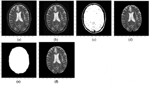
Figure 4. Steps of skull stripping for dimmed image: (a) original image, (b) applying AGC, (c) binarization, (d) connected component, (e) the created mask and (f) skull stripping
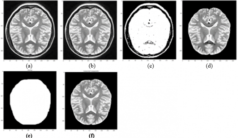
Figure 5. Steps of skull stripping for bright image: (a) original image, (b) applying AGC, (c) binarization, (d) connected component, (e)the created mask and (f) skull stripping
The proposed method is tested using two publicly available datasets: Brain MRI Images for Brain Tumor Detection [24] and 3,060 JPG-formatted brain MRI images from the Br35H::Brain Tumor Detection 2020 dataset [25], which were offered on Kaggle. the first dataset was separated into two folders, yes, no. containing 155 of tumor brain MRI images, 95 no tumor brain MRI images, respectively. The second dataset was separated into three folders, yes, no, and prediction, containing 1,500 of tumor brain MRI images, 1,500 no tumor brain MRI images, and 60 tumor brain MRI images and no tumor brain MRI images, respectively. The proposed algorithm for skull stripping is implemented and evaluated using Python on Google Collaboratory Eq. (9) of the accuracy is considered an evaluation metric for the proposed method [26]. as it was calculated manually by counting the number of successful images and dividing it by the total number of images.
Accuracy $=\frac{\text { no.of } \text { succesful.images }}{\text { total no.of } \text { images }} \ \times 100 \%$ (9)
The algorithm suggested in this research may correctly extract the brain and remove non-brain tissues with 96% accuracy.
We noted from the related work that they merely said that they had good results without mentioning the accuracy of their methodology. While our approach has a 96% accuracy rate.
The proposed method has been compared with Mouli’s skull-stripping method [11] which uses otsu thresholding and morphological operation. The proposed method in this article succeeded in stripping the skull in many images, while the other method failed. Figure 6 illustrates the failure of Mouli's strategy in some images, as it failed to reduce the skull in the images in the case the skull is very close to the brain, the skull is thick and some images have lost parts of it. all these cases may have affected the success of future operations like Tumor segmentation and classification. While our method worked well.
The proposed method has been compared with Lina’s skull-stripping method [21] which uses gamma correction with a gamma value 2.5, otsu thresholding, and morphological operation. The proposed method in this article succeeded in stripping the skull in many images, while the other method failed. Figure 7 illustrates how Lina's strategy, which employs a gamma value of 2.5 for all images, fails in some images. The gamma value for each image must be unique based on the image's attributes. Consequently, some photos have lost portions of them, which affects future processes like tumor segmentation and classification. While our method is successful.
(a) (b) (c)
Figure 6. Column (a) shows the initial images, column, (b) shows the results of Mouli’s skull-stripping method, and column, (c) shows the result of the proposed method in this paper
(a) (b) (c)
Figure 7. Column (a) shows the initial images, column, (b) shows the results of Lina’s skull-stripping method [20], and column, (c) shows the result of the proposed method in this paper
Figure 8. Samples of skull stripping process on no tumor brain images
Figure 9. Samples of the skull stripping process on tumor brain images
As shown in Figures 8 and 9, the suggested method was successful in removing the skull and undesirable tissue from a variety of brain images, including those that were dark or bright, the skull bone was thick or thin, the bone was close to the brain, and the bone was far from the brain. Figure 8 shows some samples from the mentioned above dataset for no tumor brain images, while Figure 9 is for images with tumors.
In this study, a skull stripping method is devised by enhancing the contrast of the brain image using adaptive gamma correction (AGC). The AGC’s parameters are modified dynamically dependent on the properties of the input image. we successfully extract the brain and remove non-brain tissues with 96% accuracy. The proposed method's performance was compared with that of related works. It was discovered that the suggested procedure was successful in removing the skull from numerous photos whereas other ways failed. The proposed method works with 2D images. Future work will focus on stripping the skull from 3D images using machine learning [27, 28], precisely separating the tumor from the brain, and defining the correct size, shape, and stage of the tumor based on the features extracted.
[1] Jaafar, H.I., Mohamed, Z., Abidin, A.F.Z., Ab Ghani, Z. (2012). PSO-tuned PID controller for a nonlinear gantry crane system. In 2012 IEEE International Conference on Control System, Computing and Engineering, Penang, Malaysia, pp. 515-519. https://doi.org/10.1109/ICCSCE.2012.6487200
[2] Ramli, L., Mohamed, Z., Abdullahi, A.M., Jaafar, H.I., Lazim, I.M. (2017). Control strategies for crane systems: A comprehensive review. Mechanical Systems and Signal Processing, 95: 1-23. https://doi.org/10.1016/j.ymssp.2017.03.015
[3] Lee, H.H., Cho, S.K. (2001). A new fuzzy-logic anti-swing control for industrial three-dimensional overhead cranes. Proceedings 2001 ICRA. IEEE International Conference on Robotics and Automation (Cat. No.01CH37164), Korea (South). https://doi.org/10.1109/ROBOT.2001.933070
[4] Chang, C.Y., Chiang, K.H., Hsu, S.W. (2005). Fuzzy controller for the 3-D overhead crane system. In 2005 IEEE International Conference on Robotics and Biomimetics-ROBIO, pp. 724-729. https://doi.org/10.1109/ROBIO.2005.246358
[5] Önen, Ü., Çakan, A. (2017). Anti-swing control of an overhead crane by using genetic algorithm based LQR. International Journal of Engineering and Computer Science, 6(6). https://doi.org/10.18535/ijecs/v6i6.12
[6] Zhou, Q., Wang, K., Xiong, X., Zhao, J. (2021). Optimization of bridge crane control system using fuzzy PID control and speed control of frequency converter. Journal of Physics: Conference Series, 1802(3): 032007. https://doi.org/10.1088/1742-6596/1802/3/032007
[7] Matsuo, T., Yoshino, R., Suemitsu, H., Nakano, K. (2004). Nominal performance recovery by PID+ Q controller and its application to antisway control of crane lifter with visual feedback. IEEE Transactions on Control Systems Technology, 12(1): 156-166. https://doi.org/10.1109/TCST.2003.821964
[8] Abdullah, M., Amin, A.A., Iqbal, S., Mahmood-ul-Hasan, K. (2021). Swing up and stabilization control of rotary inverted pendulum based on energy balance, fuzzy logic, and LQR controllers. Measurement and Control, 54(9-10): 1356-1370. https://doi.org/10.1177/00202940211035406
[9] Karaman, S., Frazzoli, E. (2011). Sampling-based algorithms for optimal motion planning. The International Journal of Robotics Research, 30(7): 846-894. https://doi.org/10.1177/0278364911406761
[10] Park, M.S., Chwa, D., Hong, S.K. (2008). Antisway tracking control of overhead cranes with system uncertainty and actuator nonlinearity using an adaptive fuzzy sliding-mode control. IEEE Transactions on Industrial Electronics, 55(11): 3972-3984. https://doi.org/10.1109/TIE.2008.2004385
[11] Solihin, M.I., Chuan, C.Y., Astuti, W. (2020). Optimization of fuzzy logic controller parameters using modern meta-heuristic algorithm for gantry crane system (GCS). Materials Today: Proceedings, 29: 168-172. https://doi.org/10.1016/j.matpr.2020.05.641
[12] Esleman, E.A., Önal, G., Kalyoncu, M. (2021). Optimal PID and fuzzy logic based position controller design of an overhead crane using the Bees Algorithm. SN Applied Sciences, 3(10): 1-13.
[13] Jaafar, H.I., Latif, N.A., Kassim, A.M., Abidin, A.F.Z., Hussien, S.Y.S., Aras, M.S.M. (2015). Motion control of nonlinear gantry crane system via priority-based fitness scheme in firefly algorithm. In AIP Conference Proceedings, 1660(1): 070031. https://doi.org/10.1063/1.4915749
[14] Suvorov, V.A., Bahrami, M.R., Akchurin, E.E., Chukalkin, I.A., Ermakov, S.A., Kan, S.A. (2021). Anti sway tuned control of gantry cranes. SN Applied Sciences, 3(8): 1-10. https://doi.org/10.1007/s42452-021-04719-w
[15] Solihin, M.I., Kamal, M.A.S., Legowo, A. (2008). Optimal PID controller tuning of automatic gantry crane using PSO algorithm. In 2008 5th International Symposium on Mechatronics and Its Applications, Amman, Jordan, pp. 1-5. https://doi.org/10.1109/ISMA.2008.4648804
[16] Ranjbari, L., Shirdel, A.H., Aslahi-Shahri, M., Anbari, S., Ebrahimi, A., Darvishi, M., Alizadeh, M., Rahmani, R., Seyedmahmoudian, M. (2015). Designing precision fuzzy controller for load swing of an overhead crane. Neural Computing and Applications, 26(7): 1555-1560. https://doi.org/10.1007/s00521-015-1825-z
[17] Jaafar, H.I., Mohamed, Z., Jamian, J.J., Abidin, A.F.Z., Kassim, A.M., Ab Ghani, Z. (2013). Dynamic behaviour of a nonlinear gantry crane system. Procedia Technology, 11: 419-425. https://doi.org/10.1016/j.protcy.2013.12.211
[18] Tuba, M., Bacanin, N. (2014). JPEG quantization tables selection by the firefly algorithm. In 2014 International Conference on Multimedia Computing and Systems (ICMCS), Marrakech, Morocco, pp. 153-158. https://doi.org/10.1109/ICMCS.2014.6911315
[19] Zubair, A.F., Mansor, M.S.A. (2019). Embedding firefly algorithm in optimization of CAPP turning machining parameters for cutting tool selections. Computers & Industrial Engineering, 135: 317-325. https://doi.org/10.1016/j.cie.2019.06.006
[20] Anshory, I., Hadidjaja, D., Jakaria, R.B. (2020). Bldc motor: Modeling and optimization speed control using firefly algorithm. Dinamik, 25(2): 51-58.
[21] Kayalvizhi, E., Karthikeyan, A., Arunarasi, J. (2015). An optimal energy management system for electric vehicles using firefly optimization algorithm based dynamic EDF scheduling. International Journal of Engineering and Technology, 7(4).
[22] Nedic, N., Stojanovic, V., Djordjevic, V. (2015). Optimal control of hydraulically driven parallel robot platform based on firefly algorithm. Nonlinear Dynamics, 82(3): 1457-1473. https://doi.org/10.1007/s11071-015-2252-5
[23] Carbas, S. (2020). Enhanced firefly algorithm for optimum steel construction design. In Applications of Firefly Algorithm and Its Variants, Springer, Singapore, 119-146. https://doi.org/10.1007/978-981-15-0306-1_6
[24] Yang, X.S. (2010). Nature-Inspired Metaheuristic Algorithms. Luniver Press.
[25] Yang, X.S. (200). Firefly Algorithms for Multimodal Optimization. In: Watanabe, O., Zeugmann, T. (eds) Stochastic Algorithms: Foundations and Applications. SAGA 2009. Lecture Notes in Computer Science, vol 5792. Springer, Berlin, Heidelberg. https://doi.org/10.1007/978-3-642-04944-6_14
[26] Okubanjo, A., Oyetola, O., Adekomaya, O. (2018). Vision based control of gantry crane system. Eskişehir Technical University Journal of Science and Technology A-Applied Sciences and Engineering, 19(4): 1023-1032. https://doi.org/10.18038/aubtda.420980
[27] Deniz, F.N. (2022). An approach to preserve optimality in implementation of fractional order PID controllers considering approximation methods. In 2022 International Symposium on Multidisciplinary Studies and Innovative Technologies (ISMSIT), Ankara, Turkey, pp. 402-407. https://doi.org/10.1109/ISMSIT56059.2022.9932718
[28] Azmi, N.I.M., Yahya, N.M., Fu, H.J., Yusoff, W.A.W. (2019). Optimization of the PID-PD parameters of the overhead crane control system by using PSO algorithm. In MATEC Web of Conferences, 255: 04001. https://doi.org/10.1051/matecconf/201925504001