Diana Basim Abdulhameed Al-Qaysi* | Nooralden Abdulkarem Jasim Al-Tulaibawi
© 2022 IIETA. This article is published by IIETA and is licensed under the CC BY 4.0 license (http://creativecommons.org/licenses/by/4.0/).
OPEN ACCESS
This study's objective is to evaluate the antibacterial activity of ethanol extracts of Salvia officinalis L., which have been employed as traditional medicines by local healers, against two multidrug-resistant bacteria, Escherichia coli and Staphylococcus aureus. In the present investigation, fifty samples were collected from burn patients, and isolates were identified from smears taken from the burn department in hospitals, including the floors, walls, light sources, and beds at the Alsaader Hospital in Missan City, using morphological, cultural, and the VITEK 2 Compact device. Besides, the antibiotic sensitivity test for Staphylococcus aureus and Escherichia coli is tested against seven antibiotics. The results show that this isolate showed resistance to most antibiotics used in the experiment, and therefore it is regarded as MDR. Agar well diffusion methods are employed to detect the antibacterial susceptibility test versus Staphylococcus aureus and Escherichia coli at four concentrations: 62.5, 125, 250, and 500 mg/ml. The results show that the alcoholic extract had high inhibitory activity against Staphylococcus aureus and E. coli at all concentrations compared to all seven tested antibiotics. The results also reveal the inhibition zone diameter of the extract against the growth of bacteria increased significantly with concentration increase. At 500 mg/ml, the highest inhibitory zone was 41.66 mm, while at 62.5 mg/ml, it was 30.66 mm.
antibacterial activity, Salvia officinalis, compact device, pathogenic bacteria, medicinal plants
The situation is exacerbated by the advent of drug resistance to several antibiotics. According to current findings, Staphylococcus aureus (S. aureus) and Escherichia coli (E. coli) are not only multidrug-resistant pathogens [1, 2], but also pan drug-resistant and broad-spectrum drug-resistant bacteria [3].
It has been discovered that they are becoming gradually resistant to an increasing number of antimicrobial medicines, according to Lowy [1]. Multidrug resistance in E. coli climbed from 7.2% in the 1950s to 63.6% in the 2000s, with ampicillin, sulfamate, and tetracycline resistance rising at a particularly rapid rate [4]. Bereket et al. [5] reported that S. aureus and E. coli are also responsible for the majority of nosocomial infections. The problem is further worsened, according to the study, by the growth of difficult-to-treat multidrug-resistant microorganisms in the hospital environment. On the other hand, the discovery of novel antimicrobial medications is of the utmost importance in view of evidence of the rapid global spread of resistant clinical isolates. Despite this, the history of quick and widespread resistance to newly released antimicrobial medications shows that even new antimicrobial drug families will have a short lifespan [6, 7].
Medicinal plants would be a great source for a variety of medications [8]. Phytochemicals produced in the plant's secondary metabolism have been found to have antibacterial characteristics, which have been used to treat a variety of bacteria [9, 10]. Secondary metabolites found in plants exhibit antibacterial activities when tested in vitro [11, 12].
Pavi et al. [13] investigated the use of supercritical CO2 extraction to obtain extracts of sage rich in these compounds. The yield of carnosic acid and carnosol was examined in relation to pressure, temperature, and CO2 flow rate. Sulaiman et al. [14] reported that the purchased oil lacked antimicrobial activity against any of the tested pathogens, whereas the extracted oil of S. officinalis inhibited the growth of all tested microorganisms with the exception of MDR K. pneumonia. Joshi et al. [15] demonstrated that the bacterial inhibitory activity of essential oil and three different extracts was evaluated using the agar well disc diffusion method to determine its antibiotic potential. In addition, the essential oil of S. officinalis L. leaves demonstrated potent antimicrobial activity against gram-positive Staphylococcus aureus, gram-negative Klebsiella pueunmoniae, and E. coli bacteria. Yaseen et al. [16] investigated the antibacterial activity of green synthetic nanoparticles of Salvia officinalis aqueous leaf extract loaded with silver nitrate. Their findings demonstrated that different concentrations of green synthetic nanoparticles are capable of inhibiting all bacterial isolates with zones of inhibition greater than those observed with ready-to-use silver nanoparticles. Bouteldja et al. [17] investigated the valorization of Salvia officinalis L. (Lamiaceae), a medicinal plant known for its traditional use, by phytochemical characterization and evaluation of the antioxidant and antibacterial activity of their extracts. They reported that the antibacterial activity of S. officinalis methanolic extract demonstrated a strong ability to inhibit B. subtilis, M. luteus, E. coli, and S. aureus.
The importance of the research lies in knowing how effective ethanol extracts of Salvia officinalis L. as traditional treatments. Therefore, the purpose of this study is to see how effective ethanol extracts of Salvia officinalis L. are against two multidrug-resistant bacteria, Escherichia coli and Staphylococcus aureus, which have been employed as traditional treatments by local healers. Fifty samples were collected from burn patients, and isolates were identified using morphological, cultural, and the VITEK 2 Compact devices from smears taken from the burn department in hospitals in Missan City.
2.1 Collection of bacterial isolates
Pure cultures of Staphylococcus aureus and E. coli were used. The cultures of pathogenic bacteria were obtained from patients who attended Al-Sadder hospital in Missan City. The bacteria were activated and subcultured three successive times on nutrient agar.
2.2 Identification of isolates
Bacterial isolates were diagnosed according to cultural and morphological Characteristics and by using VITEK 2 Compact.
Below some test details and indicators:
1. Microscopic & cultural characteristics
It is including:
(a) Microscopic properties: Gram's stain was used to examine the isolated bacteria for studying the microscopic properties.
(b) Cultural characteristics: Morphological colonies characteristics were recorded on the media that are used (MacConkey agar, blood agar and Mannitol salt agar) for primary identification of isolates.
2. Biochemical tests
It is including:
(i) Oxidase Test: The culture across oxidase disk was smeared by a sterile wooden applicator stick. In a positive reaction, the bacterial growth becomes dark purple immediately while oxidase-negative organisms will remain colorless or will turn purple after 10 seconds.
(ii) Catalase Test (Hydrogen Peroxide 3%): An amount of growth was suspended in a drop of hydrogen peroxide 3% on slide and observed the evolution of bubble.
(iii) Indole Production Test: Peptone water was inoculated with a young bacterial colony at 37℃ for 24-48 hr. A few drops of Kovacs reagent were added to each tube. Formation of pink ring indicates a positive test.
(iv) Methyl Red Test: MR-VP broth media was inoculated with a young bacterial isolate incubated at 37℃ for 24 hours. Five drops of methyl red solution were added, mixed, and the result is read immediately. A positive result incubated when a bright red was appeared.
(v) Vogues-Proskauer Test: MR-VP (medium was prepared by dissolving 1g glucose, 1g Peptone and 1g K2 HPO4, in 100 ml of D.W and pH was adjusted to 7 and sterilized by autoclave at 121℃ For 15 min, and is used to detect the complete and partial hydrolysis of glucose. Broth was inoculated with a young bacterial isolate and incubated at 37℃ for 24- 48 hours. One ml of 40% KOH solution and 3 ml of 5% solution of α -naphthol were added to each tube. A positive reaction was indicated by appearance of a pink color in 15-20 minutes.
(vi) Simmon's Citrate Test: Simmon’s citrate slant was inoculated with a young colony and incubated at 37℃ for 24-48 hr. The appearance of blue color of growth indicates a positive result.
2.3 Antibiotic discs
Seven antibiotics discs were used for antibiotic sensitivity test, as shown in Table 1.
2.4 Plants collection and authentication
The plant Salvia officinalis in Figure 1 has been chosen based on its use in folk medicine. In particular, we selected plants that have been used for the treatment of purulent wounds, burns, infections, and inflammations. Leaves, flowers, and whole plants were harvested in the flowering period and collected from Basrah city, and they were identified by the Center of Marine Sciences.
2.5 Preparation of plants samples
In the current investigation, the plant was washed with tap water, let dry at room temperature, and then ground into a powder using an electric grinder. The results of this investigation are presented below. Until they were needed, the powdered plants were kept in plastic tubes and kept at a temperature of 4 degrees Celsius in a refrigerator [18].
Table 1. Antibiotics discs their Con., Manufacturing Company and diameter of inhibition zones cited by [19]
|
No. |
Antibiotics |
Con. ml/disc |
Symbol |
Manufacturing company |
R |
Intermediate |
S |
|
1. |
Amikacin |
10 |
(AK) |
Bioanalyse |
≤ 12 |
13-16 |
≥ 17 |
|
2. |
Ampicillin |
10 |
(AM) |
Bioanalyse |
≤ 13 |
14-16 |
≥ 17 |
|
3. |
Chloramphenicol |
30 |
(C) |
Bioanalyse |
≤ 12 |
13-17 |
≥ 18 |
|
4. |
Imipenem |
10 |
(IPM) |
Bioanalyse |
≤ 13 |
14-15 |
≥ 16 |
|
5. |
Penicillin |
10 |
P)) |
Bioanalyse |
≤ 11 |
12-21 |
≥ 22 |
|
6. |
Tetracycline |
30 |
(T) |
Bioanalyse |
≤ 11 |
12-14 |
≥ 15 |
|
7. |
Vancomycin |
30 |
(VA) |
Bioanalyse |
≤ 14 |
15-16 |
≥ 17 |
Con. = concentration, S= Sensitive, R= Resistance
Figure 1. Salvia officinalis
2.6 Preparation of plant extract
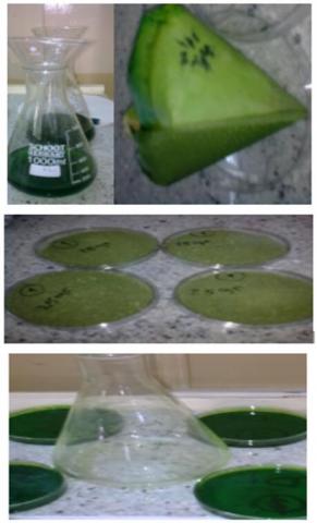
In a flask with a capacity of 500 milliliters, a suspension containing 20 grammes of finely powdered material and 400 milliliters of ethanol with a concentration of 96% was refluxed for 24 hours at room temperature while being stirred by a Magnatic stirrer plate [20, 21]. The extract was then filtered using filter paper type Whattman, No. 1, and dried at 25℃ at room temperature as shown in Figure 2. After that, it is weighed and stored in the refrigerator until it is utilized.
Figure 2. Flow chart of sample preparation process
2.7 Concentration of plants extracts
The stock solution for the extract was generated by dissolving 500 mg of dried extract in 1 ml of ethanol for alcohol extract, resulting in a final concentration of 500 mg/ml; from this stock solution, additional concentrations of 62.5, 125, and 250 mg/ml were prepared and employed against bacteria [22, 23].
2.8 Antibiotic sensitivity test
This part includes some steps. With the assistance of a sterile wire loop, the tips of around four to five individual colonies of the organism that was going to be researched were removed from the initial culture and placed in a test tube that contained ten milliliters of Mueller-Hinton medium [24].
A slightly turbid bacterial suspension is formed after a 2- to 5-hour incubation at 37 degrees Celsius. The turbidity was compared to a McFarland No. 0.5 tube. A sterile cotton swab was dipped into the standard bacterial suspension after adjusting the density of the inoculums for 15 minutes. The excess fluid was eliminated by vigorously rotating the brush inside the tube above the fluid level [19]. To achieve uniform distribution of the inoculums, the swab was streaked in two separate planes onto the dry surface of a Mueller-Hinton plate. The plate lids were replaced, and the inoculation plates were set aside for 3 to 5 minutes on a flat, level surface to absorb any excess moisture. After selecting the discs, they were placed on the inoculation plate, and then using sterile forceps, they were gently pressed into the agar. After 15 minutes, the inoculation plates were inverted and incubated at 37 degrees Celsius for 18 to 24 hours. After the incubation period was complete, the diameters of the entire inhibition zone were determined by utilizing reflected light and a ruler. The end point was the location when no visible growth was seen, measured to the nearest mm. Table 1 shows how the diameter of the inhibition zone for each antimicrobial drug was turned into sensitive and resistant categories using an interpretation chart [25].
2.9 Antibacterial activity measurement
By replacing antibiotic discs with holes of 8 mm diameter drilled with a corky bore and carefully filling with 100 l from samples under consideration of extracts using a micropipette, the antibacterial activity of extracts was tested employing the agar well diffusion method. To serve as a control, ethanol was introduced to one hole in the cultured media, and the Petri dishes were incubated at 37℃ for 24 hours. The inhibition zone was determined using a ruler, and the procedure was done three times [26].
3.1 Antibiotic susceptibility of staphylococcus aureus and Escherichia coli
The disc diffusion method is employed to assess susceptibility of staphylococcus aureus and Escherichia coli to seven different antibiotics [18].
According to the results of the bioassays described in Table 2, staphylococcus aureus has exhibited resistance to commonly used antibiotics such as ampicillin, penicillin, penicillin G, imipenem, and tetracyclin, whereas Vancomycin explained the width of the inhibitory zone against pathogenic bacteria. Sosa et al. [27] discovered that bacterial resistance to -lactamase was usually caused by the -lactamase enzyme hydrolyzing the antibiotic or altering cell wall permeability, and that this resistance could also be caused by the antimicrobial minimizing its interaction with the target site (Penicilline Binding Protien), which represented surface proteins responsible for cell wall synthesis.
Table 2. Antibiotic sensitivity test of S. aureus and E. coli
|
No. |
Antibiotics |
symbol |
Inhibition zone(mm) |
Inhibition zone(mm) |
|
1. |
Amikacin |
(AK) |
Of s.aureus |
Of E. coli |
|
2. |
Ampicillin |
(AM) |
16 |
10 |
|
3. |
Chloramphenicol |
(C) |
- |
15 |
|
4. |
Imipenem |
(IPM) |
18 |
25 |
|
5. |
Penicillin |
(P) |
- |
22 |
|
6. |
Tetracycline |
(T) |
- |
12 |
|
7. |
Vancomycin |
(VA) |
10 |
- |
Chloramphenicol was effective against S. aureus, although Vancomycin was more effective. Finally, S. aureus displayed intermediate resistance to Amikacin, owing to the development of changed enzymes and the loss of the outer membrane hole [28]. Amikacin, Penicillin, Tetracycline, and Vancomycin resistance was found in E. coli, although it was susceptible to Chloramphenicol and Imipenem medicines.
This study revealed the emergence of antibiotic resistance; resistance to some antibiotics may arise as a result of the extensive and often indiscriminate use of commercial antimicrobial medications frequently used to treat infectious illnesses [29].
Inactivation or modification of the antimicrobial agent by enzymatic means, bypassing of pathways, decreased absorption (lower intracellular concentration of antimicrobial agent by membrane permeability reduction or active efflux pump), alteration or overproduction of the target enzyme [30], or the presence of plasmid conferring resistance [31] can all cause bacterial resistance to various drugs.
3.2 Phytochemical screening of Salvia officinalis L.
The results of phytochemical screening of an alcoholic extract of Salvia officinalis were shown in Table 3.
The extracts of Salvia officinalis L. give positive test for glaycosides, aponines, flavonoids, resins, alkaloids, and phenols as the other studies find [32-34] While give negative test for tannins and coumarin. Flavonoides compounds are high antioxidant activity, phenols are antimicrobial activities, antibacterial, fungistatic and virustatic [35].
Table 3. Phytochemical screening of Salvia officinalis L. extract
|
Active compounds |
Result |
|
Tannins |
+ |
|
Glaycosides |
+ |
|
Saponines |
- |
|
Flavonoides |
+ |
|
Resins |
+ |
|
Coumarin |
_ |
|
Alkaloids |
- |
|
Phenols |
+ |
3.3 Antibacterial activity of Salvia officinalis versus pathogenic bacteria (S. aureus and E. coli)
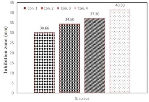
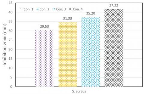
The alcoholic extract of Salvia officinalis listed in Tables 4 and 5 was tested under four concentrations against two multidrug resistant bacteria, S. aureus and E. coli. The findings reveal a broad range of antibacterial activity against the pathogens studied.
As mentioned early, the antibacterial efficacy of Salvia officinalis extract was determined via the agar well diffusion method in current investigation.
The susceptibility pattern to the alcoholic extract on pathogenic bacteria (S. aureus and E. coli) of Salvia officinalis expressed maximum inhibitory zone at 500 mg/ml, which was (40.50 mm) of S. aureus and inhibition zone of E. coli was (37.33 mm), but low 62.5 mg/ml was (30.66 mm) of S. aureus and inhibition zone of E. coli was (37.33 mm (29.50 mm) as shown in Figures 3 and 4. Most studies looking into the antibacterial effects of essential oils verified that they are more potent versus gram-positive bacteria than gram-negative bacteria [36-39]. The findings were consistent with those of [40, 41], who found that an alcoholic extract of Salvia officinalis had potent antibacterial action against both gramme positive and gramme negative microorganisms. This research validated our findings.
Several studies on the antibacterial capabilities of the Salvia genus reveal a wide range of results, depending on the microorganisms' susceptibility and the efficacy of the studied chemicals. Essential oil-rich Salvia species (such as S. officinalis) with volatile monoterpenoid as a significant ingredient have been shown to be antibacterial [41].
Gram-positive bacteria are more sensitive to S. officinalis essential oil than other bacteria, according to Kamatou et al. [42]. Other than the structure of their cellular walls, gram-positive and gram-negative microorganisms are distinct from one another in a number of other ways. The primary distinction lies in the presence of lipoproteins and lipopolysaccharides in gram-negative bacteria, which act as a barrier to hydrophobic compounds. Gram-positive microorganisms do not have these components.
Table 4. The inhibition zone of alcoholic extract on the growth of S. aureus
|
No. |
Con. mg/ml |
Inhibition zone (mm) |
|
1. |
62.5 |
30.66 |
|
2. |
125 |
34.50 |
|
3. |
250 |
37.20 |
|
4. |
500 |
40.50 |
|
5. |
Control |
_ |
Figure 3. Influence of alcoholic extract concentration on inhibition zone of S.aureus
Con. 1= 62.5, Con. 2= 125, Con. 3= 250, Con. 4= 500 mg/ml
Table 5. The inhibition zone of alcoholic extract on the growth of E. coli
|
No. |
Con. mg/ml |
Inhibition zone (mm) |
|
1. |
62.5 |
29.50 |
|
2. |
125 |
31.33 |
|
3. |
250 |
35.20 |
|
4. |
500 |
37.33 |
|
5. |
Control |
_ |
Figure 4. Effect of alcoholic extract concentration on inhibition zone of E. coli
Con. 1= 62.5, Con. 2= 125, Con. 3= 250, Con. 4= 500 mg/ml
The purpose of this investigation is to study the antibacterial activity of ethanol extracts of Salvia officinalis L., which have been employed by local healers as traditional remedies, against two multidrug-resistant bacteria, S. aureus and E. coli. Antibiotic resistance was found in this isolate, and as a result, it has been labelled MDR according to the research. In comparison to all seven antibiotics tested, the alcoholic extract demonstrated the highest inhibitory action against S. aureus and E. coli at all doses. This extract's inhibitory zone diameter grew dramatically with increasing concentration, according to the findings. It was 41.66 mm at 500 mg/ml and 30.66 mm when the concentration was 62.5 mg/ml. Therefore, the findings of this study back up local healers' usage of Salvia officinalis in their traditional practises. Besides, the antibacterial activity of alcoholic extracts was stronger against G + ve and G - ve, whereas G + ve was the most sensitive to G - ve. More research is needed to determine the efficacy of this medicinal plant against additional microorganisms in various agro-ecological environments, as well as their safety levels and phytochemical compositions. Additional research is needed to investigate the other characteristics of antimicrobial efficacy, such as in vivo efficacy.
[1] Lowy, F.D. (2003). Antimicrobial resistance: the example of Staphylococcus aureus. The Journal of Clinical Investigation, 111(9): 1265-1273. https://doi.org/10.1172/jci18535
[2] Strateva, T., Yordanov, D. (2009). Pseudomonas aeruginosa–a phenomenon of bacterial resistance. Journal of Medical Microbiology, 58(9): 1133-1148. http://dx.doi.org/10.1099/jmm.0.009142-0
[3] Magiorakos, A.P., Srinivasan, A., Carey, R.B., Carmeli, Y., Falagas, M.E., Giske, C.G., Monnet, D.L. (2012). Multidrug-resistant, extensively drug-resistant and pandrug-resistant bacteria: an international expert proposal for interim standard definitions for acquired resistance. Clinical Microbiology and Infection, 18(3): 268-281. https://doi.org/10.1111/j.1469-0691.2011.03570.x
[4] Tadesse, D.A., Zhao, S., Tong, E., Ayers, S., Singh, A., Bartholomew, M.J., McDermott, P.F. (2012). Antimicrobial drug resistance in Escherichia coli from humans and food animals, United States, 1950–2002. Emerging Infectious Diseases, 18(5): 741. http://dx.doi.org/10.3201/eid1805.111153
[5] Bereket, W., Hemalatha, K., Getenet, B., Wondwossen, T., Solomon, A., Zeynudin, A., Kannan, S. (2012). Update on bacterial nosocomial infections. Eur Rev Med Pharmacol Sci, 16(8): 1039-44.
[6] Medina, E., Pieper, D.H. (2016). Tackling threats and future problems of multidrug-resistant bacteria. How to Overcome the Antibiotic Crisis, pp. 3-33. https://doi.org/10.1007/82_2016_492
[7] Bavaro, D.F., Belati, A., Diella, L., Stufano, M., Romanelli, F., Scalone, L., Saracino, A. (2021). Cefiderocol-based combination therapy for “difficult-to-treat” Gram-negative severe infections: Real-life case series and future perspectives. Antibiotics, 10(6): 652. https://doi.org/10.3390/antibiotics10060652
[8] Manandhar, S., Luitel, S., Dahal, R.K. (2019). In vitro antimicrobial activity of some medicinal plants against human pathogenic bacteria. Journal of Tropical Medicine. https://doi.org/10.1155%2F2019%2F1895340
[9] Kowalczyk, T., Wieczfinska, J., Skała, E., Śliwiński, T., Sitarek, P. (2020). Transgenesis as a tool for the efficient production of selected secondary metabolites from plant in vitro cultures. Plants, 9(2): 132. http://dx.doi.org/10.3390/plants9020132
[10] Kamaruzaman, N.H., Noor, N.N.M., Mohamed, R.M.S.R., Al-Gheethi, A., Ponnusamy, S.K., Sharma, A., Vo, D.V.N. (2022). Applicability of bio-synthesized nanoparticles in fungal secondary metabolites products and plant extracts for eliminating antibiotic-resistant bacteria risks in non-clinical environments. Environmental Research, 209: 112831. https://doi.org/10.1016/j.envres.2022.112831
[11] Leylaie, S., Zafari, D. (2018). Antiproliferative and antimicrobial activities of secondary metabolites and phylogenetic study of endophytic Trichoderma species from Vinca plants. Frontiers in Microbiology, 9: 1484. https://doi.org/10.3389/fmicb.2018.01484
[12] Djeussi, D.E., Noumedem, J.A., Seukep, J.A., Fankam, A.G., Voukeng, I.K., Tankeo, S.B., Kuete, V. (2013). Antibacterial activities of selected edible plants extracts against multidrug-resistant Gram-negative bacteria. BMC Complementary and Alternative Medicine, 13(1): 1-8. http://dx.doi.org/10.1186/1472-6882-13-164
[13] Pavić, V., Jakovljević, M., Molnar, M., Jokić, S. (2019). Extraction of carnosic acid and carnosol from sage (Salvia officinalis L.) leaves by supercritical fluid extraction and their antioxidant and antibacterial activity. Plants, 8(1): 16. http://dx.doi.org/10.3390/plants8010016
[14] Sulaiman, A.M., Abdulaziz, A.S., Almutawea, A.M., Alansari, S.A., Aldoseri, F.M., Bekhit, S.A., Bekhit, A.A. (2022). Evaluation of the antibacterial effect of salvia officinalis essential oil and its synergistic effect with meropenem. Letters in Applied NanoBioScience, https://doi.org/10.33263/lianbs122.044
[15] Joshi, S., Pandey, R.D., Bhattarai, R., Gharti, B.B. (2021). Antimicrobial activity of essential oil and crude organic extracts of Salvia officinalis L. leaves from Nepal. International Journal of Innovative Science and Research Technology, 6(2). https://doi.org/10.1186/s40529-015-0096-4
[16] Yaseen, S.M., Hussein, A.A., Al-Ezzy, R.M. (2019). Antibacterial activity of silver nanoparticles using Salvia officinalis extract on some pathogenic bacteria. Journal of Pharmacy and Pharmacology, 7: 237-248. https://doi.org/10.17265/2328-2150%2F2019.05.003
[17] Bouteldja, R., Doucene, R., Aggad, H., Abdi, F.Z., Belkhodja, H., Abdali, M., Abaid, S. (2021). Phytochemical characterization, antioxidant and antibacterial activity of Salvia officinalis (L.) extracts from the Tiaret region. European Journal of Biological Research, 11(3): 356-366. http://dx.doi.org/10.5281/zenodo.5129324
[18] CLSI. (2011). Performance Standards for antimicrobial susceptibility testing; twenty-first informational supplement. M100-S21, Clinical and Laboratory Standards Institute, 31(1):172.
[19] Harborne, J.B. (1984). Methods of plant analysis. In Phytochemical methods. Springer, Dordrecht, pp. 1-36.
[20] Ahmad, I., Mehmood, Z., Mohammad, F. (1998). Screening of some Indian medicinal plants for their antimicrobial properties. Journal of ethnopharmacology, 62(2): 183-193. https://doi.org/10.1016/S0378-8741(98)00055-5
[21] Al-Jboriy, K.A., Al-Anasary, B.S., Ali, H.S.A. (2010). Study of the sensitivity of some pathogenic isolated from respiratory infection in human against same plant extracts. Al-Anbar Journal of Veterinary Sciences, 3(2).
[22] Nwachukwu, E., Uzoeto, H.J.O. (2010). Antimicrobial activities of leaf of Vitex doniana and Cajanus cajan on some bacteria. Researcher, 2(3): 37-47.
[23] Tripathi, Y.B., Tiwari, O.P., Nagwani, S., Mishra, B. (2009). Pharmacokinetic-interaction of Vitex negundo Linn. & paracetamol. Indian Journal of Medical Research, 130(4): 479.
[24] Pollack, R.A., Findlay, L., Mondschein, W., Modesto, R.R. (2018). Laboratory Exercises in Microbiology. John Wiley & Sons.
[25] Atlas, R.M., Brown, A.E., Parks, L.C. (1995). Laboratory Manual of Experimental Microbiology, Mosby. Mosby-Year Book, Inc. (January 1, 1900).
[26] Egharevba, H.O., Kunle, O.F., Iliya, I., Orji, P.N., Abdullahi, M.S., Okwute, S.K., Okogun, J.I. (2010). Phytochemical analysis and antimicrobial activity of Punica granatum L. (fruit bark and leaves). New York Science Journal, 3(3): 91-98.
[27] Sosa, A.D.J., Byarugaba, D.K., Amábile-Cuevas, C.F., Hsueh, P.R., Kariuki, S., Okeke, I.N. (Eds.). (2010). Antimicrobial Resistance in Developing Countries. New York: Springer, pp. 3-7.
[28] Muhammad, I., Uzma, M., Yasmin, B., Mehmood, Q., Habib, B. (2011). Prevalence of antimicrobial resistance and integrons in Escherichia coli from Punjab, Pakistan. Brazilian Journal of Microbiology, 42(2): 462-466. https://doi.org/10.1590/s1517-83822011000200008
[29] Evans, W. (2009). Trease and Evans' Pharmacognosy. Elsevier Health Sciences.
[30] Adomi, P.O. (2006). Antibacterial activity of aqueous and ethanol extracts of the stem bark of Alstonia boonei and Morinda lucida. Scientific Research and Essays, 1(2): 050-053. https://doi.org/10.5897/SRE.9000188
[31] Coates, A., Hu, Y., Bax, R., Page, C. (2002). The future challenges facing the development of new antimicrobial drugs. Nature Reviews Drug Discovery, 1(11): 895-910. https://doi.org/10.1038/nrd940
[32] Neoji, U., Saumya, R., Mishra, P.K., Raju, K.C. (2008). Lipid content and in vitro antimicrobial activity of oil seeds of some indian medical plants. Curr. Res. Bacteriol, 1(1): 1-6. https://dx.doi.org/10.3923/crb.2008.1.6
[33] Avato, P., Fortunato, I.M., Ruta, C., D’Elia, R. (2005). Glandular hairs and essential oils in micropropagated plants of Salvia officinalis L. Plant Science, 169(1): 29-36. https://doi.org/10.1016/j.plantsci.2005.02.004
[34] Stanojević, D., Čomić, L., Stefanović, O., Solujić-Sukdolak, S. (2010). In vitro synergistic antibacterial activity of Salvia officinalis L. and some preservatives. Archives of Biological Sciences, 62(1): 167-174. http://dx.doi.org/10.2298/ABS1001167S
[35] Reverchon, E., Taddeo, R., Porta, G.D. (1995). Extraction of sage oil by supercritical CO2: Influence of some process parameters. The Journal of Supercritical Fluids, 8(4): 302-309. https://doi.org/10.1016/0896-8446(95)90005-5
[36] Sobahi, T.R., Mogib, M.A. (2001). GC/MS Analysis of the Volatile Constituents of Artemesia monosperma. Science, 13(1). http://dx.doi.org/10.4197/Sci.13-1.9
[37] Mohamed, A.H., Sayed, E.M., Hegazy, M. E.F., Helaly, S.E., Esmail, A. M., Salaheldin, N. (2010). Chemical constituents and biological activities of Artemisia herba-alba. Records of Natural Products, 4(1). http://dx.doi.org/10.13140/RG.2.1.2544.8806
[38] Randrianarivelo, R., Sarter, S., Odoux, E., Brat, P., Lebrun, M., Romestand, B., Danthu, P. (2009). Composition and antimicrobial activity of essential oils of Cinnamosma fragrans. Food Chemistry, 114(2): 680-684. https://doi.org/10.1016/j.foodchem.2008.10.007
[39] AM, A.Z., Altalhi, A.D., El-Fattah, R.A. (2008). Fungal control of pathogenic fungi isolated from some wild plants in Taif Governorate, Saudi Arabia. Malaysian Journal of Microbiology, 30-39. http://dx.doi.org/10.21161/mjm.01908
[40] Doudi, M., Yahyaabadi, S., Mosafa, E. (2014). In-vitro antibacterial properties of sage (Salvia officinalis) ethanol extract against multidrug resistant Staphylococcus aureus, Escherichia coli, Pseudomonas aeruginosa and Klebsiella pneumoniae. Zahedan Journal of Research in Medical Sciences, 16(10).
[41] Nadir, M., Rasheed, M., Sherwani, S.K., Kazmi, S., Ahmad, V.U. (2013). Chemical and antimicrobial studies on the essential oil from Salvia santolinifolia Boiss. Pakistan Journal of Pharmaceutical Sciences, 26(1): 39-52.
[42] Kamatou, G.P.P., Viljoen, A.M., Gono-Bwalya, A.B., van Zyl, R.L., Van Vuuren, S.F., Lourens, A.C.U., Steenkamp, P. (2005). The in vitro pharmacological activities and a chemical investigation of three South African Salvia species. Journal of Ethnopharmacology, 102(3): 382-390. https://doi.org/10.1016/j.jep.2005.06.034